Visual Attention: fMRI Investigation of Object-based Attentional Control

0 views·09:59 min
1. Participant recruitment
- Recruit 20 participants.
- Participants should be right-handed and have no history of neurological or psychological disorders.
- Participants should have normal or corrected-to-normal vision to ensure that they will be able to see the visual cues properly.
- Participants should not have metal in their body. This is an important safety requirement due to the high magnetic field involved in fMRI.
- Participants should not suffer from claustrophobia, since the fMRI requires lying in the small space of the scanner bore.
2. Pre-scan procedures
- Fill out pre-scan paperwork.
- When participants come in for their fMRI scan, instruct them to first fill out a metal screen form to make sure they have no counter-indications for MRI, an incidental-findings form giving consent for their scan to be looked at by a radiologist, and a consent form detailing the risks and benefits of the study.
- Prepare participants to go in the scanner by removing all metal from their body, including belts, wallets, phones, hair clips, coins, and all jewelry.
3. Provide instructions for the participant.
- Tell the participant that in the scanner, they will see images of faces and houses.
- For the initial localizer runs, instruct participants that they will passively view faces and houses.
- Explain to the participants that in the task runs, they will see a face and a house superimposed on each other. Their task will be to pay attention to either the house or the face, as follows.
- When the task first begins, they will be told via text instructions whether to pay attention to houses or faces.
- Participants will have four runs where they are instructed to pay attention to houses, and four runs where they are instructed to pay attention to faces.
- The schedule of face and house runs will be random for each subject.
- Stress to the participant the importance of keeping their head still throughout the scan.
4. Put the participant in the scanner.
- Give the participant ear plugs to protect their ears from the noise of the scanner and ear phones to wear so they can hear the experimenter during the scan, and have them lie down on the bed with their head in the coil.
- Give the participant the emergency squeeze ball and instruct them to squeeze it in case of emergency during the scan.
- Use foam pads to secure the participants head in the coil to avoid excess movement during the scan, and remind the participant that it is very important to stay as still as possible during the scan, as even the smallest movements blur the images.
5. Data collection
- Collect a high-resolution anatomical scan.
- Begin functional scanning.
- Synchronize the start of stimulus presentation with the start of the scanner.
- Present pictures via a laptop connected to a projector. The participant has a mirror above their eyes, reflecting a screen at the back of the scanner bore.
- Present each participant with two localizer runs, in which they passively see blocks of faces and houses. These localizer runs are used to identify the FFA and PPA in each individual participant.
- Present each set of faces and houses in a 30-s block, with 20 s of fixation between blocks. Show each stimulus for 750 ms, followed by a 250-ms inter-stimulus interval, in which only a fixation cross is present on the screen. Repeat blocks in alternating order to obtain five blocks of faces and five blocks of houses.
- Present each participant with eight functional runs of the attention task.
- Begin each run with a superimposed face and house (Figure 1) in the center of the screen, and an instruction to begin by attending to either the face or the house. The initial attention target will be random from run to run.
- Each run will contain 300 pairs of superimposed faces and houses. Pairs of faces and houses are repeated between runs, but not within runs.
- Show each superimposed stimulus pair for one second. Every second, replace the superimposed house and face with a new superimposed house and face.
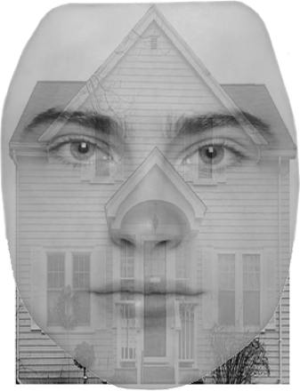
Figure 1. Face stimulus and house stimulus superimposed together. Each stimulus presented was a superimposed face and house. The participant was instructed to focus on either the face or the house.
6. Post-scan procedures
- Bring the participant out of the scanner.
- Debrief the participant.
7. Data analysis
- Preprocess the data.
- Perform motion correction to reduce motion artifacts.
- Perform temporal filtering to remove signal drifts.
- Smooth the data to increase signal-to-noise ratio.
- Model the data for each participant.
- Create a model of what the expected hemodynamic response should be for each task condition (faces and houses) in the localizer scan.
- Fit the data to this model, resulting in a statistical map, where the value at each voxel represents the extent to which that voxel was involved in the task condition.
- Identify the clusters for each subject that correspond to FFA and PPA based on their anatomical locations. The FFA will include all contiguous voxels in the mid-fusiform gyrus (which responds significantly more to faces than to houses) and the PPA included all voxels in the parahippocampal gyrus that responded more significantly to houses than to faces. To establish these regions of interest, a minimum significance threshold of p < 10-6 was used for each voxel.
- Use each individual's FFA and PPA masks as an ROI to extract signal activations during the four focus-on-faces runs and the four focus-on-houses runs.
- Quantify percent signal change for face-focus and house-focus conditions in FFA and PPA for each subject.
- Create a model of what the expected hemodynamic response should be for each task condition (faces and houses) in the localizer scan.
- Perform a two-way analysis of variance (ANOVA) on percent signal change values to test for differences among the conditions. The factors in this test are ROI (PPA vs. FFA) and attention (faces vs. houses).
Source: Laboratories of Jonas T. Kaplan and Sarah I. Gimbel— University of Southern California
The human visual system is incredibly sophisticated and…
Chapters in this video
0:00
Overview
1:23
Experimental Design
3:35
Running the Experiment
5:37
Data Analysis and Results
7:50
Applications
9:28
Summary
Copyright © 2026 MyJoVE Corporation. All rights reserved.