-1::1
Simple Hit Counter
Skip to content

Products

Solutions

×
×
Sign In

EN

EN - EnglishCN - 简体中文DE - DeutschES - EspañolKR - 한국어IT - ItalianoFR - FrançaisPT - Português do BrasilPL - PolskiHE - עִבְרִיתRU - РусскийJA - 日本語TR - TürkçeAR - العربية
Sign In Start Free Trial

RESEARCH

JoVE Journal

Peer reviewed scientific video journal

Behavior
Biochemistry
Bioengineering
Biology
Cancer Research
Chemistry
Developmental Biology
View All
JoVE Encyclopedia of Experiments

Video encyclopedia of advanced research methods

Biological Techniques
Biology
Cancer Research
Immunology
Neuroscience
Microbiology
JoVE Visualize

Visualizing science through experiment videos

EDUCATION

JoVE Core

Video textbooks for undergraduate courses

Analytical Chemistry
Anatomy and Physiology
Biology
Cell Biology
Chemistry
Civil Engineering
Electrical Engineering
View All
JoVE Science Education

Visual demonstrations of key scientific experiments

Advanced Biology
Basic Biology
Chemistry
View All
JoVE Lab Manual

Videos of experiments for undergraduate lab courses

Biology
Chemistry

BUSINESS

JoVE Business

Video textbooks for business education

Accounting
Finance
Macroeconomics
Marketing
Microeconomics

OTHERS

JoVE Quiz

Interactive video based quizzes for formative assessments

Authors

Teaching Faculty

Librarians

K12 Schools

Products

RESEARCH

JoVE Journal

Peer reviewed scientific video journal

JoVE Encyclopedia of Experiments

Video encyclopedia of advanced research methods

JoVE Visualize

Visualizing science through experiment videos

EDUCATION

JoVE Core

Video textbooks for undergraduates

JoVE Science Education

Visual demonstrations of key scientific experiments

JoVE Lab Manual

Videos of experiments for undergraduate lab courses

BUSINESS

JoVE Business

Video textbooks for business education

OTHERS

JoVE Quiz

Interactive video based quizzes for formative assessments

Solutions

Authors
Teaching Faculty
Librarians
K12 Schools

Language

English

EN

English

CN

简体中文

DE

Deutsch

ES

Español

KR

한국어

IT

Italiano

FR

Français

PT

Português do Brasil

PL

Polski

HE

עִבְרִית

RU

Русский

JA

日本語

TR

Türkçe

AR

العربية

    Menu

    JoVE Journal

    Behavior

    Biochemistry

    Bioengineering

    Biology

    Cancer Research

    Chemistry

    Developmental Biology

    Engineering

    Environment

    Genetics

    Immunology and Infection

    Medicine

    Neuroscience

    Menu

    JoVE Encyclopedia of Experiments

    Biological Techniques

    Biology

    Cancer Research

    Immunology

    Neuroscience

    Microbiology

    Menu

    JoVE Core

    Analytical Chemistry

    Anatomy and Physiology

    Biology

    Cell Biology

    Chemistry

    Civil Engineering

    Electrical Engineering

    Introduction to Psychology

    Mechanical Engineering

    Medical-Surgical Nursing

    View All

    Menu

    JoVE Science Education

    Advanced Biology

    Basic Biology

    Chemistry

    Clinical Skills

    Engineering

    Environmental Sciences

    Physics

    Psychology

    View All

    Menu

    JoVE Lab Manual

    Biology

    Chemistry

    Menu

    JoVE Business

    Accounting

    Finance

    Macroeconomics

    Marketing

    Microeconomics

Start Free Trial
Loading...
Home
JoVE Journal
Genetics
Ultralow Input Genome Sequencing Library Preparation from a Single Tardigrade Specimen
JoVE Journal
Genetics
Author Produced
A subscription to JoVE is required to view this content.  Sign in or start your free trial.
JoVE Journal Genetics
Ultralow Input Genome Sequencing Library Preparation from a Single Tardigrade Specimen

Ultralow Input Genome Sequencing Library Preparation from a Single Tardigrade Specimen

Full Text
9,783 Views
10:28 min
July 15, 2018

DOI: 10.3791/57615-v

Yuki Yoshida1,2, Sayuri Konno1,3, Ryousuke Nishino1,3, Yumi Murai1,3, Masaru Tomita1,2,3, Kazuharu Arakawa1,2,3

1Institute for Advanced Biosciences,Keio University, 2Systems Biology Program, Graduate School of Media and Governance,Keio University, 3Faculty of Environment and Information Studies,Keio University

Summary

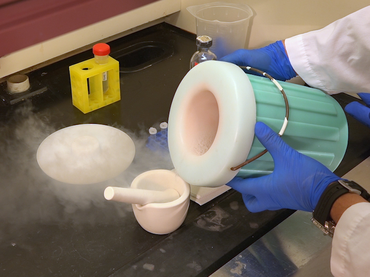
Contamination during the genomic sequencing of microscopic organisms remains a large problem. Here, we show a method to sequence the genome of a tardigrade from a single specimen with as little as 50 pg of genomic DNA without whole genome amplification to minimize the risk of contamination.

Transcript

The goal of this protocol is to sequence the genome of a microscopic organism called tardigrades. We have established a method to sequence the genome of a tardigrade, Hypsibius dujardini, from a single specimen with as low as 50 picograms of genomic DNA with a whole genome application. After isolating a single tardigrade, we minimize bacterial contamination by using antibiotics and visual inspection.

We have also utilized two homogenization methods. First and most commonly used in C.elegans using freeze and thaw cycles, and second, a manual crushing of the tardigrade with a pipette tip. DNA is then used to construct a sequencing library and then is sequenced in a MiSeq Instrument.

An overall summary of the protocol. After isolation of a single individual, it is subjected to three freeze and thaw cycles for homogenization. The genomic DNA is extracted and purified and is fragmented by sonication.

Then a sequencing library is constructed, and after validation of library size distribution, it is sequenced with a sequencing instrument. Prepare 2%agarose gel using distilled water as the solvent in a 90 millimeter plastic culture dish, and 10 millimeters of 1%penicillin streptomycin with distilled water. The gel can be stored for two to three weeks in an incubator set at 18 degrees.

Collect a single tardigrade and place on the prepared agar plate, and wash with distilled water two to three times to remove remaining particles. Place the single tardigrade in penicillin streptomycin antibiotics for two to six hours to remove bacterial contamination, and place the decontaminated animal on a clean slide glass using a P10 pipette. Observe the tardigrade under a microscope at 500 times magnification, and confirm that there are no remaining bacteria.

Collect individual using a P10 pipette with maximum of five microliters of liquid, and place into a low-binding PCR tube, and remove excess liquid as much as possible. Homogenization and DNA extraction. Homogenize the animal to obtain genomic DNA with one of the following methods.

Homogenization with freeze and thaw cycles. Immediately following step 2.5, add 100 microliters of lysis buffer to the PCR tube containing the tardigrade. Place the PCR tube in liquid nitrogen for 10 minutes, and move to a heat block, warm to 37 degrees for 10 minutes.

Repeat this step three times. Manual crushing. Under a stereo microscope, crush the individual with a P10 pipette tip by pressing the animal against the PCR tube wall, and immediately add 100 microliters of lysis buffer.

Incubate for 30 minutes at room temperature for lysis to occur. Transfer the complete volume of the lysis mixture to a clean 1.5 milliliters low-binding microtube. Add 100 microliters of lysis buffer to the low-binding PCR tube, which is used for homogenization and is now empty.

And after pipetting, transfer the mixture to 1.5 low-binding microtube. Repeat this step twice. Add 300 microliters of lysis buffer to a low-binding PCR tube, and after pipetting, move the mixture to 1.5 milliliter low-binding microtube.

Add the total of 600 microliters lysis mixture to spin column placed in the collection tube, and centrifuge at 10, 000 G for one minute. Reapply the flow through to the column, and centrifuge at 10, 000 G for one minute. This step is critical to ensure that most of the genomic DNA is bound to the column.

Add 500 microliters of wash buffer to the spin column, and centrifuge at 10, 000 G for one minute. Transfer the spin column to a clean 1.5 milliliter microtube. Apply 20 microliters of 10 millimolar first ACL to the spin column and wait for five minutes at room temperature.

Centrifuge at 10, 000 G for one minute. The dilution buffer must not contain EDTA, for it interferes with laboratory preparation enzymes. Reapply the flow through to the spin column, and after five minutes incubation at room temperature, centrifuge for one minute at 10, 000 G.Sequencing library construction.

DNA fragmentation. Transfer 15 microliters of genomic DNA elute To a 15 microliter microtube for DNA fragmentation, and centrifuge for one minute using a tabletop centrifuge. Fragment the genomic DNA to 550 base pairs.

After through pipetting, transfer 10 microliters of fragmented DNA mixture to a clean, low-binding PCR tube. Experiment can be stopped here. Preserve DNA at four or minus 20 degrees.

Sequencing library construction. It is absolutely critical to use the specified kit in the following procedures, due to low-input DNA. Prepare the reagents required, following the manufacturer's protocol, and construct the sequencing library without any modifications.

After applying library amplification mixture to a library synthesis mixture, perform PCR reaction in a thermal cycler. The PCR reaction was conducted as shown. Purification of the PCR reaction.

Add 50 microliters of magnetic beads and pipette 10 times, and centrifuge briefly with a tabletop micro centrifuge. Incubate for two minutes at room temperature. Incubate on magnetic stand for five minutes, or until the solution becomes completely clear, and remove the supernatant.

Follow the manufacturer's protocol. Transfer the supernatant without disturbing the pellet into a new low-binding PCR tube. DNA quality check and quantification, and sequencing.

Validation of DNA library size distribution. Add three microliters of sample water with one microliter of sequencing library, and mix thoroughly for one minute with a vortex, and briefly centrifuge with a tabletop centrifuge. Conduct electrophoresis and validate the library size distribution with associated software.

The main fragment peak should be broadly ranging from about 300 to 1, 000 base pairs. DNA quantification. Add 796 microliters of the solution buffer and four microliters of the fluorescence reagent and mix thoroughly.

Dispense 190 microliters of the working solution to two assay tubes, and 197 microliters to one assay tube. Add 10 microliters of standards with known concentrations of DNA to each assay tube containing 190 microliters of working solution, and three microliters of the prepared library to assay tube containing 197 microliters of working solution. Vortex briefly, and centrifuge on the tabletop centrifuge.

Quantify the DNA using a fluorometer with three microliter settings. Sequencing of the DNA library. Prepare the sequencing library based on the manufacturer's protocol.

Set the reagent cassette and flow cell into the sequencing instrument, and enter the sequencing running information, following the manufacturer's protocol. Run sequencing. Represented results.

Exposure of contaminants in high-quality DNA extraction remains to be the critical point in our protocol. To visually examine if they're contaminant, we have incubated the tardigrade in antibiotics, which are penicillin and streptomycin, and also examined the individual under a 500 times microscope. As shown here, visible microbes around the tardigrades are completely illuminated.

Following efficient homogenization, DNA extraction, fragmentation, and library preparation. The size distribution of the library is validated using a high sensitive electrophoresis for quality control. The purple and green lines indicate upper and lower markers at 1, 500 and 25 base pairs respectively.

Lane L is a ladder marker, lane S and N1 to N4 are five replicates of the same experiment, all starting from a single tardigrade specimen. As it can be seen from the broad uniform and distribution between 200 and 1, 000 base pairs, our protocol is highly reproducible, even with the ultra-low input. Here's the result of fast QC, for synchronized reads obtained from a single sequencing run.

Reads are obtained as spare ends of 300 base pairs, where forward and reverse reads are shown in top and bottom, respectively. The distribution shown here is typical of 300 pair end reads, with very high quality base calling above Q30 for first 200 base pairs, and gradually declining to the end. So this method uses only a single individual for one sample.

There is no requirements of massive amounts of animals, such as those required in previous tardigrade genome sequencing projects. Culturing methods for most tardigrades have not yet been established. Therefore, enabling genomics sequencing from a single individual, such as those directly from the fieldwork, will have a large impact on tardigrade molecular biology, and possibly for other small animals.

Our DNA sequencing methods makes it possible to analyze other numerous microscopic organisms that have not yet been sequenced, such as rare hogweed species, nematodes, water firs, and other options, by connecting the part of the zones and a wider public area, furthering the setting where various biological mechanisms may be possible.

Explore More Videos

TardigradeGenome SequencingSingle SpecimenUltralow InputGenomic DNAHomogenizationFreeze-thawPipette CrushingLibrary PreparationMiSeq SequencingBacterial ContaminationAntibioticsAgarose GelMicroscopyLysis BufferDNA Extraction

Related Videos

Automated Gel Size Selection to Improve the Quality of Next-generation Sequencing Libraries Prepared from Environmental Water Samples

13:26

Automated Gel Size Selection to Improve the Quality of Next-generation Sequencing Libraries Prepared from Environmental Water Samples

Related Videos

10.7K Views

Mapping Genome-wide Accessible Chromatin in Primary Human T Lymphocytes by ATAC-Seq

09:08

Mapping Genome-wide Accessible Chromatin in Primary Human T Lymphocytes by ATAC-Seq

Related Videos

18.2K Views

Robust DNA Isolation and High-throughput Sequencing Library Construction for Herbarium Specimens

13:03

Robust DNA Isolation and High-throughput Sequencing Library Construction for Herbarium Specimens

Related Videos

10.9K Views

Generation of Genome-wide Chromatin Conformation Capture Libraries from Tightly Staged Early Drosophila Embryos

10:35

Generation of Genome-wide Chromatin Conformation Capture Libraries from Tightly Staged Early Drosophila Embryos

Related Videos

20.9K Views

Genome-wide Quantification of Translation in Budding Yeast by Ribosome Profiling

12:57

Genome-wide Quantification of Translation in Budding Yeast by Ribosome Profiling

Related Videos

11.7K Views

Hybrid De Novo Genome Assembly for the Generation of Complete Genomes of Urinary Bacteria using Short- and Long-read Sequencing Technologies

12:08

Hybrid De Novo Genome Assembly for the Generation of Complete Genomes of Urinary Bacteria using Short- and Long-read Sequencing Technologies

Related Videos

5.4K Views

An Integrated Workflow to Study the Promoter-Centric Spatio-Temporal Genome Architecture in Scarce Cell Populations

11:36

An Integrated Workflow to Study the Promoter-Centric Spatio-Temporal Genome Architecture in Scarce Cell Populations

Related Videos

2.6K Views

Ultra-long Read Sequencing for Whole Genomic DNA Analysis

10:34

Ultra-long Read Sequencing for Whole Genomic DNA Analysis

Related Videos

23.3K Views

Isolation of Region-specific Microglia from One Adult Mouse Brain Hemisphere for Deep Single-cell RNA Sequencing

09:49

Isolation of Region-specific Microglia from One Adult Mouse Brain Hemisphere for Deep Single-cell RNA Sequencing

Related Videos

11.2K Views

Generating Transposon Insertion Libraries in Gram-Negative Bacteria for High-Throughput Sequencing

08:19

Generating Transposon Insertion Libraries in Gram-Negative Bacteria for High-Throughput Sequencing

Related Videos

10.9K Views

JoVE logo
Contact Us Recommend to Library
Research
  • JoVE Journal
  • JoVE Encyclopedia of Experiments
  • JoVE Visualize
Business
  • JoVE Business
Education
  • JoVE Core
  • JoVE Science Education
  • JoVE Lab Manual
  • JoVE Quizzes
Solutions
  • Authors
  • Teaching Faculty
  • Librarians
  • K12 Schools
About JoVE
  • Overview
  • Leadership
Others
  • JoVE Newsletters
  • JoVE Help Center
  • Blogs
  • Site Maps
Contact Us Recommend to Library
JoVE logo

Copyright © 2025 MyJoVE Corporation. All rights reserved

Privacy Terms of Use Policies
WeChat QR code