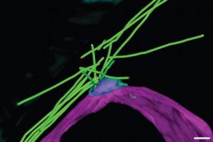
 An international group of scientists led by the University of Leicester have observed structural changes in cellular architecture during the differentiation and formation of gametes. For the first time, scientist have visualized the architecture of microtubules in the model organism.
Using yeast as a model organism, scientists discovered the protein responsible for microtubule structures, which they are calling the microtubule organizing centre, or MTOC. When found in cells undergoing sexual differentiation, this protein is called the radial microtubule organizing centre, or rMTOC. Collaborating with the Paterson Institute for Cancer Research in Manchester, scientists also discovered the protein responsible for rMTOC development, the Hrs1/Mcp6 protein. By understanding the regulation of microtubule production using these two proteins, scientists gain a more thorough understanding of cell differentiation.
Microtubules are a fundamental protein structure in almost all cells. They serve as scaffolding for other cellular structures to build upon, and they also provide production of distinct shapes for cells. For example, microtubules allow neurons to have long axons or sperm cells to develop the tails that allow them to swim. Microtubules also serve as “roads” for motor proteins to move on. Identification of microtubules regulators is crucial, as microtubules serve in many cellular processes that, when occurring atypically, could cause conditions like trisomy 21 (Down Syndrome) or lissencephaly. Furthermore, mitotic inhibitors, a treatment that helps prevent tumor growth, rely on disrupting microtubules. The discovery of rMTOC could help provide therapies that enhance these cancer treatments.
Read the press release concerning the discovery here.

