

ERRATUM NOTICE
Important: There has been an erratum issued for this article. Read more …
Summary
Contact hypersensitivity (CHS) is a murine experimental model of allergic contact dermatitis (ACD). CHS is based on sensitization with reactive hapten by painting the shaved skin of the chest and abdomen, with a subsequent ear skin challenge with a diluted hapten, causing a swelling reaction that is assessed in various ways.
Abstract
Contact hypersensitivity (CHS) is an experimental model of allergic contact dermatitis (ACD) that can be studied in mice. This study aims to present an objective laboratory method that may help to study the CHS reaction in mice, which can be measured and quantified by various tests. To induce CHS, on day "0", mice were sensitized on a previously shaved spot by abdominal skin painting with the hapten 2,4,6-trinitrochlorobenzene (TNCB) in an acetone-ethanol mixture, whereas negative control mice were sham sensitized with vehicle alone-acetone-ethanol mixture. On day "4", the baseline ear thickness was measured with a micrometer prior to the elicitation of CHS (challenge) by painting both ears with diluted TNCB both in the test and control groups. After 24 h, the ear swelling was measured with a micrometer. CHS is an example of a T cell-mediated immune response that causes swelling in inflamed tissue, peaking 24 h after the skin challenge with the same hapten. An increase in ear edema correlated with augmented ear weight, myeloperoxidase (MPO) activity, pro-inflammatory cytokine concentration in the ear extracts, increased thickening of the edematous dermis in the histological examination, and ear vascular permeability. There was also an increase in the concentration of TNP-specific IgG1 antibodies in the sera of the test group when compared with the control mice. Additionally, CHS can be successfully transferred with the CHS-effector cells obtained from donors previously sensitized with TNCB. The CHS-effector cells were administered intravenously into naïve recipient mice, which were subsequently challenged with the same diluted hapten. Ear swelling was measured with a micrometer 24 h later.
Introduction
Allergic contact dermatitis (ACD) is a common skin inflammatory disease in industrialized countries caused by a type IV hypersensitivity reaction resulting from exposure to low molecular weight chemicals called haptens. The substances causing contact sensitization in humans include heavy metal ions (chromium, nickel, iron, cobalt), turpentine, fragrances, dyes, and preservatives present in cosmetics (e.g., p-phenylenediamine), some drugs (e.g., neomycin, benzocaine), β-lactam antibiotics (i.e., penicillin), chemicals produced by plants (pentadecacatechol, a substance present in poison ivy), as well as hydroquinone-used in the photographic industry1,2. ACD etiological agents are very high as over 100,000 chemicals are used in industry alone, and 2,000 new ones are synthesized each year. To date, more than 3,700 molecules have been identified that may be contact haptens/allergens3. The contact hypersensitivity reaction (CHS) is an experimental model of ACD that can be studied in mice, guinea pigs, and rats and can be induced by the topical skin application of reactive chemical haptens dissolved in organic solvents4,5,6. This study aims to describe an objective laboratory method that may help to study the CHS reaction in mice, which can be measured and quantified by various tests.
The CHS consists of sensitization (induction) and effector (challenge) phases. In animal models, haptens first bind covalently to proteins in the body to create neoantigens. During the sensitization phase, activated keratinocytes promote the migration and maturation of skin dendritic cells (sDCs) by producing pro-inflammatory cytokines-tumor necrosis factor α (TNF-α) and interleukin 1β (IL-1β)7. Epidermal Langerhans cells (LCs) present antigens during the CHS induction and effector phases8. LCs exposed to hapten during sensitization promote the induction of both regulatory and effector cells9. Increasing evidence from several studies suggests that CHS responses can be mediated by either CD4+ MHC class II-restricted Th1 cells, locally releasing interferon-γ (IFN-γ) to employ a characteristic inflammatory infiltrate, CD8+ MHC class I-restricted Tc1 lymphocytes that can also release IFN-γ but mostly mediate cytotoxic damage to keratinocytes, and now also interleukin 17 (IL-17)-producing Th17 cells10,11.
Several different CHS models employing various species12,13,14 and haptens have been developed (a detailed comparison of different haptens, solvents, and time of application is summarized in Table 1). The mouse, a frequently used laboratory species, offers a few advantages in studying CHS. There are more strains, knockouts (KO), and transgenic animals among mice compared to other species, which makes them a very attractive animal15. In addition, the CHS model requires many animals, and mice are more economical here. Animal models do not mimic ACD in all aspects; in particular, they show crusting and desquamation, which is not common for humans16,17. The features of chronic disease are challenging to reproduce, mainly because the described model does not assume the application of the hapten for a long period of time. However, it has been confirmed here that many of the significant aspects of ACD are reproduced. It has also been shown that, as in humans, these features are associated with local allergic reactions. The choice of hapten, its solvent, and its application outlined in this protocol were dictated by the fact that the results have been confirmed by numerous in vitro tests and that it was tested and modified in the laboratory for many years until the current version was established. Murine models allow for the analysis of the cell subsets or cytokines that are involved in the development of ACD and are essential for preclinical assessments of new treatments.
Subscription Required. Please recommend JoVE to your librarian.
Protocol
All experiments presented in this article were conducted according to the guidelines of the 1st Local Ethical Committee on Animal Testing in Krakow. All the procedures described were performed according to the local recommendations, especially regarding using ketamine/xylazine as an anesthetic, using both sides of the ears to apply the substance/hapten, cutting off the ear, and collecting blood by eyeball removal. BALB/c (haplotype H-2d), CBA/J (H-2k), and C57BL/6 (H-2b) male and female mice, 6-12 weeks old, were used for the present study (see Table of Materials). For statistical significance, it is best if each group of mice consists of 10-12 animals.
1. Animal preparation
- Clean the operating table with a 70% ethanol solution before and after all procedures. If using mice that require sterile conditions, perform all operations in a biosafety cabinet.
2. Marking mice for identification
- Label the mice by shaving the skin with a razor blade: #0 - unmarked, #2 - on the right front paw, #3 - on the right side, #4 - on the right hind paw, #5 - at the base of the tail, #6 - on the left hind paw, #7 - on the left side, #8 - on the left front paw.
NOTE: Due to the induced reaction, mice cannot be marked classically by ear punching or tagging. The mice were not anesthetized while labeling.
3. Induction of CHS
NOTE: This procedure is depicted in Figure 1.
- Perform the sensitization (induction) following the steps below.
- On day "0", shave mice on the chest and abdomen (square 2 cm x 2 cm) by applying grey soap with water and shaving with a razor blade.
NOTE: Before applying hapten, wait for 6 h so that the skin is not irritated. - Prepare 5% hapten: 2,4,6-trinitrochlorobenzene (TNCB, see Table of Materials) in an acetone-ethanol mixture (ratio 1:3) or vehicle alone (acetone-ethanol). Prepare solutions just before use in a glass vial and protect it from light by covering the vial with aluminum foil.
- On the same day, sensitize the mice by applying 150 µL of 5% hapten on the previously shaved spot. In the group of control mice, apply the vehicle alone to assess the non-specific inflammatory reaction. Before putting the animal back into the cage, wait for 30 s, letting the hapten dry.
CAUTION: Use gloves; TNCB causes a severe allergic reaction in most people.
- On day "0", shave mice on the chest and abdomen (square 2 cm x 2 cm) by applying grey soap with water and shaving with a razor blade.
- Perform elicitation (challenge) and ear swelling measurement.
- On day "4", prepare 0.4% hapten: TNCB in an acetone: olive oil mixture (ratio 1:1). Prepare the solution just before use in a glass vial and protect it from light by covering the vial with aluminum foil.
- Anesthetize the mice with an intraperitoneal (i.p.) injection of a mixture of ketamine (90-120 mg/kg) and xylazine (5-10 mg/kg) (see Table of Materials) for deep anesthesia. Ensure the mouse is fully anesthetized for at least 5 min by toe pinch.
- Measure the ear thickness (0 h measurement, baseline) with a micrometer (see Table of Materials) by an observer unaware of the experimental groups.
- Apply 10 µL of 0.4% hapten on both sides of the ears in both groups (test and control). Before putting the animal back into the cage, wait for 30 s and allow the hapten to dry.
- On day "5", 24 h after hapten application, repeat steps 3.2.2-3.2.3 for the 24 h measurement.
- Evaluate the CHS response by calculating the difference in auricle thickness before and after the challenge with the hapten: 24 h ear thickness (µm) - 0 h ear thickness (µm). Count each ear as a separate measurement. Next, express the ear swelling in micrometers (µm) ± standard error of the mean (SEM) (Table 2, Figure 3).
4. Ear biopsies
- Directly after the 24 h measurement of ear thickness (when the mouse is still under deep anesthesia), cut off the ears as close to the skull as possible with scissors. Collect the biopsies from the distal side of the ears by making a 6 mm diameter punch using a biopsy punch (see Table of Materials).
- Measure the ear weight (step 4.2) and additionally perform one of the following tests on the same ear biopsy: myeloperoxidase (MPO) assay (step 4.3) or in vitro measurement of cytokine concentration in the ear extracts (e.g., IFN-γ, IL-17A, TNF-α [step 4.4]).
NOTE: Cut the ears before blood collection. After this procedure, the mice must be euthanized (e.g., by cervical dislocation).
- Measure the ear weight (step 4.2) and additionally perform one of the following tests on the same ear biopsy: myeloperoxidase (MPO) assay (step 4.3) or in vitro measurement of cytokine concentration in the ear extracts (e.g., IFN-γ, IL-17A, TNF-α [step 4.4]).
- Measure the weight of each ear biopsy on the analytical balance and express it in milligrams (mg) (Figure 4).
- Perform an MPO assay following the steps below.
- Prepare homogenization buffer by dissolving 0.5% hexadecyltrimethylammonium bromide in 50 mmol phosphorate buffer KH2PO4/Na2HPO4 and adjusting the pH to 6.0 (use at room temperature, RT).
- Homogenize the biopsies in 2 mL microcentrifuge tubes with 500 µL of prepared buffer for 10 min using a homogenizer with 5 mm diameter stainless steel beads (add two beads/vial) (see Table of Materials). Next, cool down the sample for 15 min at 4 °C and homogenize it for an additional 10 min.
NOTE: Microcentrifuge tubes have a round bottom so that the beads can easily move. - Freeze the homogenates at −20 °C for 30 min. Thaw and vortex (ensure that the samples are thawed). Repeat this procedure 3x.
- Centrifuge the homogenates at 3,000 x g for 30 min at 4 °C. Harvest the supernatants with a pipette. Express the MPO activity in units (U) per 1 mg of protein.
NOTE: The protocol can be paused here. The samples are stable at −20 °C for 3 months. - To measure MPO activity, perform an enzymatic reaction by mixing 20 µL of supernatant and 200 µL of MPO substrate (0.167 mg/mL of ortho-dianisine dihydrochloride in 50 mmol of KH2PO4/Na2HPO4 buffer with 5 x 10⁻⁴% H202) and add into 96-well flat-bottom plates. Incubate the plates for 20 min at RT.
- Prepare the standard curve by using 20 µL of the MPO standard at concentrations from 0.008 U up to 0.5 U in 200 µL of the MPO substrate. Prepare the blank sample with the MPO substrate alone.
CAUTION: Use a mask while working with ortho-dianisine dihydrochloride.
NOTE: The plates must be made of polypropylene, which has a lower binding capacity so proteins or DNA will not bind. - Measure the optical density (OD) at a wavelength of λ = 460 nm. The enzymatic reaction is stable for 10 min. Read the MPO activity in tested samples from the standard curve.
- To measure protein concentration, use 20 µL of the supernatant, perform a test with a Bicinchoninic acid kit for protein determination (see Table of Materials), and measure the OD at λ = 562 nm (Figure 5).
- Perform in vitro measurement of cytokines in the ear extract.
- Homogenize the ear biopsies at RT in 2 mL microcentrifuge tubes with 500 µL of tissue protein extraction reagent (T-PER) for 10 min using a homogenizer with 5 mm diameter stainless steel beads (add two beads/vial). Next, cool down the sample for 15 min at 4 °C and homogenize it for an additional 10 min.
NOTE: Microcentrifuge tubes have a round bottom so that the beads can easily move. - Centrifuge the homogenates at 3,000 x g for 30 min at 4 °C.
NOTE: The protocol can be paused here. The samples are stable at −80 °C for 6 months. - Assess the cytokine levels using a commercially available ELISA set (e.g., IFN-γ) (see Table of Materials) following the manufacturer's instructions (Figure 6).
- Homogenize the ear biopsies at RT in 2 mL microcentrifuge tubes with 500 µL of tissue protein extraction reagent (T-PER) for 10 min using a homogenizer with 5 mm diameter stainless steel beads (add two beads/vial). Next, cool down the sample for 15 min at 4 °C and homogenize it for an additional 10 min.
5. Histology of ear tissue
- Directly after the 24 h measurement of ear thickness, when the mouse is still deeply anesthetized, cut off the ears as close to the skull as possible with the scissors (step 4.1).
NOTE: After this procedure, the mice must be euthanized (e.g., by cervical dislocation). - Perform paraffin embedding of the tissue blocks following the steps below.
- Directly after removal, place the ear into ~10 mL of 10% formalin for 24 h.
- Place the ears into a tissue processing cassette. Put the cassettes in an automated processor (see Table of Materials) for dehydration cycles (alcohol 70%, 90%, 100%, 30 min each at RT), clearing cycles (xylene 3x, 30 min each at RT), and wax infiltration cycles (paraffin 3x, 30 min each at 56 °C).
- Remove the cassettes from the automated processor, and hold onto the warming plate until required. Fill the wax mold with warm wax (from the dispenser).
- Remove the sections from the cassette with warmed forceps and place them in the mold; next, place the cassette base on the top of the mold and then fill with more wax. Place it in chilled water on a cold plate for 30 min so that the paraffin solidifies to form a block containing the specimen.
- Use a rotary microtome (see Table of Materials) to cut sections ~5 µm thick. Float the sections in a warm bath to flatten them. Pick up the sections onto a glass microscope slide. Allow them to dry at RT to ensure the sections adhere to the slide.
NOTE: Use slides that eliminate the need to apply adhesive materials or protein coatings to prevent the loss of tissue sections during staining.
- Perform hematoxylin and eosin (H&E) staining.
- Prepare 17 staining dishes with the following: xylene (four dishes), 100% ethanol (absolute alcohol) (four dishes), 90% ethanol, 80% ethanol, 70% ethanol, 50% ethanol, PBS (three dishes), hematoxylin solution, eosin solution. Transfer the slides from one dish to the next as per the below steps, and perform all at RT.
NOTE: The procedure in each dish may be repeated approximately 10x (e.g., if a 20-slides dish is used, 200 stains can be made without changing the fluids). - Deparaffinize the sections by incubation at 65 °C for 30 min in the incubator. Immerse the slides in xylene for 30 min. Repeat 1x in new xylene for 30 min.
- Immerse the slides in 100% ethanol for 5 min. Repeat 1x in new 100% ethanol for 5 min. Immerse the slides in the ethanol row, 90%, 80%, 70%, and 50%, for 2 min in each dilution.
- Immerse the slides in phosphate-buffered saline (PBS) for 5 min. Wipe away excess liquid from around the tissue and back side of slides.
- Stain the sections in hematoxylin solution (see Table of Materials) for 7-8 min. Wash in running water for 30 s from the reverse side so as not to damage the sections. Repeat step 5.3.4.
- Stain the sections with eosin solution (see Table of Materials) for 30 s. Wash as mentioned in step 5.3.5 and then repeat step 5.3.4.
- Immerse the slides in 100 % ethanol (absolute alcohol) for 2 min. Repeat 1x in new 100% ethanol for 2 min.
- Immerse the slides in xylene for 5 min. Repeat 1x in new xylene for 5 min.
- Let the sections air dry for 15 min at RT. Add a drop of mounting medium (see Table of Materials) on a coverslip and then place it at the top of the section.
- Prepare 17 staining dishes with the following: xylene (four dishes), 100% ethanol (absolute alcohol) (four dishes), 90% ethanol, 80% ethanol, 70% ethanol, 50% ethanol, PBS (three dishes), hematoxylin solution, eosin solution. Transfer the slides from one dish to the next as per the below steps, and perform all at RT.
- Examine the section under a light microscope under a magnification of 20x or 40x, and capture images (Figure 7).
6. Vascular permeability test
NOTE: Alternately to ear thickness measurement, a vascular permeability test can be performed.
- Sensitize mice on day "0" (steps 3.1.1-3.1.3), and then, on day "4", anesthetize the mice (step 3.2.2) and directly apply hapten on the ears (step 3.2.4), omitting the 0 h ear measurement.
- At 23 h post-challenge, anesthetize the mice (step 3.2.2).
- Inject intravenously (i.v.) 8.3 µL/g body weight of 1% Evans blue dye (see Table of Materials) in Dulbecco′s Phosphate-Buffered Saline (DPBS).
- Anesthetize the mice again-deep anesthesia (step 3.2.2)-1 h after Evans blue injection.
- Collect ear biopsies (step 4.1).
NOTE: After this procedure, the mice must be euthanized (e.g., by cervical dislocation). - Extract the dye from the tissue, place ear punches into the tubes containing 1 mL of formamide, and incubate at 37 °C in the atmosphere of 5% CO2 for 18 h.
- Centrifuge the biopsies at 3,000 x g for 3 min at RT. Collect the supernatants with a pipette.
- Measure the OD at a wavelength of λ = 565 nm in 96-well flat-bottom plates against a blank containing formamide. The color is stable for 24 h. Read the concentration of the test samples from the standard curve (use the concentrations of Evans blue ranging from 0.2-30 µg of dye/mL formamide) (Figure 8).
NOTE: The plates must be made of polypropylene, which has a lower binding capacity so proteins or DNA will not bind.
7. Serum collection and anti-TNP immunoglobulin (IgG1) antibody measurement
- After collecting ear biopsies (step 4.1), when the mouse is still under deep anesthesia, remove the eyeball with tweezers, put gentle pressure on the mouse, and collect blood from the retro-orbital sinus into the tube (vial with gel for obtaining serum, see Table of Materials). An alternative method of collecting blood might be to puncture the heart with a syringe and collect the blood.
NOTE: The blood must be collected after removing the ears. The same method of bleeding must be used across an entire study due to potential differences in blood parameters18. After this procedure, mice must be euthanized (e.g., by cervical dislocation). - Invert a minimum of 6x, wait 30 min for the blood to clot, and then centrifuge at 1,300-2,000 x g for 10 min at RT.
NOTE: The protocol can be paused here. The sample is stable at −20 °C for 6 months. - Coat a 96-well flat-bottom plate with 50 µL of bovine serum albumin conjugated with 2,4,6-trinitrophenyl (TNP-BSA) dissolved in DPBS at concertation of 10 µg/mL. Next, coat the second plate with bovine serum albumin (BSA) alone dissolved in DPBS (background) at a concentration of 10 µg/mL. Incubate overnight at 4 °C.
NOTE: Mouse serum contains antibodies (Abs) against BSA, so the samples need to be tested on both plates, and next, a calculation must be done (OD TNP-BSA - OD BSA). - Wash the plates with 300 µL of DPBS containing 0.05% Tween 20. Repeat 3x.
- Prepare assay diluent (AD): DPBS containing 1% BSA. Block the wells with AD for 1 h at RT. Wash again (step 7.4).
- Prepare an internal standard (iSTD): sensitize the mice with TNCB (steps 3.1.1-3.1.3), and, 10 days after sensitization, collect and poll the serum from all donors (step 7.1 and step 7.2).
- Add to the plate 50 µL of graded concentrations of iSTD diluted with AD for making the standard curve (described in Table 3). Add to the plate 50 µL of serum samples. Test each sample and standard curve on both plates (TNP-BSA and BSA coated). Incubate for 2 h at RT. Wash the plates (step 7.4).
- Add 50 µL of biotinylated anti-mouse IgG1 monoclonal antibody (mAb, see Table of Materials) diluted 1:250 with AD and incubate for 1 h at RT. Wash again (step 7.4).
- Add 50 µL of horseradish peroxidase streptavidin (HRD streptavidin, see Table of Materials) diluted 1:2000 with AD, and incubate for 30 min at RT in the dark. Wash the plate (step 7.4).
- Add 50 µL of TMB substrate (see Table of Materials) and incubate for 30 min at RT in the dark.
- Stop the enzymatic reaction by adding 25 µL of 1 M H2SO4; the reaction is stable for 30 min.
- Measure the OD at a wavelength of λ = 450 nm and a background of 570 nm (the background must be subtracted from every 450 nm measurement). When presenting the results, subtract the BSA measurements from the TNP-BSA for samples and the standard curve. Then, calculate the unit (U) of antibodies according to the standard curve (Figure 9).
8. Adoptive transfer of the CHS-effector cells
NOTE: This procedure is depicted in Figure 2.
- Donors (at the ratio of one donor:1 recipient): sensitize mice with TNCB on day "0" (steps 3.1.1-3.1.3).
- On day "4", anesthetize the mice-deep anesthesia (step 3.2.2).
- After disinfecting the skin, isolate the axillary and inguinal lymph nodes (ALNs) and spleens (SPLs) with forceps. Pool together the ALNs in one vial and the SPLs in another.
NOTE: There is one axillary lymph node behind the pectoral muscle in each axilla. One inguinal lymph node in the hip region is situated next to three blood vessels. The spleen is located on the left side of the body behind the intestine and stomach19. Make sure that the operating area is disinfected, including skin and hair. Work with sterile tools, including sterile gloves, in a biosafety cabinet to maintain sterile conditions. After this procedure, the mice must be euthanized (e.g., by cervical dislocation). - Mash tissue between the frosted ends of two microscope slides. Pass the cell suspension through a cell strainer with a pore size of 70 µm (see Table of Materials).
- Wash the cells with DPBS supplemented with 1% fetal bovine serum (FBS). Centrifuge at 300 x g for 10 min at 4 °C. Decant the supernatant and resuspend the remaining cell pellet in 1-5.0 mL of DPBS.
- Count the live cells using a hemocytometer20 with Trypan blue, and mix 10 µL of the cell suspension with 90-990 µL (depending on the cell number) of Trypan blue. Take into account the dilution when calculating the cell number (10x-100x).
- Prepare a mixture of ALNs and SPLs (ratio 1:1): 8.0 x 10⁶ up to 7.0 x 10⁷/mouse in 200 µL of DPBS.
- Recipients (naïve syngeneic mice): anesthetize naïve recipient mice (step 3.2.2) and inject i.v. with a prepared mixture of the CHS-effector cells (step 8.7). In the control group of mice, do not inject any cells.
- Measure the ear thickness prior to (0 h) and after (24 h) the challenge (steps 3.2.1-3.2.6) (Figure 10).
- Additionally, perform tests on isolated CHS-effector cells (e.g., cell phenotyping or the measurement of produced cytokines by the CHS-effector cells by flow cytometry). Cell cultures can also be established, and the ability of the CHS-effector cells to proliferate in the presence of the antigen or the amount of secreted cytokines in the culture supernatants can be assessed21,22 (data not presented).
NOTE: Performing additional tests requires correspondingly more cell donors.
Subscription Required. Please recommend JoVE to your librarian.
Representative Results
For CHS induction, the animals were sensitized via skin painting (abdominal) with 150 µL of 5% TNCB or sham sensitized with the vehicle alone. On day "4", the ear swelling responses of both ears were induced by contact painting (challenge) with 10 µL of 0.4% TNCB in both mice previously contact sensitized with TNCB (test group) and the control group mice (sham sensitized). The presented data depict that the mice sensitized with TNCB and challenged 4 days later developed significantly increased ear swelling compared with the sham-sensitized ones similarly challenged (Figure 3, Table 2, test vs. control group). The ear swelling results were completely validated in further studies, highlighting that an increase in ear edema determined with a micrometer agreed with augmented ear weight (Figure 4), MPO activity (Figure 5), IFN-γ concentration in the ear extracts (Figure 6), increased thickening of the edematous dermis in the histological examination (Figure 7), and ear vascular permeability (Figure 8). An increase in the concentration of TNP-specific IgG1 antibodies was also found in the sera of the test mice when compared with the control animals (Figure 9).
As an example of T cell-mediated immune response, CHS can also be transferred into naïve syngeneic recipient mice. Donors were sensitized by TNCB application, and subsequently, the CHS-effector cells were administered i.v. into the naïve recipient mice, which were challenged with the hapten and tested for CHS 24 h later (Figure 10). The animals that received the CHS-effector cells from donors previously sensitized with TNCB showed significantly increased ear swelling compared to animals that were challenged only (did not receive any cells).
The CHS reaction has a complex mechanism and involves various cells. Antigen presentation and T/B cell activation occur in the peripheral lymph organs (e.g., ALNs and SPL). It was determined that CHS-effector cells depleted of CD4+ but not CD8+ cells prior to adoptive cell transfer resulted in the absence of the CHS reaction in the recipient mice. Those cells were found to be positive for IFN-γ (T-box transcription factor TBX21, Tbet+) and IL-17A (retinoic acid receptor-related orphan nuclear receptor gamma, RORγT+) (Supplementary Figure 1).
The presented results from the representative experiments were performed on C57BL/6 and CBA/J male and female mice at 8-12 weeks of age. Following the 3R rules in the use of animals23, especially reduction, for the purposes of this article, the results of experiments are shown on small groups of animals. Data in the graphs are shown as mean ± SEM. Statistical significance was set at p < 0.05. The graphs were drawn using Prism software (see Table of Materials).

Figure 1: Induction of CHS. Sensitization, challenge, and ear measurement. Abbreviations: CHS = contact hypersensitivity reaction; TNCB = 2,4,6-trinitrochlorobenzene. Please click here to view a larger version of this figure.

Figure 2: Adoptive transfer of the CHS-effector cells. Abbreviations: ALNs =axillary and inguinal lymph nodes; CHS = contact hypersensitivity reaction; i.v. = intravenously; SPLs = spleens; TNCB = 2,4,6-trinitrochlorobenzene. Please click here to view a larger version of this figure.

Figure 3: Representative evaluation of CHS to TNCB by the measurement of ear swelling with a micrometer. Mice were TNCB (test group) or sham (control group) sensitized and subsequently challenged. The thickness of the auricle was measured before and after the challenge, and differences in ear swelling were calculated by subtracting the 0 h ear thickness (µm) from the 24 h ear thickness (µm). Ear swelling was expressed as mean ± SEM, ****p < 0.0001, n = 10 mice/group (data from Table 2). Abbreviations: CHS = contact hypersensitivity reaction; SEM = standard error of the mean; TNCB = 2,4,6-trinitrochlorobenzene. Please click here to view a larger version of this figure.

Figure 4: Representative evaluation of CHS by the measurement of ear weight. Ear weight is one of the parameters that corresponds with ear swelling. Mice were TNCB (test group) or sham (control group) sensitized and subsequently challenged. At 24 h after the challenge, 6 mm diameter punches were taken from the removed ears. The punches were weighed on an analytical laboratory balance. Ear weight was expressed in milligrams (mg) as mean ± SEM, ***p < 0.001, n = 10 mice/group. Abbreviations: CHS = contact hypersensitivity reaction; SEM = standard error of the mean; TNCB = 2,4,6-trinitrochlorobenzene. Please click here to view a larger version of this figure.

Figure 5: Representative evaluation of MPO activity. Increased MPO activity in tissue extracts correlates with ear inflammation. TNCB-sensitized mice (test group) and sham-sensitized mice (control group) were challenged. At 24 h post-challenge, the ears were removed, and 6 mm diameter punches of the ear were extracted and processed. MPO activity is expressed in U per protein content (U/g of protein). Results displayed as mean ± SEM, **p < 0.01, n = 5-6 mice/ group. Abbreviations: CHS = contact hypersensitivity reaction; MPO = myeloperoxidase; SEM = standard error of the mean; TNCB = 2,4,6-trinitrochlorobenzene; U = units. Please click here to view a larger version of this figure.

Figure 6: Representative evaluation of cytokine production-IFN-γ concentration in ear extracts. Mice were TNCB (test group) or sham (control group) sensitized and subsequently challenged. At 24 h post-challenge, the ears were removed, and 6 mm diameter punches of the ear were taken. The concentration of IFN-γ was determined in tissue homogenates by ELISA. Results shown as mean ± SEM, *p < 0.05, n = 5 mice/group. Abbreviations: CHS = contact hypersensitivity reaction; IFN-γ = interferon gamma; SEM = standard error of the mean; TNCB = 2,4,6-trinitrochlorobenzene. Please click here to view a larger version of this figure.

Figure 7: Representative histology of the ear tissue. Hematoxylin and eosin staining. Mice were TNCB (test group) or sham (control group) sensitized and subsequently challenged. (A-C) Histological examination in the test group manifested in a significantly increased concentration of inflammatory cells (mononuclear and polymorphonuclear cells), mainly in the dermis, with microabscess formation in the epidermis. Thickening of the edematous dermis and a thickened, hyperplastic epidermis were also noticed. (D-E) Control group. Please click here to view a larger version of this figure.

Figure 8: Representative vascular permeability test. Observed ear tissue edema is a result of increased vascular permeability. To determine changes in vascular permeability, mice were TNCB (test group) or sham (control group) sensitized and then challenged 4 days later. At 23 h after the challenge, Evans blue was injected, and, 1 h after Evans blue injection, the animals were euthanized, and 6 mm diameter punches of the ear were made. Results shown as mean ± SEM, **p < 0.01, n = 5 mice/group. Abbreviations: CHS = contact hypersensitivity reaction; SEM = standard error of the mean; TNCB = 2,4,6-trinitrochlorobenzene. Please click here to view a larger version of this figure.

Figure 9: Representative anti-TNP IgG1 antibody measurement. The concentration of anti-TNP IgG1 antibodies in serum was measured 24 h after the challenge with hapten TNCB in sham sensitized (control) and in TNCB-sensitized (test group) mice. The collected sera were tested for antibody concentration by ELISA. Results shown as mean ± SEM, ***p < 0.001, n = 10 mice/group. Abbreviations: CHS = contact hypersensitivity reaction; IgG1 = immunoglobulin G subclass 1; SEM = standard error of the mean; TNCB = 2,4,6-trinitrochlorobenzene. Please click here to view a larger version of this figure.

Figure 10: Representative adoptive transfer of the CHS-effector cells. The CHS-effector cells were obtained from donors that were sensitized with TNCB. Next, the collected immune cells were injected, i.v., into naïve syngeneic recipients, which were challenged for elicitation of the CHS effector phase. The control group of mice did not receive any cells prior to the challenge. The thickness of the auricle was measured before and after the challenge. Results shown as mean ± SEM, ***p < 0.001, n = 7 mice/group. Abbreviations: CHS = contact hypersensitivity reaction; SEM = standard error of the mean; TNCB = 2,4,6-trinitrochlorobenzene. Please click here to view a larger version of this figure.
| Mouse strain | Sensitization solution (dose) on shaved abdomen |
Elicitation solution (dose) on both sides of ear/ s |
Sensitization / elicitation day | Refs | |||||||||||
| BALB/c (H-2d); C57BL/6 (H-2b) TCRδ-/-, β2m-/-, CD1d-/- (B10 PL (H-2u background) |
25 μL of 0.5 % DNFB in acetone-olive oil mixture (ratio 4:1) | 5 μL of 0.1 % DNFB in acetone-olive oil mixture (ratio 4:1) | 0 / 5 | 22 | |||||||||||
| C57BL/6 (H-2b) | 50 μL of 0.5 % DNFB in acetone-olive oil mixture (ratio 4:1) | 25 μL of 0.2 % DNFB in acetone-olive oil mixture (ratio 4:1) | 0 / 5 | 32 | |||||||||||
| C57BL/6 (H2b); IL-17A-/- (C57BL/6 background) | 150 μL of 5% TNCB in acetone-ethanol mixture (ratio 1:3) | 10 μL of 0.4 % TNCB in olive oil-acetone mixture (ratio 1:1) | 0 / 4 | 33 | |||||||||||
| CBA/J (H-2k); C57BL/6 (H-2b) TLR2-/-, MyD88-/-, IL-17A-/- (C57BL/6 background) |
150 μL of 5 % TNP-Cl (TNCB) in an acetone-ethanol mixture (ratio 1:3) | 10 μl of 0.4 % TNP-Cl (TNCB) in olive oil-acetone mixture (ratio 1:1) | 0 / 4 | 21 | |||||||||||
| C57BL/6 (H-2b); BALB/c (H-2d) | 25 μL of 1 % TNCB in an acetone | 10 μL of 0.1 or 0.2 % TNCB in an acetone (and higher up to 1 %) | 0 / 7 | 34 | |||||||||||
| C57BL/6 (H-2b); TLR2-/-/ TLR4-/- (double-knockout mice on C57BL/6 background) |
100 μL of 3 % TNCB in an acetone | 20 μL of 1 % TNCB in an acetone (just on the back side of ears) | 0 / 5 | 31 | |||||||||||
| C57BL/6 (H-2b) MHC class II-deficient mice (C57BL/6 background) |
100 μL of 3 % TNCB in acetone-olive oil mixture (ratio 4:1) | 20 μL of 0.5 or 1 % TNCB in acetone-olive oil mixture (ratio 4:1) | 0 / 6 | 35 | |||||||||||
| C57BL/6 (H-2b) | 100 μL of 7 % TNCB in an acetone | 20 μL 1 % TNCB in an acetone | 0 / 5 | 36 | |||||||||||
| C57BL/6 (H-2b) | 100 μL 3 % OX in ethanol | 20 μL 1 % OX in an ethanol | 0 / 5 | ||||||||||||
| C57BL/6 (H-2b); CD4-/-, CD8-/- (C57BL/6 background) | 25 μL of 0.5 % DNFB in acetone-olive oil (ratio 4:1) | 10 μL of 0.2 % DNFB in acetone-olive oil (ratio 4:1) | 0 / 5 | 10 | |||||||||||
| C57BL/6 (H-2b); CD4-/-, CD8-/- (C57BL/6 background) | 150 μL of 3 % OX in alcohol-acetone (ratio 3:1) | 10 μL 1% OX in alcohol-acetone (ratio 3:1) | 0 / 5 | ||||||||||||
| C57BL/6 (H-2b); C3H/HeN (H-2k); TLR4-/- (C3H/HeJ background); MyD88-/- (C57BL/6 background) | 100 mg/ ear of 10 % NiCl2 in white petrolatum on the dorsal side of both ears | 10 % NiCl2 in white petrolatum | 0,1,2 / 23, 24 | 37 | |||||||||||
| NOD (H-2g7) MyD88-/- (NOD background) | 400 μL of 0.5 % FITC in acetone and dibutyl phthalate | 10 μL of 0.1 % FITC in acetone and dibutyl phthalate | 0 / 5 | 25 | |||||||||||
Table 1: Comparison of the CHS model in various studies. Abbreviations: DNFB = 1-fluoro-2,4-dinitrobenzene; FITC = fluorescein isothiocyanate; NiCl2 = nickel (II) chloride; TNCB = 2,4,6-trinitrochlorobenzene; TNP-Cl = trinitrophenyl chloride; OX = oxazolone. Please click here to download this Table.
| Control group (negative) | Test group (CHS reaction) | ||||||
| Mouse # ear: L, R | 0 h ear thickness [μm] | 24 h ear thickness [μm] | 24 h – 0 h ear thickness [μm] | Mouse # ear: L, R | 0 h ear thickness [μm] | 24 h ear thickness [μm] | 24 h – 0 h ear thickness [μm] |
| 1 L | 365 | 380 | 15 | 1 L | 345 | 427.5 | 82.5 |
| 1 R | 335 | 380 | 45 | 1 R | 340 | 455 | 115 |
| 2 L | 345 | 355 | 10 | 2 L | 355 | 475 | 120 |
| 2 R | 327.5 | 352.5 | 25 | 2 R | 342.5 | 457.5 | 115 |
| 3 L | 340 | 370 | 30 | 3 L | 340 | 460 | 120 |
| 3 R | 325 | 355 | 30 | 3 R | 345 | 495 | 150 |
| 4 L | 335 | 380 | 45 | 4 L | 357.5 | 432.5 | 75 |
| 4 R | 340 | 350 | 10 | 4 R | 335 | 402.5 | 67.5 |
| 5 L | 350 | 380 | 30 | 5 L | 335 | 387.5 | 52.5 |
| 5 R | 337.5 | 360 | 22.5 | 5 R | 335 | 425 | 90 |
| 6 L | 335 | 365 | 30 | 6 L | 350 | 430 | 80 |
| 6 R | 340 | 375 | 35 | 6 R | 342.5 | 405 | 62.5 |
| 7 L | 345 | 337.5 | 0 | 7 L | 340 | 502.5 | 162.5 |
| 7 R | 345 | 335 | 0 | 7 R | 327.5 | 447.5 | 120 |
| 8 L | 370 | 380 | 10 | 8 L | 327.5 | 515 | 187.5 |
| 8 R | 375 | 355 | 0 | 8 R | 327.5 | 540 | 212.5 |
| 9 L | 385 | 370 | 0 | 9 L | 330 | 415 | 85 |
| 9 R | 342.5 | 362.5 | 20 | 9 R | 327.5 | 390 | 62.5 |
| 10 L | 307.5 | 340 | 32.5 | 10 L | 337.5 | 445 | 107.5 |
| 10 R | 325 | 350 | 25 | 10 R | 352.5 | 455 | 102.5 |
| Mean | 20.75 | Mean | 108.5 | ||||
| ± SEM | 3.245 | ± SEM | 9.565 | ||||
Table 2: Representative example of calculating the difference in ear thickness in the effector phase of CHS. Calculating the difference in the thickness of the auricle before and after the challenge with the hapten: 24 h ear thickness (µm) - 0 h ear thickness (µm). Each ear counts as a separate measurement. Ear swelling expressed in micrometers (µm) ± SEM, n = 20. Abbreviations: L = left; R = right. Please click here to download this Table.
| iSTD dilution with AD | anti-TNP IgG1 Ab (U/mL) |
| 100x | 250 |
| 200x | 125 |
| 400x | 62.5 |
| 800x | 31.25 |
| 1600x | 15.63 |
| 3200x | 7.8 |
| only AD | 0 |
Table 3: Preparation of the different concentrations of iSTD for the standard curve for anti-TNP IgG1 Ab measurement. The 100x iSTD dilution was assumed to be 250 U of anti-TNP IgG1 Ab. Abbreviations: Ab = antibody; AD = assay diluent; iSTD = internal standard; IgG1 = immunoglobulin G subclass 1; TNP = 2,4,6-trinitrophenyl; U = units. Please click here to download this Table.
Supplementary Figure 1: The phenotype of CHS-effector cells. The CHS-effector cells were obtained from donors' ALNs and SPLs, which were previously sensitized with TNCB. (A) Employing the MACS technique, the CHS-effector cells (whole ALNs and SPLs) were depleted of either CD4+ or CD8+ cells. Subsequently, adoptive cell transfer was conducted prior to the elicitation of the CHS effector phase. Ear swelling was expressed as mean ± SEM. (B-E) Using a flow cytometry technique, the CHS-effector and naïve (obtained from naïve mice) cells were stained for IFN-γ, Tbet, IL-17A, and RORγt prior to analysis. Cells were gated for the TCRβ+CD4+population. Results shown as mean ± SEM, ***p < 0.001, **p < 0.01, * p < 0.05, n = 4-6 mice/group. Abbreviations: ALNs = axillary and inguinal lymph nodes; CD4 = cluster of differentiation 4; CHS = contact hypersensitivity reaction; IFN-γ = interferon gamma; IL = interleukin; MACS = magnetic-activated cell sorting; ns = non significant; RORγt = retinoic-acid-receptor-related orphan nuclear receptor gamma; SEM = standard error of the mean; SPLs = spleens; Tbet = T-box transcription factor TBX21; TCRβ = T cell receptor beta; TNCB = 2,4,6-trinitrochlorobenzene. Please click here to download this File.
Subscription Required. Please recommend JoVE to your librarian.
Discussion
CHS is induced via haptens, which bind to self-protein antigens in the skin, creating neoantigens. CHS is mediated by local extravascular recruitment of circulating antigen-specific CHS-effector T cells, which results in swelling in the challenged tissue, peaking 24 h after exposure of the secondary skin to the same hapten6. The swelling of the tissue is mainly caused by the infiltration of leukocytes and leukocyte-dependent fibrin deposition24. These changes can be detected with a micrometer measuring the ear swelling of hapten-sensitized and challenged versus sham-sensitized and challenged mice.
CHS can also be determined by the comparison of ear weight. Then, the ear punches utilized for ear weight determination can be used for further tests. The cell infiltrates in the inflamed ears consist of cytokine-producing T lymphocytes and MPO-positive neutrophils. In the ear tissue homogenates, various parameters can be measured, such as MPO activity or the concentration of pro-inflammatory cytokines (e.g., TNF-α, IFN-γ, IL-17A, or others) using the ELISA test or the expression of the cytokine mRNA in the skin using the qPCR test25. Additionally, vessel permeability changes can be evaluated using the Evans blue test21,26.
In the inflamed ear tissue, the changes observed can also be complemented with in vitro tests, highlighting T cell proliferation and cytokine production. This can be readily accomplished by culturing ALNs in the presence of hapten-conjugated protein antigens22,27. The evaluation of cytokine secretion by ALNs isolated from sensitized but not challenged mice provides details regarding cytokine production by the CHS-effector cells in the place of their induction.
Limitations
Many micrometers measure ear swelling with differing accuracy. For example, the lowest pressure is exerted by a spring caliper, so it seems that the results will best reflect the actual thickness ear measurement. However, micrometers that exert more pressure, such as Mitutoyo, are likely to compress more tightly the fluid that accumulates in the ears in the early phase of edema formation. The biphasic nature of CHS can be visualized more clearly using such micrometers because more pressure is exerted. This is more difficult when using a spring caliper with light pressure28. Nevertheless, in some studies, the thickness of the ears was measured with a caliper29. Also, the observer's experience ensures accurate measurements, which can be influenced by subjective feelings, even if the observer is unaware of the experimental groups.
Alternative methods have been described here that can help confirm the measurements of ear swelling with a micrometer, making the data presented more reliable and less subjective. However, these alternative strategies for assessing CHS can only be used at one point in time. Micrometer measurements can be repeated at various time points, allowing for studying the CHS kinetics.
Modifications
The protocol for studying the CHS reaction we use in our laboratory differs significantly from those used in other laboratories, including the dose of hapten and the solvent composition used for both elicitation and sensitization, as well as the time point at which the reaction is assessed. The ear thickness can be measured at different time points (e.g., 2 h, 24 h, 48 h, and 72 h after the challenge)10,30. Table 1 shows different experimental models, particularly the differences in the animal strain, hapten, and the sensitization/challenge times used in various studies10,21,22,26,31,32,33,34,35,36,37. The CHS protocol might also be conducted without previous shaving of the abdomen of the mice.
The next difference concerns the execution of the elicitation (challenge) itself. As is typical, the sensitization is done by painting shaved mice's abdomen skin with the hapten or vehicle alone. Subsequently, in both groups, one ear is challenged with diluted hapten on each ear side. As a control, the opposite ear is painted with an identical amount of vehicle alone10,31.
Critical steps
The most critical moment is to trigger CHS, as the test results depend on it. (1) Hapten solution is very volatile and light sensitive, so it must be tightly closed and protected from light, and when in use, it must be quickly applied to the skin of the animals so it will not evaporate. (2) After applying the hapten to the skin of the abdomen, it must be dried before the animal returns to the cage because mice can smear it against the bedding or apply it to the ears with their paws (then the measurement of the thickness of the auricle may be inadequate). (3) Various methods of labeling animals, such as marking mouse tails with a solvent-resistant marker, are also known. However, the dyes present in the marker might (not studied) become a hapten and trigger CHS. Therefore, in this study, a marking method was chosen that does not affect the reaction. (4) Choosing a shaving method is very important as it can irritate the skin. In this protocol, a gray soap was used because it has soothing properties; it accelerates the treatment of minor cuts and festering wounds, which is helped by its antibacterial effects. It soothes swelling and inflammation of the skin.
Further investigations and standardization of the experimental procedures, including the animal use (strain and sex), are required to compare the results from different studies.
Many different environmental factors and new substances with biological functions can be tested in the CHS model. This model can be useful if researchers want to show if tested factors modulate T cell-dependent immune responses.
Subscription Required. Please recommend JoVE to your librarian.
Disclosures
The authors have nothing to disclose.
Acknowledgments
This study was supported by subvention SUBZ.A020.22.060 of the Medical University in Wroclaw, Poland, and by grants from the Ministry of Science and Higher Education N N401 545940 to MS and IP2012 0443 72 to MMS.
Materials
| Name | Company | Catalog Number | Comments |
| 70% ethanol | Merck KGaA, Darmstadt, Germany | 65350-M | for surface disinfection |
| 96-well flat-bottom plates, polypropylene | Greiner Bio-One GmbH, Kremsmunster, Austria | 655101 | for MPO and Evans blue measurement - plates should be made of polypropylene, that has a lower binding capacity so proteins or DNA will not bind |
| Acetone (ACS reagent, ≥99.5%) | Merck KGaA, Darmstadt, Germany | 179124 | |
| Aluminum foil | Merck KGaA, Darmstadt, Germany | Z185140 | |
| Analytical balance | Sartorius Weighing Technology GmbH, Goettingen, Germany | PRACTUM224-1s, 29105177 | |
| Automated tissue processor | MediMeas Instruments, Sarsehri, Haryana, India | MTP-E-212 | automatically prepare tissue samples by fixing, dehydrating, clearing, and infiltrating them with paraffin |
| BD Vacutainer SST II Advance (tube with gel for obtaining serum) | Becton Dickinson (BD), Franklin Lakes, NJ, USA | BD 366882 | |
| Bicinchoninic acid kit for protein determination | Merck KGaA, Darmstadt, Germany | BCA1-1KT | |
| Biotin Rat Anti-Mouse IgG1 | Becton Dickinson (BD Biosciences), Franklin Lakes, NJ, USA | 553441 | |
| BSA (bovine serum albumine) | Merck KGaA, Darmstadt, Germany | A9418 | protein assays & analysis, 2 mg/mL |
| Cell strainer, pore size 70 μm | BIOLOGIX, China | 15-1070 | suitable for 50 mL tubes |
| Coverslip | VWR, Radnor, Pennsylvania, United States | 631-1583 | 24 mm, but it possible to use different size |
| Disposable pipettes capacity: 5 mL, 10 mL, 25 mL | Merck KGaA, Darmstadt, Germany | Z740301, Z740302, Z740303 | |
| DPBS (Dulbecco′s phosphate buffered saline) | ThermoFisher Scientific, Waltham, Massachusetts, USA | 14190144 | no calcium, no magnesium, mammalian cell culture |
| DPX Mountant for histology | Merck KGaA, Darmstadt, Germany | 6522 | mounting media for H-E, might be used some other e.g, Canada balsam |
| Dumont 5 tweezers - straight | Animalab, Poznan, Poland | 11251-10FST | surgical instruments for procedures on mice (should be steriled) |
| Dumont 7 tweezers - bent | Animalab, Poznan, Poland | 11272-50FST | surgical instruments for procedures on mice (should be steriled) |
| Eosin Y solution, alcoholic | Merck KGaA, Darmstadt, Germany | HT110116 | |
| Eppendorf Safe-Lock Tubes 1.5 mL | Eppenforf, Germany | 3,01,20,086 | polypropylene |
| Eppendorf Safe-Lock Tubes, 2.0 mL | Eppenforf, Germany | 3,01,20,094 | polypropylene, round bottom (the homogenization beads can easily move) |
| Ethanol 100% (absolute alkohol) | Merck KGaA, Darmstadt, Germany | 1.07017 | |
| Ethanol 96% | Merck KGaA, Darmstadt, Germany | 1.59010 | |
| Evans blue | Merck KGaA, Darmstadt, Germany | E2129 | |
| FBS (fetal bovine serum) | ThermoFisher Scientific, Waltham, Massachusetts, USA | A3160802 | |
| Formalin solution, neutral buffered, 10% | Merck KGaA, Darmstadt, Germany | HT501128 | |
| Formamide 99.5% (GC) | Merck KGaA, Darmstadt, Germany | F7503 | |
| Freezer -20 °C | Bosch, Germany | GSN54AW30 | |
| Fridge +4 °C / freezer -20 °C | Bosch, Germany | KGV36V10 | mammalian Cell Culture, qualified, Brazil, 10 x 50 mL |
| Glass microskope slides, SuperFrost Plus | VWR, Radnor, Pennsylvania, United States | 631-0108, 631-0446, 631-0447, 631-0448, 631-0449 | Slides that eliminates the need to apply adhesive materials or protein coatings, to preventing any tissue sections loss during staining. |
| Graph Pad Prism | GraphPad Software Inc. | v. 9.4.0 | |
| Grey soap | Pollena Ostrzeszów, Producent Chemii Gospodarczej Sp. Z.o.o. , Sp. K., Poland | Bialy jelen soap bar | grey Soap Bar Natural Hypoallergenic. Generally available product |
| H2SO4 (sulfuric acid) 1 mol/l (1 M) | Merck KGaA, Darmstadt, Germany | 1.60313 | |
| Harris hematoxylin solution | Merck KGaA, Darmstadt, Germany | HHS16 | |
| Hemocytometer | VWR, Avantor, U.S.A | 612-5719 | manual counting chamber is recommend, which is more accurate |
| Hexadecyltrimethylammonium bromide | Merck KGaA, Darmstadt, Germany | H5882 | |
| Homogenizer | QIAGEN Hilden, Germany | Tissue Lyser LT, SN 23.1001/05234 | homogenizer with stainless steel beads (diameter 5 mm) for 2 mL centrifuge tubes |
| Horseradish peroxidase streptavidin (HRP streptavidin) | Vector Laboratories, Inc., Burlingame, CA, USA | SA-5004-1 | |
| Hydrogen peroxide solution (H202) | Merck KGaA, Darmstadt, Germany | H1009 | |
| Incubator Heracell 150i | Thermo Electron LED Gmbh, Germany | 41071068 | 37 oC in the atmosphere of 5 % CO2, and 65 0C for deparaffinization the sections for histology |
| Ketamine 100 mg/mL, solution for injection | Biowet Pulawy Sp. z o.o., Pulawy, Poland | cat.# not avaliable | |
| KH2PO4 (potassium dihydrogen phosphate) 99.995% anhydrous basis | Merck KGaA, Darmstadt, Germany | 1.05108 | |
| Laboratory Centrifuge | Heraeus Megafuge 1.0R, Thermo Scientific, Germany | B00013899 | speed to 300 x g, with cooling to 4 0C |
| Laboratory Centrifuge | Heraeus Fresco 21, Thermo Scientific, Germany | 75002425 | speed to 3,000 x g, with cooling to 4 0C |
| Mask (FFP2) | VWR, Radnor, Pennsylvania, United States | 111-0917 | for working with ortho-dianisine dihydrochloride |
| Mice | Breeding unit of the Chair of Biomedical Sciences, Faculty of Health Sciences, Jagiellonian University Medical College, Krakow, Poland | CBA/J, C57BL/6 | |
| Micrometer | Mitutoyo, Tokyo, Japan | 193-111 | digit Outside Micrometer, Ratchet Stop, 0-25mm Range, 0.001mm Graduation, +/-0.002mm Accuracy, https://shop.mitutoyo.pl/web/mitutoyo/pl_PL/all/all/Mikrometr%20analogowy%20/PR/193-111/index.xhtml |
| microplate, 96 well, microlon, high binding (for ELISA test) | Greiner Bio-One GmbH, Kremsmunster, Austria | 655061 | with maxi-sorp binding surfaces for reliable and reproducible results in colormetric assays |
| Microscope with objectives | Leica Microsystems CMS GmbH, Germany | DM1000, 294011-082007 | histology presented in the paper was performed under ThermoFisher Scientific EVOS M5000 Imaging System, with objectives: FL 20X LWDPH, 0.40NA/3.1WD and FL 40X LWDPH 0.65NA/1.79WD |
| Myeloperoxidase from human leukocytes (MPO standard) | Merck KGaA, Darmstadt, Germany | M6908 | |
| Na2HPO4 x 7 H2O (sodium phosphate dibasic heptahydrate) | Merck KGaA, Darmstadt, Germany | S9390 | |
| Olive-oil | Merck KGaA, Darmstadt, Germany | 75343 | pure, natural |
| OptEIA Mouse IFN-γ ELISA Set | Becton Dickinson (BD Biosciences), Franklin Lakes, NJ, USA | 555138 | |
| Ortho-dianisine dihydrochloride | Merck KGaA, Darmstadt, Germany | D3252 | |
| Paraffin wax | Merck KGaA, Darmstadt, Germany | 76242 | beads, waxy solid |
| PBS (phosphate buffered saline) | ThermoFisher Scientific, Waltham, Massachusetts, USA | 20012027 | pH 7.2, mammalian cell culture |
| ph meter | Elmetron, Poland | CP-401 | |
| Pipettes, variable volume with tips | Merck KGaA, Darmstadt, Germany | EP3123000900-1EA | 3-pack, Option 1, 0.5-10 uL/10-100 uL/100-1000 uL, includes epT.I.P.S. |
| Razor blade | VWR, Radnor, Pennsylvania, United States | PERS94-0462 | scraper and cutter blades, single edge, aluminium spine, 100 blades per box, individually wrapped |
| Rotary microtome | MRC Laboratory-Instruments, Essex, CM20 2HU UK | HIS-202A | |
| Scissors - straight, sharp / sharp | Animalab, Poznan, Poland | 14060-10FST | Surgical instruments for procedures on mice (should be steriled) |
| Screw cap (open top) | Merck KGaA, Darmstadt, Germany | 27056 | black polypropylene hole cap, for use with 22 mL vial with 20-400 thread |
| Spectrophotometer | BioTek Instruments, U.S.A | 201446 | universal microplate spectrophotometer: λ range: 200 - 999 nm, absorbance measurement range: 0.000 - 4.000 Abs |
| Staining dish 20 slides with rack | Merck KGaA, Darmstadt, Germany | S6141 | e.g. 20 slide staining dishes complete with covers, slide rack and handle |
| Sterile Disposable Biopsy Punch 6mm | Integra LifeSciences, Princeton, NJ, USA | 33-36 | |
| Surgical scissors | Animalab, Poznan, Poland | 52138-46 | surgical instruments for procedures on mice (should be steriled) |
| Tissue processing cassettes | Merck KGaA, Darmstadt, Germany | Z672122 | tissue processing/ embedding cassettes with lid |
| TMB Substrate Reagent Set | Becton Dickinson (BD Biosciences), Franklin Lakes, NJ, USA | 555214 | |
| TNCB (2,4,6-trinitrochlorobenzene) | Tokyo Chemical Industry CO., LTD, Japan | C0307 | |
| TNP-BSA (bovine serum albumin conjugated with 2,4,6-trinitrophenyl) | Biosearch Technologies LGC, Petaluma, CA, USA | T-5050 | |
| T-PER (tissue protein extration reagent) | ThermoFisher Scientific, Waltham, Massachusetts, USA | 78510 | |
| Tubes 15 mL sterile | Merck KGaA, Darmstadt, Germany | CLS430055 (Corning) | polypropylene, conical bottom |
| Tubes 50 mL, sterile | Merck KGaA, Darmstadt, Germany | CLS430290 (Corning) | polypropylene, conical bottom |
| Tween 20 | Merck KGaA, Darmstadt, Germany | P1379 | |
| Vials, screw top, clear glass (vial only) 22 mL | Merck KGaA, Darmstadt, Germany | 27173 | for the preparation of hapten, screwed on so that it does not evaporate |
| Water bath | AJL Electronic, Poland | LW102 | |
| Wax (paraffin) dispenser | VWR, Radnor, Pennsylvania, United States | 114-8737 | |
| Xylazine (xylapan 20 mg/mL) solution for injection | Vetoquinol Biowet Sp. z o.o., Gorzow Wielkopolski, Poland | cat.# not avaliable | |
| Xylene (histological grade) | Merck KGaA, Darmstadt, Germany | 534056 |
References
- Nosbaum, A., Vocanson, M., Rozieres, A., Hennino, A., Nicolas, J. F. Allergic and irritant contact dermatitis. European Journal of Dermatology. 19 (4), 325-332 (2009).
- Hertl, M., et al. Predominance of epidermal CD8+ T lymphocytes in bullous cutaneous reactions caused by beta-lactam antibiotics. The Journal of Investigative Dermatology. 101 (6), 794-799 (1993).
- Martin, S. F. T lymphocyte-mediated immune responses to chemical haptens and metal ions: implications for allergic and autoimmune disease. International Archives of Allergy and Immunology. 134 (3), 186-198 (2004).
- Takeyoshi, M., Iida, K., Suzuki, K., Yamazaki, S. Skin sensitization potency of isoeugenol and its dimers evaluated by a non-radioisotopic modification of the local lymph node assay and guinea pig maximization test. Journal of Applied Toxicology. 28 (4), 530-534 (2008).
- Nakamura, K., Aizawa, M. Studies on the genetic control of picryl chloride contact hypersensitivity reaction in inbred rats. Transplantation Proceedings. 13 (2), 1400-1403 (1981).
- Asherson, G. L., Ptak, W. Contact and delayed hypersensitivity in the mouse. I. Active sensitization and passive transfer. Immunology. 15 (3), 405-416 (1968).
- Honda, T., Egawa, G., Grabbe, S., Kabashima, K. Update of immune events in the murine contact hypersensitivity model: toward the understanding of allergic contact dermatitis. The Journal of Investigative Dermatology. 133 (2), 303-315 (2013).
- Kaplan, D. H., Jenison, M. C., Saeland, S., Shlomchik, W. D., Shlomchik, M. J. Epidermal langerhans cell-deficient mice develop enhanced contact hypersensitivity. Immunity. 23 (6), 611-620 (2005).
- Wang, L., et al. Langerin expressing cells promote skin immune responses under defined conditions. Journal of Immunology. 180 (7), 4722-4727 (2008).
- Wang, B., et al. CD4+ Th1 and CD8+ type 1 cytotoxic T cells both play a crucial role in the full development of contact hypersensitivity. Journal of Immunology. 165 (12), 6783-6790 (2000).
- Mori, T., et al. Cutaneous hypersensitivities to hapten are controlled by IFN-gamma-upregulated keratinocyte Th1 chemokines and IFN-gamma-downregulated langerhans cell Th2 chemokines. The Journal of Investigative Dermatology. 128 (7), 1719-1727 (2008).
- Peszkowski, M. J., Warfvinge, G., Larsson, A. Allergic and irritant contact responses to DNFB in BN and LEW rat strains with different TH1/TH2 profiles. Acta Dermato-Venereologica. 74 (5), 371-374 (1994).
- Henningsen, S. J., Mickell, J., Zachariae, H. Plasma kinins in dinitrochlorobenzene contact dermatitis of guinea-pigs. Acta Allergologica. 25 (5), 327-331 (1970).
- Maibach, H. I., Maguire, H. C. Elicitation of delayed hypersensitivity (DNCB contact dermatitis) in markedly panleukopenic guinea pigs. The Journal of Investigative Dermatology. 41, 123-127 (1963).
- Martel, B. C., Lovato, P., Bäumer, W., Olivry, T. Translational animal models of atopic dermatitis for preclinical studies. The Yale Journal of Biology and Medicine. 90 (3), 389-402 (2017).
- Jin, H., He, R., Oyoshi, M., Geha, R. S.
- Li, Y. Z., Lu, X. Y., Jiang, W., Li, L. F. Anti-inflammatory effect of qingpeng ointment in atopic dermatitis-like murine model. Evidence-Based Complementary and Alternative. 2013, 907016 (2013).
- Hoggatt, J., Hoggatt, A. F., Tate, T. A., Fortman, J., Pelus, L. M. Bleeding the laboratory mouse: Not all methods are equal. Experimental Hematology. 44 (2), 132-137 (2016).
- Bedoya, S. K., Wilson, T. D., Collins, E. L., Lau, K., Larkin, J. Isolation and th17 differentiation of naïve CD4 T lymphocytes. Journal of Visualized Experiments. (79), e50765 (2013).
- Vlab, A. Hemocytometer - Counting of Cells. Amrita University. , Available from: https://www.youtube.com/watch?v=MKS0KM3lr90 (2011).
- Majewska-Szczepanik, M., Strzepa, A., Marcińska, K., Wen, L., Szczepanik, M. Epicutaneous immunization with TNP-Ig and Zymosan induces TCRαβ+ CD4+ contrasuppressor cells that reverse skin-induced suppression via IL-17A. International Archives of Allergy and Immunology. 164 (2), 122-136 (2014).
- Majewska-Szczepanik, M., Zemelka-Wiącek, M., Ptak, W., Wen, L., Szczepanik, M. Epicutaneous immunization with DNP-BSA induces CD4+ CD25+ Treg cells that inhibit Tc1-mediated CS. Immunology and Cell Biology. 90 (8), 784-795 (2012).
- Directive 2010/63 / EU of the European Parliament and of the Council of 22 September 2010 on the protection of animals used for scientific purposes. Official Journal of the European Union. , Available from: https://eur-lex.europa.eu/LexUriServ/LexUriServ.do?uri=OJ:L:2010:276:0033:0079:En:PDF (2010).
- Colvin, R. B., Dvorak, H. F. Role of the clotting system in cell-mediated hypersensitivity. II. Kinetics of fibrinogen/fibrin accumulation and vascular permeability changes in tuberculin and cutaneous basophil hypersensitivity reactions. Journal of Immunology. 114, 377-387 (1975).
- Szczepanik, M., et al. Regulation of contact sensitivity in non-obese diabetic (NOD) mice by innate immunity. Contact Dermatitis. 79 (4), 197-207 (2018).
- Askenase, P. W., Majewska-Szczepanik, M., Kerfoot, S., Szczepanik, M. Participation of iNKT cells in the early and late components of Tc1-mediated DNFB contact sensitivity: Cooperative role of γδ-T cells. Scandinavian Journal of Immunology. 73 (5), 465-477 (2011).
- Zemelka-Wiącek, M., Majewska-Szczepanik, M., Ptak, W., Szczepanik, M. Epicutaneous immunization with protein antigen induces antigen-non-specific suppression of CD8 T cell mediated contact sensitivity. Pharmacological Reports. 64 (6), 1485-1496 (2012).
- Van Loveren, H., et al. Use of micrometers and calipers to measure various components of delayed-type hypersensitivity ear swelling reactions in mice. Journal of Immunological Methods. 67 (2), 311-319 (1984).
- Tsuji, R. F., et al. B cell-dependent T cell responses: IgM antibodies are required to elicit contact sensitivity. The Journal of Experimental Medicine. 196 (10), 1277-1290 (2002).
- Campos, R. A., et al. Cutaneous immunization rapidly activates liver invariant Valpha14 NKT cells stimulating B-1 B cells to initiate T cell recruitment for elicitation of contact sensitivity. The Journal of Experimental Medicine. 198 (12), 1785-1796 (2003).
- Rühl-Muth, A. C., Maler, M. D., Esser, P. R., Martin, S. F. Feeding of a fat-enriched diet causes the loss of resistance to contact hypersensitivity. Contact Dermatitis. 85 (4), 398-406 (2021).
- Bour, H., et al. histocompatibility complex class I-restricted CD8+ T cells and class II-restricted CD4+ T cells, respectively, mediate and regulate contact sensitivity to dinitrofluorobenzene. European Journal of Immunology. 25 (11), 3006-3010 (1995).
- Majewska-Szczepanik, M., et al. Obesity aggravates contact hypersensitivity reaction in mice. Contact Dermatitis. 87 (1), 28-39 (2022).
- Katagiri, K., Arakawa, S., Kurahashi, R., Hatano, Y. Impaired contact hypersensitivity in diet-induced obese mice. Journal of Dermatological Science. 46 (2), 117-126 (2007).
- Bouloc, A., Cavani, A., Katz, S. I. Contact hypersensitivity in MHC class II-deficient mice depends on CD8 T lymphocytes primed by immunostimulating Langerhans cells. The Journal of Investigative Dermatology. 111 (1), 44-49 (1998).
- Martin, S., et al. Peptide immunization indicates that CD8+ T cells are the dominant effector cells in trinitrophenyl-specific contact hypersensitivity. The Journal of Investigative Dermatology. 115 (2), 260-266 (2000).
- Vennegaard, M. T., et al. Epicutaneous exposure to nickel induces nickel allergy in mice via a MyD88-dependent and interleukin-1-dependent pathway. Contact Dermatitis. 71 (4), 224-232 (2014).
Tags
Contact Hypersensitivity Allergic Contact Dermatitis Murine Model Laboratory Method Ear Edema Cellular Mechanisms T-cell Dependent Immune Responses New Therapies Shaving Mouse Skin Labeling The Paw Sensitizing Mice Hapten Application Ear Thickness MeasurementErratum
Formal Correction: Erratum: Contact Hypersensitivity as a Murine Model of Allergic Contact Dermatitis
Posted by JoVE Editors on 01/19/2023.
Citeable Link.
An erratum was issued for: Contact Hypersensitivity as a Murine Model of Allergic Contact Dermatitis. The Protocol and Representative Results sections were updated.
Step 3.2.1 of the Protocol has been updated from:
On day "4", prepare 0.4% hapten: TNCB in an acetone-ethanol mixture (ratio 1:1). Prepare the solution just before use in a glass vial and protect it from light by covering the vial with aluminum foil.
to:
On day "4", prepare 0.4% hapten: TNCB in an acetone: olive oil mixture (ratio 1:1). Prepare the solution just before use in a glass vial and protect it from light by covering the vial with aluminum foil.
Figure 1 of the Representative Results has been updated from:
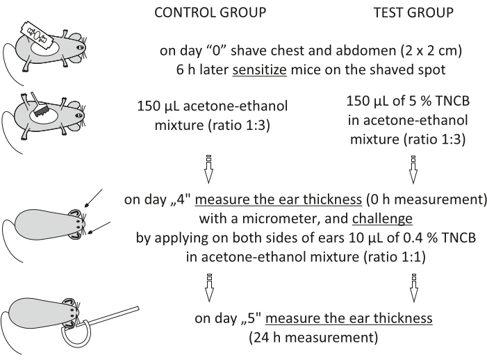
Figure 1: Induction of CHS. Sensitization, challenge, and ear measurement. Abbreviations: CHS = contact hypersensitivity reaction; TNCB = 2,4,6-trinitrochlorobenzene. Please click here to view a larger version of this figure.
to:
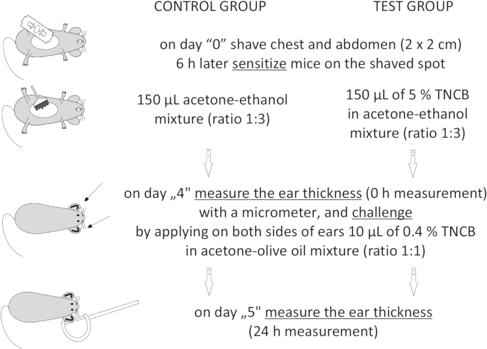
Figure 1: Induction of CHS. Sensitization, challenge, and ear measurement. Abbreviations: CHS = contact hypersensitivity reaction; TNCB = 2,4,6-trinitrochlorobenzene. Please click here to view a larger version of this figure.
Figure 2 of the Representative Results has been updated from:
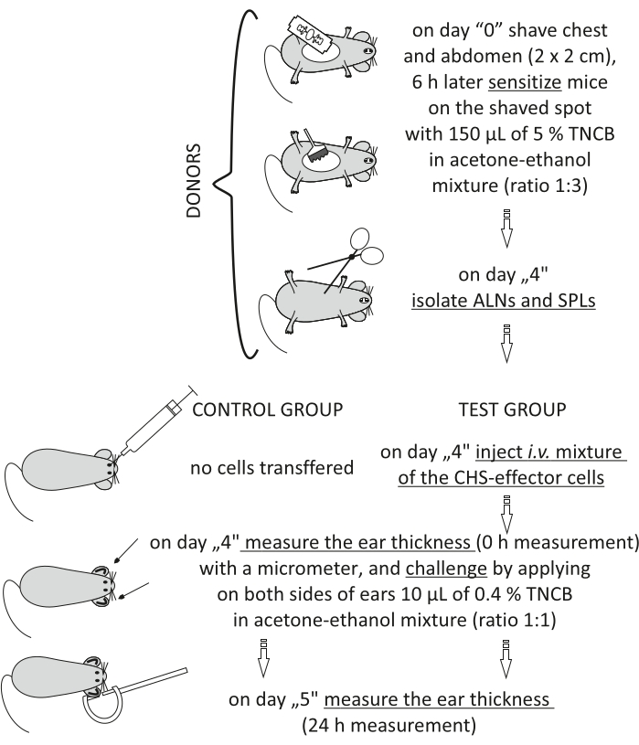
Figure 2: Adoptive transfer of the CHS-effector cells. Abbreviations: ALNs =axillary and inguinal lymph nodes; CHS = contact hypersensitivity reaction; i.v. = intravenously; SPLs = spleens; TNCB = 2,4,6-trinitrochlorobenzene. Please click here to view a larger version of this figure.
to:
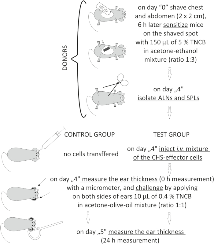
Figure 2: Adoptive transfer of the CHS-effector cells. Abbreviations: ALNs =axillary and inguinal lymph nodes; CHS = contact hypersensitivity reaction; i.v. = intravenously; SPLs = spleens; TNCB = 2,4,6-trinitrochlorobenzene. Please click here to view a larger version of this figure.
