


Overview
Source: Laboratories of Dr. Ian Pepper and Dr. Charles Gerba - The University of Arizona
Demonstrating Author: Bradley Schmitz
Fungi are heterotrophic eukaryotic organisms, and with the exception of yeasts, are aerobic. They are abundant in surface soils and are important for their role in nutrient cycling and the decomposition of organic matter and organic contaminants. White rot fungi (phanerochaete chryosporium) for example, (Figure 1) are known to degrade aromatics.
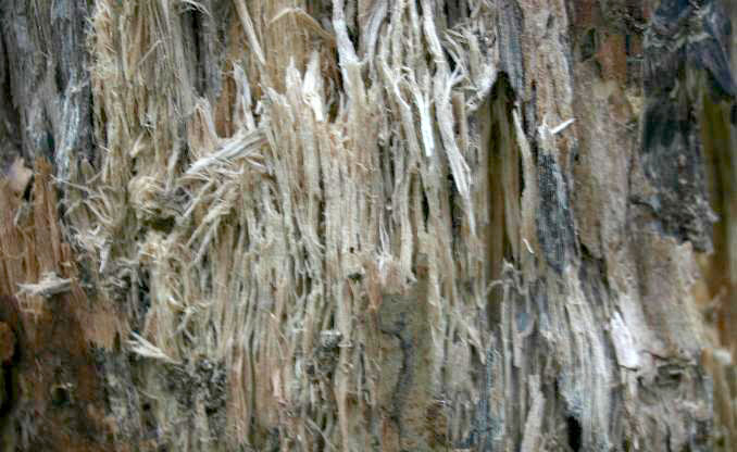
Figure 1. White rot on birch.
Principles
Soils generally contain millions of fungi per gram, so the soil is typically diluted using a dilution series. A dilution series is made by suspending a given amount of soil in a dispersing solution, such as deionized water. The aliquots of the suspensions are then transferred to fresh solution, until the suspension is diluted sufficiently to allow individual discrete fungal colonies to grow on the agar plates. (Figure 2)
After inoculation on several replicate agar plates, the plates are incubated at 25 °C. After the macroscopic fungal colonies are formed, they are counted, as shown in Figure 3. Because the assumption is that one fungal colony is derived from one organism, the term Colony Forming Units (CFUs) is used in the final analysis, with the results expressed in terms of CFUs per gram of oven dry soil.
Normal culturable fungal counts from fertile soil are in the range of 106-106 fungal “propagules” (spores, hyphae, or hyphal fragments) per gram of dry soil. Culturable plate counts have been in use for enumerating organisms since the nineteenth century. They continue to be used today, as they are inexpensive to perform, require little labor, are quick, and are fairly reproducible. However, they do suffer from a number of errors, which must be considered when evaluating the results. The most significant of these errors is the fact that many organisms will not culture on media plates.

Figure 2. Dilution and plating technique. Here, the diluted soil suspension is incorporated directly in the agar medium rather than being surface applied as in the case of spread plating. From Environmental & Pollution Science, 2nd Ed., Academic Press, San Diego, CA

Figure 3. Soil fungi isolated from a surface soil grown in a Petri dish containing Rose Bengal Agar. Photo courtesy K.L. Josephson. From Environmental & Pollution Science, 2nd Ed., Academic Press, San Diego, CA.
Subscription Required. Please recommend JoVE to your librarian.
Procedure
1. Soil Sample Preparation
- First, determine the initial moisture content of the soil by overnight drying of a known amount of the moist soil, and reweighing the dried soil. The equation to determine the initial moisture content of the soil is:

(Equation 1)
where:
MC = moisture content
W = net weight
D = dry weight - Calculate the amount of water that must be added to 25 g of soil to increase the soil moisture content to 10% on a dry weight basis.
- Next, add that amount of water to 25 g of the soil.
- Cover the container with plastic wrap and puncture the film several times with a probe to allow aeration during incubation. Secure the film with a rubber band.
- Weigh the soil and wrap, then incubate the sample at room temperature for one week.

2. Fungus Inoculation and Incubation
- After the incubation is complete, weigh the soil sample with the wrap and rubber band. Calculate the weight loss due to moisture loss.
- Calculate the new soil moisture content.
- Next, prepare a 1/10-dilution series of the soils as shown in Figure 2. This will result in dilutions of 10-1 (bottle A), 10-2 (tube B), 10-3 (tube C), 10-4 (tube D), and 10-5 (tube E) g soil per mL suspensions.
- Prepare sterile Rose Bengal-streptomycin agar plates. Both the Rose Bengal and streptomycin inhibit bacterial growth. For very fertile soils where soil microbial populations are high, the chosen dilutions should be higher, i.e., 10-3, 10-4, and 10-5, g soil per plate.
- To each of the 10-3 plates, add 0.1 mL from dilution tube 10-2, and spread over the agar surface using an ethanol-flame sterilized glass spreader. Because a quantity of 0.1 mL is plated, this makes the effective dilution 10-3. Repeat these steps for all dilutions.
- Incubate plates at room temperature for one week.
3. Colony Counting and Examination by Microscopy
- Make colony counts at one and only one dilution of each soil. The plates that are counted should have discreet countable colonies. Overgrown plates should not be counted. Likewise, plates with less than 10 colonies should not be counted. Note and describe the cultural characteristics of three different colonies.
- Prepare pressure tape (transparent tape) mounts on slides for detailed microscope study using the following procedure:
- Deposit a drop of lactophenol mounting fluid at the center of a clean glass slide
- Cut a strip of clear cellophane tape about 3 cm long from the stock roll. To avoid contaminating the adhesive surface, use forceps when handling the tape. A dissecting needle will aid in freeing the tape from the forceps.
- The adhesive side of the tape is applied to the surface of a sporulating fungus colony. Take care to avoid excessive pressure on the tape or too dense a mass of hyphae and spores will be collected.
- Remove the tape from contact with the fungus colony and apply it, adhesive side down, to the drop of mounting fluid on the glass slide. Rub the tape gently with a smooth, flat instrument to express air bubbles.
- Examine the tape mount microscopically under the oil immersion objective with oil.
- Identify two different fungal genera using the supplied fungal identification key (Figure 4).

Figure 4. Fungal identification key.
Filamentous fungi are multicellular, eukaryotic organisms often found in large numbers in soil, playing an important role in the ecosystem. Fungi can be isolated, quantified, and examined in the laboratory.
Fungi are abundant in surface soils and perform important roles in nutrient cycling and decomposition of organic matter and contaminants. Being unable to synthesize their own food they are classified as heterotrophic and are aerobic and therefore require oxygen.
To culture fungi, soil samples are diluted at known concentrations into sterile water, then plated onto agar plates and incubated. The resulting fungal colonies may then be counted and identified.
This video will illustrate the principles behind the isolation, quantification, and identification of filamentous fungi.
Soils generally contain millions of fungi per gram; therefore, serial dilutions are commonly used to prepare soil samples for analysis. A given amount of soil is added to a dispersing solution. Aliquots of the suspension are transferred to a buffer solution in series until the suspension is diluted enough to allow discrete fungal colonies to grow.
These dilutions are then plated onto agar media and incubated. Once they have formed visible fungal colonies, they can be counted. As it is assumed that each fungal colony is derived from a single organism, the term Colony Forming Units, or CFU's, is used to express the number of organisms calculated per gram of dry soil.
Most fungi grow as hyphae, which are long, branching structures that are the main mode of vegetative growth. Aggregations of hyphae are referred to as mycelium. Fungi may also exist as single celled organisms, with yeast being a well-known example.
Fungal reproduction can occur in one of several ways. Hyphae-producing fungi can reproduce asexually, by fragmenting hyphae, or from stalks with seed-like spores. Single-celled fungi may reproduce asexually by budding.
Fungi can also reproduce sexually. This is less common than asexual reproduction, and usually occurs in response to unfavorable environmental conditions. In hyphae-producing fungi, two reproductive hyphae from different fungal strains can fuse, creating a zygote. In single celled fungi, two cells of opposite strains will fuse.
Fertile soils normally contain in the range of 106 fungal "propagules" per gram of dry weight. Propagules are fungal spores, hyphae, or hyphal fragments. Using culturable plate counts is a quick, inexpensive, and reproducible technique to elucidate fungal content. However, results can be skewed by the fact that not all organisms will culture on media plates.
Rose-Bengal agar plates with antibiotics, such as streptomycin, are typically used for fungal cultures. Because bacteria are much more numerous than fungi in most soils, the ideal fungal media will select against bacterial growth while allowing the fungi to survive. Rose-Bengal dye inhibits the growth of most bacteria, and reduces the size of fungal colonies. This is ideal, and prevents overgrowth of fungal plates as well as controlling bacteria.
Now that we are familiar with the principles behind culturing filamentous fungi, let's take a look at how this is carried out in the laboratory.
Once the soil samples have been collected, bring them to the laboratory for analysis. First, calculate the moisture content of the soil. Add deionized water to bring the moisture content to 15%.
Cover the containers with plastic wrap and secure with a rubber band to limit evaporation. Puncture the film several times to allow aeration.
Weigh the soil sample and container and record. Incubate at room temperature for one week to allow growth of the naturally occurring fungi.
Re-weigh the soil samples and container and record the weight. Calculate weight loss due to moisture loss over the week. Add water to the soil, to bring the weight back to the original value.
Add 10 g of moist soil to 95 mL of distilled water and mix thoroughly. This is the 10-1 dilution since 10 g of soil has a volume of roughly 5 mL. Perform four serial dilutions of 1 mL into 9 mL blanks using water from this stock solution.
Next, collect eight sterile Rose-Bengal-streptomycin agar plates. To two of the plates, add 0.1 mL from dilution tube 10-2, and spread over the agar surface using an ethanol-flame sterilized glass spreader. Because a quantity of 0.1 mL is plated, this makes the effective dilution 10-3.
Incubate these plates in a dark cabinet at room temperature, for one week.
After one week, count the discrete colonies on each plate. Plates with 10 to 20 fungal colonies should be counted.Note and describe distinguishing features of the plates or colonies.
Take a clean glass microscope slide and deposit a drop of lactophenol mounting fluid into the center. Using forceps to avoid contamination, cut a strip of clear cellophane tape about 3 cm long. If necessary, use a dissecting needle to free the tape from the forceps.
Apply the adhesive side of the tape to the surface of a sporulating fungus colony, taking care to avoid excessive pressure. Next, apply the adhesive side of the tape with spore sample to the mounting fluid on the glass slide. Rub the tape gently with a smooth, flat instrument to remove air bubbles.
Examine the tape mount under a high-powered objective.
Fungi can be identified microscopically by examination of the fruiting bodies and spores. A fungi identification key can assist this process. Common fungi types observed include Penicillium and Aspergillus.
Using the plates, the fungi can be enumerated using the same technique as with bacterial enumeration.
Identifying and culturing fungi is useful for a variety of scientific applications.
Industries like pulp and paper producing facilities may use filamentous fungi to degrade wood in their pulping process, in a technique known as biopulping. Fungi such as white-rot may degrade wood chips efficiently, leading to decreased need for the use of chemical or mechanical pulping.
Fungi can also be used to break down other materials, including certain types of biodegradable plastics. This can be useful for agricultural or gardening applications, where biodegradable mulch films may be used to create temporary barriers to unwanted plant growth.
Fungi are also producers of antibiotics, compounds that revolutionized medicine in the 20th century and continue to be a front line defense in infection today. Penicillin, a widely used antibiotic, was isolated from the common filamentous fungi Penicillium rubens. To this day, filamentous fungi are still isolated and studied in the search for new antibiotics.
You've just watched JoVE's introduction to filamentous fungi. You should now understand how to culture filamentous fungi from soil, how to quantify them, and how to identify types of fungi commonly found in soil. Thanks for watching!
Subscription Required. Please recommend JoVE to your librarian.
Results
Colony Counts
The number of fungal colonies per gram of soil is equal to the number of colonies counted on the plate multiplied by the reciprocal of the dilution plated. For example, if 46 colonies are counted at a dilution of 10-5, then the CFU per gram of soil is 46 x 105 or 4.6 x 10⁶.
Identification of Three Different Fungal Genera
Fungi can be identified microscopically by examination of the fruiting bodies and spores. A fungi identification key can assist this process. (Figure 4) Common fungi types observed include Penicillium and Aspergillus.
Subscription Required. Please recommend JoVE to your librarian.
Applications and Summary
Dilution and plating of soil fungi can be used as an indication of the health of a soil. Normally a “healthy” fertile soil will have 105 to 106 fungi per gram of soil. It can also be utilized to isolate pure cultures of specific fungi, subsequently evaluated for specific properties, such as the ability to degrade organic compounds. These can be detrimental as in the case of white rot fungi, or beneficial when toxic organics are degraded through biodegradation. Other uses of pure cultures of fungi include the isolation of fungi for antibiotics. For example, the first antibiotic ever was penicillin, produced by the soil-borne fungus Penicillium. This was first discovered by Sir. Alexander Fleming in 1929.
Subscription Required. Please recommend JoVE to your librarian.
References
- Pepper, I.L., Gerba, C.P., Brusseau, M. Environmental & Pollution Science, 2nd Ed. Academic Press, San Diego, CA. (2006).
- Pepper, I.L., Gerba, C.P. Environmental Microbiology, A Laboratory Manual, 2nd Ed. Academic Press, Boston, MA. (2005).