Described herein is a protocol to isolate and further study the infiltrating leukocytes of the decidua basalis and decidua parietalis - the human maternal-fetal interface. This protocol maintains the integrity of cell surface markers and yields enough viable cells for downstream applications as proven by flow cytometry analysis.
Method Article
Isolation of Leukocytes from the Human Maternal-fetal Interface
In This Article
Summary
Abstract
Pregnancy is characterized by the infiltration of leukocytes in the reproductive tissues and at the maternal-fetal interface (decidua basalis and decidua parietalis). This interface is the anatomical site of contact between maternal and fetal tissues; therefore, it is an immunological site of action during pregnancy. Infiltrating leukocytes at the maternal-fetal interface play a central role in implantation, pregnancy maintenance, and timing of delivery. Therefore, phenotypic and functional characterizations of these leukocytes will provide insight into the mechanisms that lead to pregnancy disorders. Several protocols have been described in order to isolate infiltrating leukocytes from the decidua basalis and decidua parietalis; however, the lack of consistency in the reagents, enzymes, and times of incubation makes it difficult to compare these results. Described herein is a novel approach that combines the use of gentle mechanical and enzymatic dissociation techniques to preserve the viability and integrity of extracellular and intracellular markers in leukocytes isolated from the human tissues at the maternal-fetal interface. Aside from immunophenotyping, cell culture, and cell sorting, the future applications of this protocol are numerous and varied. Following this protocol, the isolated leukocytes can be used to determine DNA methylation, expression of target genes, in vitro leukocyte functionality (i.e., phagocytosis, cytotoxicity, T-cell proliferation, and plasticity, etc.), and the production of reactive oxygen species at the maternal-fetal interface. Additionally, using the described protocol, this laboratory has been able to describe new and rare leukocytes at the maternal-fetal interface.
Introduction
Pregnancy is characterized by three distinct immunological phases: 1) implantation and early placentation associated with a pro-inflammatory response (i.e., implantation resembles an ‘open wound’); 2) the second trimester and most of the third trimester of pregnancy when immune homeostasis is achieved through a predominantly anti-inflammatory state at the maternal-fetal interface; and 3) parturition, a pro-inflammatory state1-7. Immune cells play an important role in the regulation of the inflammatory response at the maternal-fetal interface where their abundance and localization change throughout pregnancy6-9.
In humans, the maternal-fetal interface represents an area of direct contact between maternal (decidua) and fetal (chorion or trophoblast) tissues. This interface includes: 1) the decidua parietalis that lines the uterine cavity not covered by the placenta and is in juxtaposition to the chorion laeve; and 2) the decidua basalis, located in the basal plate of the placenta where it is invaded by interstitial trophoblasts10 (Figure 1). The intimacy of these areas of contact creates conditions for fetal antigenic exposure to the maternal immune system11-13. Not surprisingly, leukocytes comprise up to 30-40% of the decidual cells8,9,14,15 in addition to typical stromal-type cells and glandular cells8,14,16. The role of leukocytes at the maternal-fetal interface encompasses multiple processes that include the limitation of trophoblast invasion17, remodeling of spiral arteries18,19, maintenance of maternal tolerance12,20, and initiation of labor21-26. Leukocytes of both the adaptive and innate limbs of the immune system, i.e., T cells, macrophages, neutrophils, B cells, dendritic cells, and NK cells, have been identified in the decidual tissues, and their proportions and activation status have been shown to vary spatially and temporally throughout gestation6-10,12,14,24,27-30. Perturbations in the leukocyte population and/or function are associated with spontaneous abortion31, preeclampsia32, intrauterine growth restriction32,33, and preterm labor7,24. Therefore, the study of the phenotypic characteristics and functionality of leukocytes at the human maternal-fetal interface will facilitate the elucidation of the immunological pathways dysregulated in pregnancy disorders.
One of the most powerful tools used to determine the phenotype and functional properties of leukocytes is flow cytometry, technology that allows the quantitative analysis of multiple parameters simultaneously34-36. To analyze leukocytes by flow cytometry, isolation of the leukocytes in a single-cell suspension is required. Therefore, a method to separate infiltrating leukocytes from the maternal-fetal interface is needed to study their phenotypic and functional properties.
Several methods have been described to isolate leukocytes from the human maternal-fetal interface10,14,25,27,37-39. While some apply mechanical disaggregation10,25,27,38, others use enzymatic digestion37,40 for tissue dissociation. Because mechanical disaggregation produces a lower yield and reduced viability41, and enzymatic dissociation can affect viability and cell surface marker retention42, the method described herein combines gentle mechanical dissociation with enzymatic pre-treatment to increase the yield of isolated leukocytes without compromising cell viability. A similar combination of methods has been demonstrated to be effective in the isolation of leukocytes from the decidual tissues at the maternal-fetal interface39. Therefore, the protocol described herein involves mechanical disaggregation with an automatic tissue dissociator that increases consistency while saving time and labor when compared to traditional mincing with opposing scalpels, razor blades, or surgical scissors10,28. The enzyme chosen for tissue dissociation was Accutase. Unlike commonly used collagenase43, dispase44, and trypsin45, Accutase (a cell detachment solution) combines both general proteolytic and collagenolytic activities that contribute to efficient yet gentle dissociation46,47. After dissociation, the leukocytes are enriched from the total population of the decidual cells by density gradient centrifugation. Various density gradient media have been previously utilized, the most common of which are Percoll (a suspension of colloidal silica particles)48 and Ficoll (a polymer of sucrose with a high synthetic molecular weight)49. The superior efficiency of isolation by the sucrose polymer has been previously shown50, and the protocol described herein further proves that this density gradient media produces a sufficiently high purity of mononuclear leukocytes.
Hence, the protocol described herein combines the mechanical tissue disaggregation with an automatic tissue dissociator, enzymatic digestion with a cell detachment solution, and leukocyte separation with a density gradient media (1.077 + 0.001 g/ml) to isolate leukocytes from human decidual tissues. This protocol has been proven to preserve cell surface antigens along with cell viability. The isolated leukocytes can be used for multiple applications that include immunophenotyping with flow cytometry and functional studies in vitro.
Protocol
This protocol is appropriate for leukocyte isolation from the decidua basalis and decidua parietalis in preparation for immunophenotyping by flow cytometry. Furthermore, the isolated cells can be used for cell sorting, cell culture, RNA isolation, and cytology. Before working with the samples mentioned in this protocol, human ethical approval must be obtained from the Local Research Ethics Committee and Institutional Review Boards. The collection and utilization of human samples for research purposes were approved by the Institutional Review Boards of the Eunice Kennedy Shriver National Institute of Child Health and Human Development (NICHD), National Institutes of Health (NIH), Department of Health and Human Services (DHHS, Bethesda, MD, USA) and Wayne State University (Detroit, MI, USA). Written informed consent was obtained from all pregnant women prior to the collection of tissue samples.
NOTE: While working with animal blood, cells, or hazardous agents as mentioned in this protocol, it is essential that proper biosafety and laboratory safety actions be followed.
1. Dissection of the Human Decidual Tissues
NOTE: The basal plate is the base beneath and attached to the placenta and represents the maternal surface. The chorioamniotic membranes include the amnion and the chorion. The basal plate includes the decidua basalis and the chorion includes the decidua parietalis (Figure 1).
- Decidua Basalis
- Dissect a piece of the basal plate from one cotyledon of the placenta (Figures 1A and 1B).
- Place it on a sterile cutting board with the placental villi facing upward. The side showing the placental villi is usually bloody red with hairy tissue in appearance. The basal plate is smooth and pale red in color.
- Use sharp, fine-point scissors and forceps to remove the villous tissue and blood vessels. Keep the tissue soaked in 1x PBS (Ca2+- and Mg2+- free) during the process (Figure 1C). Collect 2 to 3 of these pieces and rinse them thoroughly with 1x PBS to remove the blood (Figure 1D).
- Decidua Parietalis
- Dissect a piece of the chorioamniotic membranes (approximately 10 cm x 10 cm, Figure 1E). Place it on the board with the chorion facing upward. The chorionic side contains bloody clots and is usually light yellowish in color.
- Use fine-point forceps to remove as many of the blood clots as possible (Figure 1E). Rinse the membrane in sterile 1x PBS to clean the excess blood from the membrane until the 1x PBS runs clear.
- Use a disposable sterile cell scraper to gently scrape the decidual layer from the membrane. Apply sterile 1x PBS on the membrane while completing this process (Figure 1F). Collect the decidual tissues and place them in a 50 ml plastic tube with sterile 1x PBS (Figure 1G).
2. Mechanical Disaggregation and Enzymatic Digestion
- Wash the tissue dissected from the decidua basalis (from Step 1.1.3) or the decidua parietalis (from Step 1.2.3) with sterile 1x PBS in a 50 ml plastic tube. Collect tissue pellets by centrifugation at 300 x g for 5 min at RT.
- Aspirate the supernatant located above the tissue pellet carefully without disturbing the pellet.
NOTE: At this point, the pellets are very loose because they contain red blood cells. If the volume of the pellet is about or less than 3 ml, re-suspend the pellet in 6 ml of cell detachment solution pre-warmed to 37 °C. If the pellet is larger than 3 ml of volume, add more cell detachment solution (add about 2 times the volume of the tissue samples). - Transfer the homogenized tissues (decidua basalis + cell detachment solution or decidua parietalis + cell detachment solution) to a C tube.
- Place the C tube in the automatic dissociator and run the corresponding program.
NOTE: The program for the decidua basalis runs for 17 sec with 668 total rounds per run. The program for the decidua parietalis runs for 37 sec with 235 total rounds per run. These programs have been customized to isolate leukocytes from the decidua basalis or the decidua parietalis with good yield and cell viability. - Following the mechanical disaggregation of the tissues, digest the tissues with a commercially available cell detachment solution such as accutase; (a cocktail that contains proteolytic and collagenolytic enzymes) for 45 min at 37 °C with gentle agitation.
3. Isolation of Leukocytes
- Following incubation, add 10 ml of 1x PBS to the digestion mixture and pass it through a 100 µm cell strainer into a 50 ml plastic tube. Depending on the stickiness of the digestion mixture, one may need to use several cell strainers for one mixture.
- Fill the tube with 1x PBS and centrifuge at 300 x g for 5 min at RT. Remove the 1x PBS above the cell pellets carefully and re-suspend the pellets with 5 ml of ice-cold FACS buffer.
NOTE:To study the polymorphonuclear and mononuclear leukocytes together, go to Step 6.1; to study the mononuclear leukocytes, continue to Step 3.3. - Add 5 ml of 20% density gradient media (1.077 + 0.001 g/ml) to a 15 ml plastic tube and slowly overlay the cell suspension on top of the density gradient media. While layering the sample, it is important not to mix the density gradient media with the cell suspension.
- Centrifuge with a swing-out rotor at 500 x g for 30 min at 4 ˚C without the brake. It is very important to turn off the brake to avoid mixing the cells and losing the gradient. Leukocytes will be found in the interface between the density gradient media and the FACS buffer.
- Remove the upper layer that contains the FACS buffer and cell debris very carefully using a pipette. The band at the interface contains the decidual mononuclear cells and a portion of the polymorphonuclear leukocytes.
- Transfer the cells at the interface completely to a new 15 ml plastic tube. Add at least 3 times more volume of FACS buffer.
- Centrifuge at 300 x g for 5 min to pelletize the cells. Aspirate the supernatant and wash the cells with FACS buffer. Pelletize the cells with another centrifugation at 300 x g for 5 min at RT.
NOTE: To perform the cell culture, see Step 4. To perform cell sorting, see Step 5. To perform immunophenotyping, see Step 6.
4. Applications – Cell culture
- Re-suspend the cells from Step 3.7 in 1 ml of RPMI culture medium 1640. Count the viable cells using an automatic cell counter or a hemocytometer and a trypan blue solution 0.4%.
- Add up the amount of RPMI culture medium to make a cell concentration of 1 x 10⁶ cells/ml. Cells are now ready for culture.
5. Applications – Isolation of Macrophages for Primary Cell Culture
- To isolate CD14+ cells, magnetically label the cells from Step 3.7 with CD14 microbeads (20 µl beads per 107 cells), incubate for 15 min at 4 ˚C, and separate CD14+ cells from other types of cells by applying a magnetic field. After 2 washes with MACS buffer and removal of the magnetic field, elute the magnetically retained CD14+ cells with MACS buffer.
- Following centrifugation, re-suspend the cell pellets in RPMI culture medium 1640. The cells are ready for plating (Figure 2).
6. Applications – Immunophenotyping
- To use the cells for immunophenotyping, re-suspend the cells from Step 3.7 in 1 ml of 1x PBS. Count the viable cells using an automatic cell counter or a hemocytometer and a trypan blue solution 0.4%.
- Label the cells with a fixable viability dye (Figure 3). Wash the cells twice with FACS buffer and re-suspend the cells in stain buffer. Incubate with an FcR blocker for 15 min at 4 °C.
- Add extracellular antibodies conjugated to fluorochromes. For example, in this protocol, use anti-CD3, anti-CD19, anti-CD14, anti-CD15, and anti-CD56 antibodies (Table 1). Incubate for 30 min at 4 °C in darkness.
- After 2 washes with 1x PBS, the cells will be analyzed in 500 µl of stain buffer using a flow cytometer. A representation of the gating strategy used to analyze the leukocyte sub-populations found in the decidual tissues is shown in Figure 4.
Results
The dissection of human tissues at the maternal-fetal interface (decidua basalis and decidua parietalis) is shown in Figure 1. This procedure includes the dissection of the basal plate, which includes the decidua basalis (Figure 1A-D). The decidua basalis is obtained by removing the placental villi (fetal side) from the basal plate (Figure 1C). The decidua parietalis is collected by gently scraping the chorionic membrane (Figure 1E-F). Figure 2 shows the morphology of isolated macrophages (CD14+) collected from the decidua parietalis in a term pregnancy using magnetic cell sorting. Isolated macrophages maintained the ability to release cytokines after 3 days of culture (data not shown). The yield of viable cells isolated from the decidua basalis and decidua parietalis is shown in Figure 3, and it is greater than 90% in both. Figure 4 shows the gating strategy for analyzing polymorphonuclear and mononuclear leukocytes within the viability gate, including T cells (CD3+), neutrophils (CD15+), macrophages (CD14+), NK cells (CD56+), NKT cells (CD3+CD56+), and B cells (CD19+) at term pregnancy. Figure 5 shows neutrophils, macrophages, T cells, and B cells in isolated decidual cells in a term pregnancy. Figure 6 shows NK, NKT, and T cells in isolated decidual cells in a term pregnancy.

Figure 1. Human maternal-fetal interface: placenta and chorioamniotic membranes. (A) Basal plate is dissected from the placenta; (B) basal plate is separated from the placental villi; (C) placental villi is trimmed from the basal plate including the decidua basalis; (D) decidua basalis is rinsed in 1x PBS; (E) a fragment of the chorionic membrane is obtained and blood clots are gently removed; (F) decidua parietalis is gently scraped; and (G) decidua parietalis (dotted line) is separated from the chorionic membrane. Please click here to view a larger version of this figure.
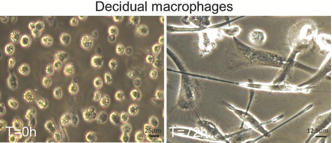
Figure 2. Decidual macrophages in culture. Macrophages (CD14+ cells) were isolated from the decidua parietalis at term pregnancy. Macrophages were plated into plastic chamber slides with RPMI1640 + 10% FBS + 1%Penicillin–Streptomycin + 50 ng/ml MCSF. Photos were taken at the time of plating (A) and after 3 days in culture (C). Please click here to view a larger version of this figure.
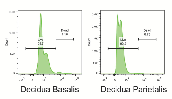
Figure 3. Total cell viability. Viability of total cells isolated from the decidua basalis and the decidua parietalis was determined following density gradient using a fixable viability dye (step 6.2). Viability of isolated cells is >95% according to the live/dead cell viability staining. Please click here to view a larger version of this figure.
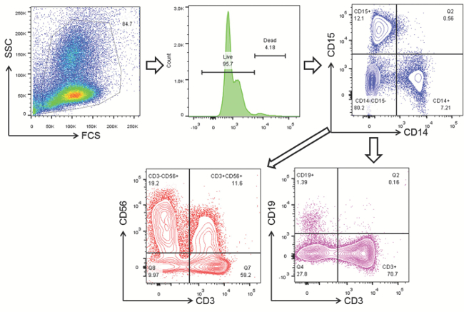
Figure 4. Gating strategy for decidual leukocyte sub-populations. Leukocytes were gated with FSC and SSC. Live cells were then gated according to the viability gate (Live). Viable cells were separated into neutrophils (CD15+) and macrophages (CD14+). The CD14-CD15- population was then further separated into NK cells (CD3-CD56+), NKT cells (CD3+CD56+), T cells (CD3+), and B cells (CD19+). Please click here to view a larger version of this figure.
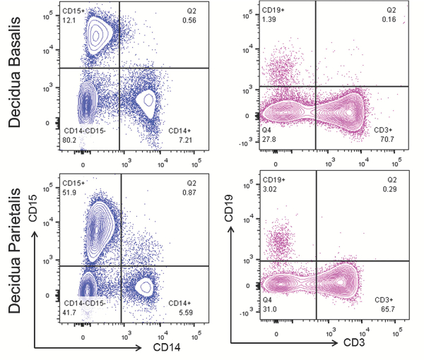
Figure 5. Leukocyte sub-populations in the decidual tissues. Neutrophils (CD15+) and macrophages (CD14+) were gated within the viability gate; T cells (CD3+) and B cells (CD19+) were gated within the CD14-CD15- gate. Please click here to view a larger version of this figure.
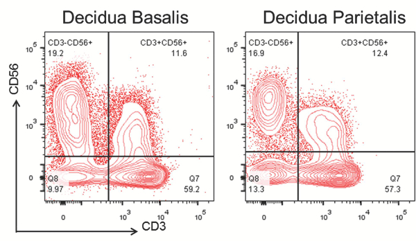
Figure 6. Rare leukocyte sub-populations in the decidual tissues. NK cells (CD56+) and NKT cells (CD56+CD3+) were gated within the CD14-CD15- gate. Please click here to view a larger version of this figure.
| Antibody list | Company | Catalog No. |
| BD Pharmingen Anti-human CD56-PE-Cy7 | BD Biosciences | 557747 |
| BD Horizon Anti-human CD15-BV605 | BD Biosciences | 562980 |
| BD Horizon Anti-human CD14-BUV395 | BD Biosciences | 563561 |
| BD Horizon Anti-human CD19-BUV737 | BD Biosciences | 564303 |
| Brilliant Violet 650 Anti-human CD3 Antibody | Biolegend | 317323 |
Table 1. List of antibodies utilized for leukocyte subset immunophenotyping
Discussion
Characterization of the functional and phenotypic properties of infiltrating leukocytes at the human maternal-fetal interface is essential to the understanding of the immune mechanisms that lead to pregnancy disorders. Several techniques have been described in order to isolate leukocytes from the human maternal-fetal interface throughout pregnancy 10,14,25,28,37,42,43. However, each of these techniques is distinct, uses different enzymes or enzyme combinations, requires different dissociation times, does not specify quantities of tissue, and, most importantly, does not always specify the viability of the isolated cells. The protocol described herein allows the isolation of infiltrating leukocytes at the human maternal-fetal interface (decidua basalis and decidua parietalis) resulting in a high yield of viability and provides detailed information about the commercial reagents, buffer preparation, tissue quantities, and incubation times validated in our laboratory.
The first critical step of the leukocyte isolation process is tissue dissociation; this step involves mechanical desegregation and/or enzymatic dissociation. Mechanical desegregation is successfully performed by scraping the chorion when isolating the decidua parietalis (Figure 1F) and by trimming the placental villi from the basal plate when isolating the decidua basalis (Figure 1C)10,28. Therefore, the protocol described herein includes both procedures as initial steps. However, it is important to consider that the basal plate (maternal tissues) and the placental villi (fetal tissues) are intimately attached, which can cause contamination of fetal leukocytes when isolating leukocytes from the decidua basalis. To avoid fetal contamination, the protocol herein recommends washing the trimmed basal plate two or three times (Figure 1D). Following cell scraping or trimming from the decidua basalis or decidua parietalis, an additional step for mechanical tissue disaggregation has been recommended39. This step is recommended because the infiltrating leukocytes at the maternal-fetal interface express cell adhesion molecules that firmly attach them to the extracellular matrix6,7. Therefore, the protocol described herein involves the mechanical disaggregation with an automatic tissue dissociator that increases consistency while saving time and labor when compared to traditional mincing with scalpels, razor blades, or surgical scissors10,28.
A second critical step in the tissue dissociation process is enzymatic dissociation. Single enzymes and a combination of different enzymes have been used to isolate infiltrating leukocytes from the decidua basalis and decidua parietalis10,14,25,28,37,42,43. In many cases, those enzymes prepared to a certain concentration by hand in the laboratory may be subject to human error. Here, instead, a ready-to-use purified collagenase/neutral protease cocktail, Accutase, has been implemented in the laboratory; as a commercial enzyme, it has been shown to provide reliable results in cell culture51. Accutase (a cell detachment solution) is known to effectively detach macrophages from culture plates without scraping and, most importantly, without losing surface antigens51. This prepared enzyme has also been used to process the digestion of human and animal nervous system tissues, resulting in viable isolated cells that remain sustainable for a long period that, over time, allows for their cell culture52. Moreover, this solution also preserves CD24 antigenicity in cells isolated from central nervous system tissues52. When compared to Liberase-1, another cocktail of collagenase and neutral protease, neither Accutase nor Liberase-1 generate free DNA aggregates; however, Accutase is gentler than Liberase-1 during tissue dissociation52. Because of this, Accutase does not dissociate cell aggregates after the digestion52. To overcome this limitation, the protocol described herein includes the automatic tissue disaggregation of the decidua basalis and decidua parietalis prior to the tissue dissociation with the solution. Accutase has also demonstrated its superiority to trypsin in the preservation of CD44, a cancer stem cell surface marker53. Our laboratory’s studies have consistently noted that the detachment solution preserves the surface antigens. Indeed, differences between pregnancy disorders and normal pregnancies have been found in the expression of several extracellular and intracellular markers in macrophages (CD14+ cells), neutrophils (CD15+ cells), T cells (CD3+ cells), and B cells (CD19+ cells), including CD80, CD86, CD163, CD209, ICAM3, CD45RO, CD45RA, CXCR3, CCR7, CCR6, CD4, CD8, CD25, FOXP3, CD24, CD38, CD27, CD38, CD43, CD23, IgM, IgD, CD5, T-bet, GATA-3, and RORγt, and in cytokine release. Therefore, the protocol described herein is optimal for immunophenotyping of infiltration leukocytes at the human maternal-fetal interface as shown in the representative results.
An important advantage of the protocol described herein is that it allows for the isolation of leukocytes with a high yield of viable cells. The representative data shows that > 90% of the isolated cells are viable. This is of great importance as this protocol has allowed the study of the functional properties of the cells isolated from human tissues at the maternal-fetal interface. For example, cultures of the decidual macrophages and the study of cytokine release under stimulation have been performed using this protocol.
To achieve successful results using the described protocol, it is important to consider the following factors: 1) tissue collection must be performed within 1-2 hr after delivery, and these tissues must be placed in a container with 1x PBS at 4°C to preserve the viability of the isolated cells; 2) mechanical tissue disaggregation must be performed using an automatic homogenizer at validated times, as described in this protocol; 3) the duration of incubation with the cell detachment solution must be less than 1 hr since its activity after this time diminishes; 4) the temperature of incubation with the cell detachment solution must be maintained at 37 °C to obtain the optimal activity of this enzyme; 5) cell pellet manipulation must be done gently with micropipettes because use of the vortex can damage the integrity of the cells (remember that isolated cells are differentiated and abrupt manipulation can easily reduce their viability); 6) buffer and centrifuge temperatures must be kept at the same temperature as the cell suspension; 7) isolated cells must be processed for immunophenotyping or used immediately as their viability reduces rapidly; and 8) when performing immunophenotyping, samples must be acquired using a flow cytometer immediately for best results.
One of the limitations of this protocol is the cost of Accutase, which is more expensive than other enzymes with similar functions, e.g., trypsin, dispase II, and collagenase. However, the advantages that Accutase displays over and above those enzymes are superior. A second limitation of the protocol is the requirement of an automatic tissue dissociator, which is not a common laboratory instrument and, therefore, can be costly. To overcome this limitation, the tissue desegregation can also be performed using small surgical scissors; however, this step cannot exceed 3-5 minutes since longer periods have been demonstrated to reduce the yield of viable cells. All dissociations must be performed by the same researcher to minimize variability.
In summary, the protocol herein offers a novel approach that combines the use of gentle mechanical and enzymatic tissue dissociation techniques to preserve the integrity of extracellular and intracellular markers in leukocytes isolated from the human tissues at the maternal-fetal interface. Aside from immunophenotyping, cell culture, and cell sorting, the future applications of this protocol are numerous and varied. We have been successfully using this protocol to isolate decidual leukocytes for in vitro studies of leukocyte functionality (i.e., cytotoxicity, T-cell proliferation and plasticity, etc.), and production of reactive oxygen species in the leukocytes isolated from decidual tissues (data not shown). Indeed, using the described protocol, our laboratory has been able to describe new and rare leukocytes at the maternal-fetal interface.
Disclosures
The authors disclose no conflicts of interest.
Acknowledgements
This work was supported by the Eunice Kennedy Shriver National Institute of Child Health and Human Development, NIH/DHHS. This work was also supported, in part, by the Wayne State University Perinatal Initiative in Maternal, Perinatal and Child Health. We gratefully acknowledge Maureen McGerty (Wayne State University) for her critical readings of the manuscript.
Materials
| Name | Company | Catalog Number | Comments |
|---|---|---|---|
| Dissection | |||
| Sterile dissection tools: surgical scissors, forceps, and fine-tip tweezers | Any vendor | 20012-027 | |
| 1X phosphate buffered saline (PBS) | Life Technologies | (1X PBS) | |
| Large and small Petri dishes | Any vendor | ||
| Dissociation | |||
| Accutase | Life Technologies | A11105-01 | (cell detachment solution) |
| Sterile 2 ml safe-lock conical tubes | Any vendor | ||
| 50 ml conical centrifuge tubes | Any vendor | ||
| 100 µm cell strainers | FALCON/Corning | 352360 | |
| 5 ml round bottom polystyrene test tubes | Any vendor | ||
| Transfer pipettes | Any vendor | ||
| C tubes | Miltenyi Biotec | 130-093-237 | |
| Cell Culture | |||
| RPMI culture medium 1640 | Life Technologies | 22400-089 | (1X) (10% FBS and 1% P/S) |
| Plastic chamber slides | Thermo Scientific | 177437 | |
| Incubator | Thermo Scientific Corporation | HEPA Class 100 | |
| Water bath | Fisher Scientific | ISOTEMP 110 | |
| Cell counter | Nexelcom | Cellometer Auto2000 | |
| Microscope | Olympus | Olympus CKX41 | |
| Cell Separation | |||
| MS columns | Miltenyi Biotec | 130-042-201 | |
| Cell separator | Miltenyi Biotec | 130-042-109 | |
| 30 μm pre-separation filters | Miltenyi Biotec | 130-041-40 | |
| Multistand | Miltenyi Biotec | 130-042-303 | |
| 15 ml safe-lock conical tubes | Any Vendor | ||
| MACS buffer | (0.5% bovine serum albumin, 2 mM EDTA and 1X PBS) | ||
| Reagents | |||
| FcR Blocking | Miltenyi Biotec | 130-059-901 | (Fc Block) |
| Anti-human cell surface antigen antibodies | BD Biosciences | (Table 1) | |
| Bovine serum albumin | Sigma | A7906 | |
| LIVE/DEAD viability dye | BD Biosciences | 564406 | |
| Lyse/Fix buffer | BD Biosciences | 346202 | |
| FACS buffer | (0.1% BSA, 0.05% Sodium Azide, and 1X PBS, pH=7.4) | ||
| Staining buffer | BD Biosciences | 554656 | |
| Trypan Blue solution 0.4% | Life Technologies | 15250-011 | |
| Ficoll | GE Healthcare | 17-1440-02 | 20% density gradient media (1.077 + 0.001 g/ml) |
| Additional Instruments | |||
| Incubator with shaker | Thermo Scientific | MAXQ 4450 | |
| Flow cytometer | BD Biosciences | LSR-Fortessa | |
| Centrifuge | Beckman Coulter | SpinChron DLX | |
| Vacuum system | Any vendor | ||
| Automatic tissue dissociator | Miltenyi Biotec | gentleMACS Dissociator |
References
- Kelly, R. W. Inflammatory mediators and parturition. Rev Reprod. 1 (2), 89-96 (1996).
- Keelan, J. A., et al. Cytokines, prostaglandins and parturition--a review. Placenta. 24, Suppl A. S33-S46 (2003).
- Romero, R., et al. Inflammation in preterm and term labour and delivery. Semin Fetal Neonatal Med. 11 (5), 317-326 (2006).
- Norman, J. E., Bollapragada, S., Yuan, M., Nelson, S. M. Inflammatory pathways in the mechanism of parturition. BMC Pregnancy Childbirth. 7, Suppl 1. S7(2007).
- Mor, G., Cardenas, I. The immune system in pregnancy: a unique complexity. Am J Reprod Immunol. 63 (6), 425-433 (2010).
- Gomez-Lopez, N., Guilbert, L. J., Olson, D. M. Invasion of the leukocytes into the fetal-maternal interface during pregnancy. J Leukoc Biol. 88 (4), 625-633 (2010).
- Gomez-Lopez, N., StLouis, D., Lehr, M. A., Sanchez-Rodriguez, E. N., Arenas-Hernandez, M. Immune cells in term and preterm labor. Cell Mol Immunol. , (2014).
- Bulmer, J. N., Morrison, L., Longfellow, M., Ritson, A., Pace, D. Granulated lymphocytes in human endometrium: histochemical and immunohistochemical studies. Hum Reprod. 6 (6), 791-798 (1991).
- Bulmer, J. N., Williams, P. J., Lash, G. E. Immune cells in the placental bed. Int J Dev Biol. 54 (2-3), 281-294 (2010).
- Sindram-Trujillo, A., Scherjon, S., Kanhai, H., Roelen, D., Claas, F. Increased T-cell activation in decidua parietalis compared to decidua basalis in uncomplicated human term pregnancy. Am J Reprod Immunol. 49 (5), 261-268 (2003).
- Red-Horse, K., et al. Trophoblast differentiation during embryo implantation and formation of the maternal-fetal interface. J Clin Invest. 114 (6), 744-754 (2004).
- Erlebacher, A. Immunology of the maternal-fetal interface. Annu Rev Immunol. 31, 387-411 (2013).
- Christiansen, O. B. Reproductive immunology. Mol Immunol. 55 (1), 8-15 (2013).
- Vince, G. S., Starkey, P. M., Jackson, M. C., Sargent, I. L., Redman, C. W. Flow cytometric characterisation of cell populations in human pregnancy decidua and isolation of decidual macrophages. J Immunol Methods. 132 (2), 181-189 (1990).
- Trundley, A., Moffett, A. Human uterine leukocytes and pregnancy. Tissue Antigens. 63 (1), 1-12 (2004).
- Richards, R. G., Brar, A. K., Frank, G. R., Hartman, S. M., Jikihara, H. Fibroblast cells from term human decidua closely resemble endometrial stromal cells: induction of prolactin and insulin-like growth factor binding protein-1 expression. Biol Reprod. 52 (3), 609-615 (1995).
- Rango, U. Fetal tolerance in human pregnancy--a crucial balance between acceptance and limitation of trophoblast invasion. Immunol Lett. 115 (1), 21-32 (2008).
- Robson, A., et al. Uterine natural killer cells initiate spiral artery remodeling in human pregnancy. FASEB J. 26 (12), 4876-4885 (2012).
- Lima, P. D., Zhang, J., Dunk, C., Lye, S. J., Anne Croy,, B, Leukocyte driven-decidual angiogenesis in early pregnancy. Cell Mol Immunol. , (2014).
- Aluvihare, V. R., Kallikourdis, M., Betz, A. G. Regulatory T cells mediate maternal tolerance to the fetus. Nat Immunol. 5 (3), 266-271 (2004).
- Thomson, A. J., et al. Leukocytes infiltrate the myometrium during human parturition: further evidence that labour is an inflammatory process. Hum Reprod. 14 (1), 229-236 (1999).
- Young, A., et al. Immunolocalization of proinflammatory cytokines in myometrium, cervix, and fetal membranes during human parturition at term. Biol Reprod. 66 (2), 445-449 (2002).
- Osman, I., et al. Leukocyte density and pro-inflammatory cytokine expression in human fetal membranes, decidua, cervix and myometrium before and during labour at term. Mol Hum Reprod. 9 (1), 41-45 (2003).
- Hamilton, S., et al. Macrophages infiltrate the human and rat decidua during term and preterm labor: evidence that decidual inflammation precedes labor. Biol Reprod. 86 (2), 39(2012).
- Gomez-Lopez, N., et al. Evidence for a role for the adaptive immune response in human term parturition. Am J Reprod Immunol. 69 (3), 212-230 (2013).
- Hamilton, S. A., Tower, C. L., Jones, R. L. Identification of chemokines associated with the recruitment of decidual leukocytes in human labour: potential novel targets for preterm labour. PLoS One. 8 (2), e56946(2013).
- Sindram-Trujillo, A. P., et al. Differential distribution of NK cells in decidua basalis compared with decidua parietalis after uncomplicated human term pregnancy. Hum Immunol. 64 (10), 921-929 (2003).
- Repnik, U., et al. Comparison of macrophage phenotype between decidua basalis and decidua parietalis by flow cytometry. Placenta. 29 (5), 405-412 (2008).
- Sanguansermsri, D., Pongcharoen, S. Pregnancy immunology: decidual immune cells. Asian Pac J Allergy Immunol. 26 (2-3), 171-181 (2008).
- Houser, B. L. Decidual macrophages and their roles at the maternal-fetal interface. Yale J Biol Med. 85 (1), 105-118 (2012).
- Tamiolakis, D., et al. Human decidual cells activity in women with spontaneous abortions of probable CMV aetiology during the first trimester of gestation. An immunohistochemical study with CMV-associated antigen. Acta Medica (Hradec Kralove). 47 (3), 195-199 (2004).
- Williams, P. J., Bulmer, J. N., Searle, R. F., Innes, B. A., Robson, S. C. Altered decidual leucocyte populations in the placental bed in pre-eclampsia and foetal growth restriction: a comparison with late normal pregnancy. Reproduction. 138 (1), 177-184 (2009).
- Eide, I. P., et al. Serious foetal growth restriction is associated with reduced proportions of natural killer cells in decidua basalis. Virchows Arch. 448 (3), 269-276 (2006).
- Brown, M., Wittwer, C. Flow cytometry: principles and clinical applications in hematology. Clin Chem. 46 (8 Pt 2), 1221-1229 (2000).
- He, H., Courtney, A. N., Wieder, E., Sastry, K. J. Multicolor flow cytometry analyses of cellular immune response in rhesus macaques. J Vis Exp. (38), (2010).
- Jaso, J. M., Wang, S. A., Jorgensen, J. L., Lin, P. Multi-color flow cytometric immunophenotyping for detection of minimal residual disease in AML: past, present and future. Bone Marrow Transplant. 49 (9), 1129-1138 (2014).
- Ritson, A., Bulmer, J. N. Isolation and functional studies of granulated lymphocytes in first trimester human decidua. Clin Exp Immunol. 77 (2), 263-268 (1989).
- Nagaeva, O., Bondestam, K., Olofsson, J., Damber, M. G., Mincheva-Nilsson, L. An optimized technique for separation of human decidual leukocytes for cellular and molecular analyses. Am J Reprod Immunol. 47 (4), 203-212 (2002).
- Male, V., Gardner, L., Moffett, A. Chapter 7. Isolation of cells from the feto-maternal interface. Curr Protoc Immunol. (UNIT 7.40), Wiley Online Library. 41-11 (2012).
- Soares, M. J., Hunt, J. S. Placenta and trophoblast: methods and protocols overview II. Methods Mol Med. 122, 3-7 (2006).
- Ritson, A., Bulmer, J. N. Extraction of leucocytes from human decidua. A comparison of dispersal techniques. J Immunol Methods. 104 (1-2), 231-236 (1987).
- White, H. D., et al. A method for the dispersal and characterization of leukocytes from the human female reproductive tract. Am J Reprod Immunol. 44 (2), 96-103 (2000).
- Maruyama, T., et al. Flow-cytometric analysis of immune cell populations in human decidua from various types of first-trimester pregnancy. Hum Immunol. 34 (3), 212-218 (1992).
- Zhang, J., et al. Isolation of lymphocytes and their innate immune characterizations from liver, intestine, lung and uterus. Cell Mol Immunol. 2 (4), 271-280 (2005).
- Carlino, C., et al. Recruitment of circulating NK cells through decidual tissues: a possible mechanism controlling NK cell accumulation in the uterus during early pregnancy. Blood. 111 (6), 3108-3115 (2008).
- Bajpai, R., Lesperance, J., Kim, M., Terskikh, A. V. Efficient propagation of single cells Accutase-dissociated human embryonic stem cells. Mol Reprod Dev. 75 (5), 818-827 (2008).
- Zhang, P., Wu, X., Hu, C., Wang, P., Li, X. Rho kinase inhibitor Y-27632 and Accutase dramatically increase mouse embryonic stem cell derivation. In Vitro Cell Dev Biol Anim. 48 (1), 30-36 (2012).
- Snegovskikh, V. V., et al. Intra-amniotic infection upregulates decidual cell vascular endothelial growth factor (VEGF) and neuropilin-1 and -2 expression: implications for infection-related preterm birth. Reprod Sci. 16 (8), 767-780 (2009).
- Oliver, C., Cowdrey, N., Abadia-Molina, A. C., Olivares, E. G. Antigen phenotype of cultured decidual stromal cells of human term decidua. J Reprod Immunol. 45 (1), 19-30 (1999).
- Chang, Y., Hsieh, P. H., Chao, C. C. The efficiency of Percoll and Ficoll density gradient media in the isolation of marrow derived human mesenchymal stem cells with osteogenic potential. Chang Gung Med J. 32 (3), 264-275 (2009).
- Gartner, S. The macrophage and HIV: basic concepts and methodologies. Methods Mol Biol. , 670-672 (2014).
- Panchision, D. M., et al. Optimized flow cytometric analysis of central nervous system tissue reveals novel functional relationships among cells expressing CD133, CD15, and CD24. Stem Cells. 25 (6), 1560-1570 (2007).
- Quan, Y., et al. Impact of cell dissociation on identification of breast cancer stem cells. Cancer Biomark. 12 (3), 125-133 (2012).
Reprints and Permissions
