Jaideep S. Talwalkar

Internal Medicine and Pediatrics

Yale School of Medicine

Jaideep S. Talwalkar

Jaideep Talwalkar is an Associate Professor of Internal Medicine and Pediatrics at Yale School of Medicine in New Haven, Connecticut. He completed his undergraduate degree from Brown University, MD from Tufts University, and post-graduate training at Yale-New Haven Hospital. His educational interests include curriculum development for residents and students. He is the Director of Clinical Skills at Yale School of Medicine and is the editor of the Yale Primary Care Pediatrics Curriculum (pcpc.yale.edu). He supervises fellows, residents and medical students in both the inpatient and outpatient settings at Yale.

Publications

Comprehensive Breast Exam

JoVE 10118

Source:
Alexandra Duncan, GTA, Praxis Clinical, New Haven, CT
Tiffany Cook, GTA, Praxis Clinical, New Haven, CT
Jaideep S. Talwalkar, MD, Internal Medicine and Pediatrics, Yale School of Medicine, New Haven, CT

Breast exams are a key part of an annual gynecological exam and are important for all patients, no matter their sex or gender expression. One out of every 8 women will be diagnosed with breast cancer; male breast cancer, though less common, has a lifetime incidence of 1 in 1000.

Breast exams can feel invasive to patients, so it is important to do everything possible to make the patients feel comfortable and empowered, rather than vulnerable. Examiners should be aware of what they are communicating, both verbally and non-verbally, and give their patients control wherever possible (for instance, always allowing them to remove their own gowns). Examiners may choose to utilize chaperones for the patients' (as well as their own) comfort. Some institutions require the use of chaperones.

While it is always important to avoid overly clinical language, certain colloquial words can cross the line from caring to overly intimate in this exam. It is helpful to avoid the words "touch" and "feel" in this exam, as this language can feel sexualized. Instead, use words like "assess," "check," or "examine."

Additionally, the best practice dictates avoiding assumptions about patients' gender, as patients with female anatomy may identify as another gender (e.g., transgender, genderqueer, etc.). This video depicts the approach to patients whose history has revealed no specific complaints or risk factors related to breast health.

In order to avoid missing potential findings, the breast exam should be performed in a systematic approach and consist of three main components: visual inspection of the breast tissue, palpation of the lymph nodes, and palpation of breast tissue.

The breast tissue extends from directly under the clavicle to around the fifth rib (or bra line). Laterally, it extends from the midaxillary line to the sternal border. The breast is viewed in four quadrants; the upper outer quadrant has the most tissue and is the location of many lymph nodes, and the tail of Spence (or axillary tail) extends to the edge of the axilla, where it attaches to the chest wall (Figure 1).

Figure 1
Figure 1. Breast anatomical landmarks.

 Physical Examinations II

Pelvic Exam II: Speculum Exam

JoVE 10141

Source:

Alexandra Duncan, GTA, Praxis Clinical, New Haven, CT

Tiffany Cook, GTA, Praxis Clinical, New Haven, CT

Jaideep S. Talwalkar, MD, Internal Medicine and Pediatrics, Yale School of Medicine, New Haven, CT

Providing comfortable speculum placement is an important skill for providers to develop, since the speculum is a necessary tool in many gynecological procedures. Patients and providers are often anxious about the speculum exam, but it is entirely possible to place a speculum without patient discomfort. It's important for the clinician to be aware of the role language plays in creating a comfortable environment; for instance, a provider should refer to the speculum "bills" rather than "blades" to avoid upsetting the patient.

There are two types of speculums: metal and plastic (Figure 1). This demonstration utilizes plastic, as plastic speculums are most commonly used in clinics for routine testing. When using a metal speculum, it's recommended to use a Graves speculum if the patient has given birth vaginally, and a Pederson speculum if the patient has not. Pederson and Graves speculums are different shapes, and both come in many different sizes (medium is used most often). Prior to placing a metal speculum, it is helpful to perform a digital cervical exam to assess for the appropriate speculum size. The depth and direction of the cervix is estimated by placing one finger into the vagina. If the patient's cervix can be located while the patient is seated, it is likely that the patient has a shallow vagina, and therefore should be most comfortable with a short metal speculum.
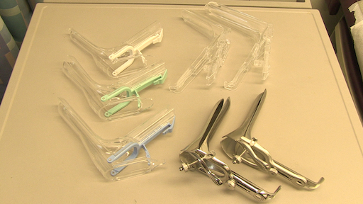
Figure 1
Figure 1. A photograph of commercially available speculums in different sizes.

Plastic speculums are all shaped like Pederson metal speculums and come in different sizes. To assess the appropriate size for a plastic speculum, the examiner places two fingers in the patient's vagina, palm down, and tries to separate the fingers: if there is no space between the fingers, a small plastic speculum should be used; if there is space between the fingers, a medium one should be used. The exam should never be performed with a large speculum (as it is significantly longer) without first determining the length of the vaginal canal.

The speculum is used to perform the Papanicolaou test as part of cervical cancer screening examinations. Cervical cancer was once the leading cause of cancer deaths for women in the United States, but in recent decades the number of cases and deaths has declined significantly1. This change is credited to the discovery made by Georgios Papanicolaou in 1928 that cervical cancer could be diagnosed by vaginal and cervical smears. The Pap test, as it is now called, detects abnormal cells in the cervix, both cancerous and pre-cancerous. Current guidelines for recommended screening intervals can be found through the U.S. Preventive Services Task Force (USPSTF) website2.

The test can be performed using either 1) a conventional glass slide and fixative with a spatula and endocervical brush (the traditional "Pap smear") or 2) the more commonly utilized liquid-based cytology with a cervical broom or a spatula and endocervical brush (Figure 2). No matter what tools are used, the samples are collected from just inside the external os and the squamocolumnar junction, or transition zone around the os (Figure 3). This video demonstrates the spatula and endocervical brush with liquid-based cytology, as the liquid preparation is a more effective technique for the detection of cervical lesions, and the spatula and endocervical brush improve specimen collection.
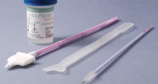
Figure 2
Figure 2. Pap smear tools. Shown in sequence are: a liquid cytology canister, cervical broom, spatula, and endocervical brush.
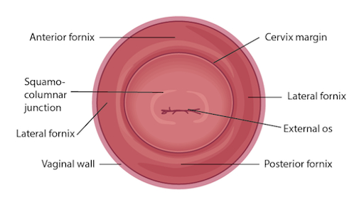
Figure 3
Figure 3. Diagram of the cervix withrelevant structures labeled.

 Physical Examinations II

Pelvic Exam I: Assessment of the External Genitalia

JoVE 10144

Source:
Alexandra Duncan, GTA, Praxis Clinical, New Haven, CT
Tiffany Cook, GTA, Praxis Clinical, New Haven, CT
Jaideep S. Talwalkar, MD, Internal Medicine and Pediatrics, Yale School of Medicine, New Haven, CT

The pelvic exam can feel invasive to patients, so it is important to do everything possible to make patients feel comfortable and empowered, rather than vulnerable. Clinicians should be aware of how they are communicating, both verbally and nonverbally, and should give their patients control whenever possible. There are many ways to do this, from how the exam table is positioned to how the patient is engaged throughout the exam. As many as 1 in 5 patients may have experienced sexual trauma; therefore, it is important to avoid triggering those patients, but it's not always possible to know who they are. The exam in this video demonstrates neutral language and techniques that can be employed with all patients to create the best experience possible.

It's important to keep the patient covered wherever possible and to minimize extraneous contact. A clinician should be careful to tuck fingers that aren't being used to examine the patient to avoid accidental contact with the clitoris or anus.

Before performing the pelvic examination, examiners should find out how knowledgeable the patients are about the exam and their own bodies, and establish the expectation that the patients can communicate questions or concerns throughout the exam. While it is always important to avoid extremely clinical language, certain colloquial words can cross the line from caring to overly intimate during this exam. It is helpful to avoid the words "touch" and "feel," which can feel sexualized in this context; instead, the words "assess," "check," "inspect," or "examine" should be used. The words "bed" and "sheet" should be avoided, and "table" and "drape" should be used instead. Also, a clinician should use the word "footrests" rather than "stirrups" to avoid connoting horses. It's a good idea to avoid telling patients to "relax," because it's a hard order for a patient to follow when anxious. Asking patients to "soften" or "release" specific muscles can be more useful, and having a patient do a Kegel exercise or Valsalva maneuver can serve as a specific relaxation technique.

Best practice dictates avoiding assumptions about patients' gender, as patients with female anatomy may identify as another gender (e.g., transgender or genderqueer). This video depicts the approach to a patient whose history has revealed no specific complaints or risk factors related to gynecological health.

The pelvic exam consists of three parts: the visual and manual exam of the external genitalia, a speculum exam, and a bimanual exam. This video covers the introduction to the pelvic exam and the external genitalia exam. To avoid missing potential findings, the external pelvic exam should be performed in a systematic approach consisting of two main components: a visual inspection of the vulva (Figure 1), and internal palpation and assessment of glands and tone.

Figure 1
Figure 1. Diagram of the vulva. A diagram showing external genitals with the structures labelled.

 Physical Examinations II

Pelvic Exam III: Bimanual and Rectovaginal Exam

JoVE 10163

Source:

Alexandra Duncan, GTA, Praxis Clinical, New Haven, CT

Tiffany Cook, GTA, Praxis Clinical, New Haven, CT

Jaideep S. Talwalkar, MD, Internal Medicine and Pediatrics, Yale School of Medicine, New Haven, CT

A bimanual exam is a thorough check of a patient's cervix, uterus, and ovaries. It can tell an experienced provider a great deal, as it may lead to the discovery of abnormalities, such as cysts, fibroids, or malignancies. However, it's useful even in the absence of such findings, as it allows the practitioner to establish an understanding of the patient's anatomy for future reference.

Performing the bimanual exam before the speculum exam can help relax patients, mentally and physically, before what is often perceived as the "most invasive" part of the exam. A practitioner already familiar with the patient's anatomy can insert a speculum more smoothly and comfortably. However, lubrication used during the bimanual exam may interfere with processing certain samples obtained during the speculum exam. Providers must be familiar with local laboratory processing requirements before committing to a specific order of examination.

This demonstration begins immediately after the end of the speculum exam; therefore, it assumes the patient has provided a history and is in the modified lithotomy position.

A rectovaginal exam is not always necessary, but it may be performed to fully assess a retroverted uterus and ovaries (this may be the only way to accomplish full assessment depending on uterine position) or to assess the rectum.

 Physical Examinations II