Large animal models have good translational values in the examination of physiology and pharmacology of neonatal asphyxia. Using newborn piglets, we develop an experimental protocol to simulate neonatal asphyxia which has advantages of studying the systemic and regional hemodynamics, oxygen transport with biochemical and pathologic pathways and correlations.
A subscription to JoVE is required to view this content. Sign in or start your free trial.
In This Article
Summary
Abstract
Annually more than 1 million neonates die worldwide as related to asphyxia. Asphyxiated neonates commonly have multi-organ failure including hypotension, perfusion deficit, hypoxic-ischemic encephalopathy, pulmonary hypertension, vasculopathic enterocolitis, renal failure and thrombo-embolic complications. Animal models are developed to help us understand the patho-physiology and pharmacology of neonatal asphyxia. In comparison to rodents and newborn lambs, the newborn piglet has been proven to be a valuable model. The newborn piglet has several advantages including similar development as that of 36-38 weeks human fetus with comparable body systems, large body size (˜1.5-2 kg at birth) that allows the instrumentation and monitoring of the animal and controls the confounding variables of hypoxia and hemodynamic derangements.
We here describe an experimental protocol to simulate neonatal asphyxia and allow us to examine the systemic and regional hemodynamic changes during the asphyxiating and reoxygenation process as well as the respective effects of interventions. Further, the model has the advantage of studying multi-organ failure or dysfunction simultaneously and the interaction with various body systems. The experimental model is a non-survival procedure that involves the surgical instrumentation of newborn piglets (1-3 day-old and 1.5-2.5 kg weight, mixed breed) to allow the establishment of mechanical ventilation, vascular (arterial and central venous) access and the placement of catheters and flow probes (Transonic Inc.) for the continuously monitoring of intra-vascular pressure and blood flow across different arteries including main pulmonary, common carotid, superior mesenteric and left renal arteries. Using these surgically instrumented piglets, after stabilization for 30-60 minutes as defined by Z<10% variation in hemodynamic parameters and normal blood gases, we commence an experimental protocol of severe hypoxemia which is induced via normocapnic alveolar hypoxia. The piglet is ventilated with 10-15% oxygen by increasing the inhaled concentration of nitrogen gas for 2h, aiming for arterial oxygen saturations of 30-40%. This degree of hypoxemia will produce clinical asphyxia with severe metabolic acidosis, systemic hypotension and cardiogenic shock with hypoperfusion to vital organs. The hypoxia is followed by reoxygenation with 100% oxygen for 0.5h and then 21% oxygen for 3.5h. Pharmacologic interventions can be introduced in due course and their effects investigated in a blinded, block-randomized fashion.
Protocol
1. Anesthesia
- Set the flow rate of the anesthetic machine at 2L/min. Connect the exhaust to vacuum suction.
- Charge face mask with anesthetic gas (Isoflurane) at 5% (˜3 min).
- Newborn piglets will be induced with inhaled Isoflurane 5% in 100% oxygen (˜3 min).
- Maintain anesthesia at 2-3% of Isoflurane. Fine adjustment of Isoflurane by 0.5% as appropriate, however, it may range from 0.5 to 5% depending on the condition of piglets.
- Once the vascular access has been established, the inhalational anesthesia can be switched to intravenous anesthesia using fentanyl (5-50 mcg/kg/h) and midazolam (200-500 mcg/kg/h) infusions. Pancuronium (50-100 mcg/kg/h) may be required to control excessive muscle movements during the surgery, whilst the ability to observe animal's state is preserved for the adjustment of anesthetic medications.
- The piglet is monitored by pulse oximetry (percutaneous oxygen saturation at 95-100%) and ECG (heart rate at 130-170 beats/min).
- The piglet's rectal temperature is maintained at 38-40°C with heating blanket and radiant warmer.
- The anesthetic state of piglet is being regularly evaluated throughout the experimental period using neurological (pupil size, tearing, body movements), behavioral (agitation), cardiovascular (tachycardia and hypertension) and respiratory (tachypnoea) parameters as appropriate. Minimal paralysis is given. Previous experience of anesthesia in piglets with and without paralysis would be useful for evaluation.
- The protocol is a non-survival procedure with euthanization of the animal at the end of experiment with an overdose of pentobarbital (100 mg/kg) intravenously.
2. Surgical placement of vascular catheters at the groin (Figure 1)
- Make a long 2-3cm incision in the right groin.
- Dissect 1cm of the right femoral venous and 1cm right femoral artery. Put two 3-0 strings around each vessel.
- Right femoral venous catheterization: Ligate the distal of the vein. Insert an Argyle catheter (3.5 or 5 French, double- lumen)(Covidien, Mansfield, MA) to 15cm and this will place at the right atrium. Tie both strings to secure the catheter. The catheter can be used for maintenance fluid and medications infusion (secondary port) and central venous/right atrial pressure measurement (primary port).
- Right femoral arterial catheterization: Ligate the distal of the artery. Lift up the proximal string to stop the blood flow. Insert an Argyle catheter (3.5 or 5 French, single-lumen) to 5cm. This will place the arterial catheter at the infra-renal aorta for continuous mean arterial pressure measurement and blood sampling. Tie both strings to secure the catheter.
- Close the skin.
3. Establish mechanical ventilation (Figure 2)
- Make a long 2-3cm horizontal incision in the neck.
- Dissect and expose 1cm of the trachea. Put two 1-0 strings around the trachea.
- Insert an endotracheal tube (3.0 or 3.5) at 1cm into the trachea. Connect to a ventilator and commence mechanical ventilation. Secure the endotracheal tube.
- Dissect and expose the common carotid artery. Encircle the vessel with a transit time ultrasound flow probe (2SB or 2RB, Transonic Systems Inc., Ithica, NY) to continuously measure the blood flow.
4. Placement of flow probes at superior mesenteric (Figure 3) and left renal (Figure 4) arteries
- Extra doses of fentanyl (5-10 mcg/kg) and acepromazine (0.01-0.02 mg/kg) are required prior to skin incision.
- Make a long subcostal-flank incision and carefully dissect muscle layers.
- Expose the abdominal aorta.
- Minimize vascular handling (vasospasm) and lymphatic injury.
- Dissect 0.5-1cm superior mesenteric artery and put a Transonic flow probe (3SB) around it.
- Dissect 0.5-1cm left renal artery and put a Transonic flow probe (2SB) around it.
- Close the skin and secure the flow probe.
5. Placement of pulmonary artery catheter (Figure 5) and flow probe (Figure 6)
- Extra doses of fentanyl (5-10 mcg/kg) and acepromazine (0.01-0.02 mg/kg) are required prior to skin incision.
- Lie the animal at the right lateral position.
- Thoracotomy at the left 4th intercostal space.
- Watch out for the internal mammary artery and vein, ligate if needed.
- Use a dental swab to press down the left lung and increase oxygen as needed.
- Open the pericardium.
- Identify the ductus arteriosus which runs from the pulmonary artery to the aorta.
- Ductus arteriosus may be ligated by placing a clip or by a thick "3-O silk" tie at its origin.
- Free the main pulmonary artery and pass a vascular sling using a thick "0" tie.
- Perform a purse string (5-0 prolene) suture at the base for the placement of pulmonary artery catheter.
- Insert a 20G Angiocath (with 3 side holes at less 1 cm from the tip of the catheter) through the purse string to a maximum of 1 cm.
- Check for free flow of venous blood.
- Connect to pressure transducer, check for pulmonary artery pressure and waveform.
- Tighten the purse string and secure the pulmonary catheter.
- Place a Transonic flow probe (6SB) around the main pulmonary artery.
- Place ultrasonic gel between the flow probe and artery to allow for optimal signal transduction.
- Cover the wound with moist saline gauze.
6. Hypoxia and reoxygenation protocol
- Decrease the inspired oxygen concentration to 10% by increasing the concentration of inhaled nitrogen gas to induce hypoxemia.
- Adjust the inspired oxygen concentration between 10% and 15% to obtain a PaO2 of 20-40 mmHg or SaO2 of 30-40% for 2h.
- Perform arterial blood analysis to assess PaCO2 and adjust ventilator rate accordingly.
- With the induction of hypoxemia, the first hour is dedicated to steadily inducing a tachycardic (and cardiac output) response.
- Continue to monitor for changes in blood flow at the common carotid, superior mesenteric and left renal arteries.
- During the second hour of hypoxia, the hypoxic stress is increased to steadily lower cardiac output to 30-40% of baseline, mean arterial pressure to 30-35 mmHg and arterial pH 6.95-7.05.
- Hypoxic stress may be prematurely terminated or extended by 15 min as appropriate.
- Increase inspired oxygen concentration abruptly to 100% abruptly by discontinuing nitrogen gas, while continuing pure oxygen.
- Monitor cardiac output, mean arterial pressure and other hemodynamic parameters for rapid recovery.
- Resuscitation with 100% oxygen can be continued for 0.5h. Following this time period, reduce the inspired oxygen concentration quickly to 21%.
- Continue reoxygenation with 21% oxygen for the remaining period of experiment. The inspired oxygen concentration can be titrated to 25% if needed.
- Fluid boluses of 10 ml/kg Ringer's lactate solution may be needed as appropriate during the experimental period. Its use has to be protocolized.
7. Representative Results:
The induction of hypoxemia in the newborn piglet over the first hour of hypoxia should increase the cardiac output (pulmonary arterial flow) to 120%-130% of baseline (Figure 7A) and heart rate (Figure 7B). Typically, cardiac output should reach its peak compensation between the first 0.5h and 1h of hypoxia. Further, blood flow should become centralized resulting in decreased mesenteric and renal perfusion but a preserved or increased common carotid arterial flow (Figure 8). During the second hour of hypoxia, there is a steady decrease of cardiac output, development of hypotension (Figure 9A), slowing of heart rate with or without arrhythmia occurred. Hypoxia should induce pulmonary hypertension with increased pulmonary artery pressure (Figure 9B), which may sometimes lower in the final 30 min of hypoxia as the cardiac output decreases.
Upon resuscitation, all hemodynamic parameters will immediately recover to normoxic baseline, except for the renal blood flow which gradually recovers over the first hour of reoxygenation. However, the hemodynamic parameters especially for the cardiac output and mean arterial pressure will gradually deteriorate over the first 2 hour of the reoxygenation to about 70-75% of normoxic baseline and 35-45 mmHg, respectively. This cardiovascular dysfunction is at least in part for myocardial stunning and warrants cardiovascular supportive therapies such as vasoactive and inotropic agents.
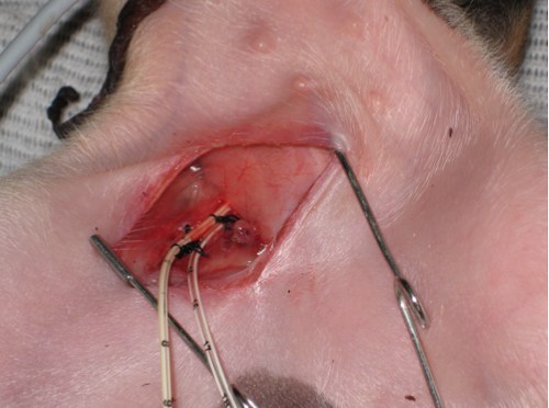
Figure 1: Groin incision with the placement of femoral arterial and venous catheters
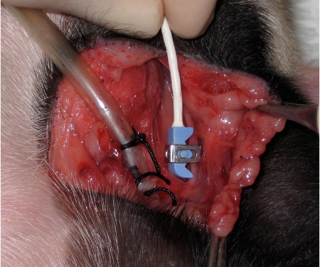
Figure 2: Neck incision with the placement of an endotracheal tube and a flow probe around the common carotid artery
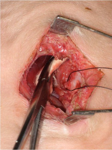
Figure 3: Flank incision with the isolation of superior mesenteric artery
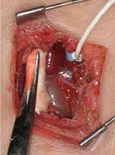
Figure 4: Flank incision with the isolation of left renal artery
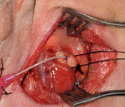
Figure 5: Thoracotomy with the placement of pulmonary artery catheter
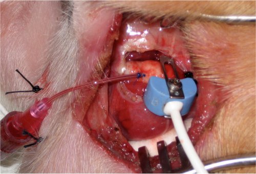
Figure 6: Thoracotomy with the placement of a Transonic flow probe around main pulmonary artery
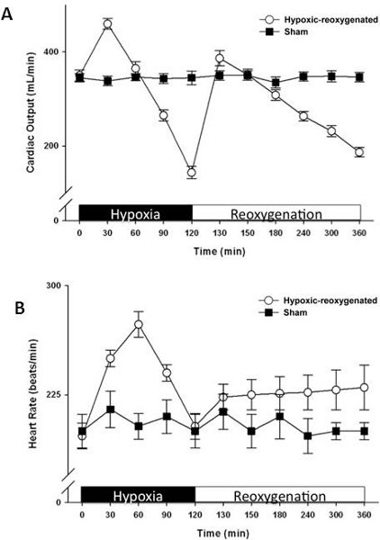
Figure 7: Temporal changes in (A) cardiac output (pulmonary arterial flow) and (B) heart rate during hypoxia and reoxygenation
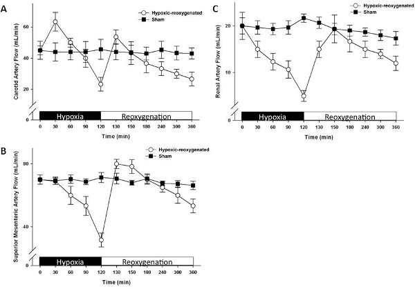
Figure 8: Temporal changes in blood flow at (A) common carotid, (B) superior mesenteric and (C) left renal arteries during hypoxia and reoxygenation
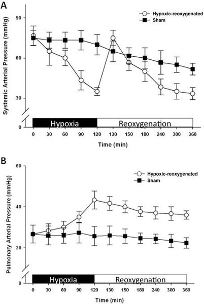
Figure 9: Temporal changes in (A) mean arterial pressure and (B) pulmonary artery pressure during hypoxia and reoxygenation
Access restricted. Please log in or start a trial to view this content.
Discussion
The current experimental protocol has an advantage to examine the systemic and regional hemodynamic changes in neonatal subjects during the hypoxia and reoxygenation process. We can also examine the respective effect of interventions used to improve the cardiovascular function during recovery. We and others have reported the experience and findings in the study of neonatal asphyxia regarding the effects in cardiovascular1, pulmonary2, neurologic3, gastrointestinal4, hepatic
Access restricted. Please log in or start a trial to view this content.
Disclosures
No conflicts of interest declared.
Acknowledgements
The authors would like to thank the Canadian Institutes of Health Research (MOP53116) and the Alberta Heritage Foundation for Medical Research for the operating grant and establishment fund, respectively, to support the development of this experimental model.
Access restricted. Please log in or start a trial to view this content.
References
- Borke, W. B. Increased myocardial matrix metalloproteinases in hypoxic newborn pigs during resuscitation: effects of oxygen and carbon dioxide. Eur. J. Clin. Invest. 34, 459-466 (2004).
- Munkeby, B. H. Resuscitation of hypoxic piglets with 100% O2 increases pulmonary metalloproteinases and IL-8. Pediatr. Res. 58, 542-548 (2005).
- Haaland, K. Posthypoxic hypothermia in newborn piglets. Pediatr. Res. 41, 505-512 (1997).
- Haase, E. Resuscitation with 100% oxygen causes intestinal glutathione oxidation and reoxygenation injury in asphyxiated newborn piglets. Ann. Surg. 240, 364-373 (2004).
- Stevens, J. Resuscitation with 21% or 100% oxygen is equally effective in restoring perfusion and oxygen metabolism in hypoxic newborn piglet liver. Shock. 27, 657-662 (2007).
- Johnson, S. T. N-acetylcysteine improves the hemodynamics and oxidative stress in hypoxic newborn pigs reoxygenated with 100% oxygen. Shock. 28, 484-490 (2007).
- Chapados, I. Plasma cortisol response to ACTH challenge in hypoxic newborn piglets resuscitated with 21% and 100% oxygen. Shock. 33, 519-525 (2010).
- Cheung, P. Y. Platelet dysfunction in asphyxiated newborn piglets resuscitated with 21% and 100% oxygen. Pediatr. Res. 59, 636-640 (2006).
- Martin-Ancel, A. Multiple organ involvement in perinatal asphyxia. J. Pediatr. 127, 786-793 (1995).
- Swindle, M. M., Smith, A. C. Comparative anatomy and physiology of the pig. Scan. J. Lab. Anim. Sci. Suppl. 25, 11-22 (1998).
- Chapados, I., Cheung, P. Y. Not all models are created equal: Animal models to study hypoxic-ischemic encephalopathy of the newborn. Neonatology. 94, 300-303 (2008).
- Liu, J. Q. Effects of post-resuscitation treatment with N-acetylcysteine on cardiac recovery in hypoxia-injured newborn pigs. PLoS ONE. 5, e15322-e15322 (2010).
- Cheung, P. Y. Cardio-renal recovery of hypoxic newborn pigs after 18%, 21% and 100% reoxygenation. Intensive Care Med. 34, 1114-1121 (2008).
- Temesvari, P. Modulation of the blood-brain barrier permeability in neonatal cytotoxic brain edema: laboratory and morphological findings obtained on newborn piglets with experimental pneumothorax. Biol. Neonate. 46, 198-208 (1984).
- Domoki, F. Reventilation with room air or 100% oxygen after asphyxia differentially affects cerebral neuropathology in newborn pigs. Acta. Paediatr. 95, 1109-1115 (2006).
- Part 15: Neonatal resuscitation: 2010 American Heart Association guidelines for cardiopulmonary resuscitation and emergency cardiovascular care. Circulation. 122, S909-S919 (2010).
Access restricted. Please log in or start a trial to view this content.
Reprints and Permissions
