Vital-dye enhanced fluorescence imaging (VFI) is a novel in vivo technique that combines high-resolution epithelial imaging with exogenous topical fluorescent contrast to highlight glandular morphology and delineate neoplasia (high grade dysplasia and cancer) in the distal esophagus.
The ability to differentiate benign metaplasia in Barrett’s Esophagus (BE) from neoplasia in vivo remains difficult as both tissue types can be flat and indistinguishable with white light imaging alone. As a result, a modality that highlights glandular architecture would be useful to discriminate neoplasia from benign epithelium in the distal esophagus. VFI is a novel technique that uses an exogenous topical fluorescent contrast agent to delineate high grade dysplasia and cancer from benign epithelium. Specifically, the fluorescent images provide spatial resolution of 50 to 100 μm and a field of view up to 2.5 cm, allowing endoscopists to visualize glandular morphology. Upon excitation, classic Barrett’s metaplasia appears as continuous, evenly-spaced glands and an overall homogenous morphology; in contrast, neoplastic tissue appears crowded with complete obliteration of the glandular framework. Here we provide an overview of the instrumentation and enumerate the protocol of this new technique. While VFI affords a gastroenterologist with the glandular architecture of suspicious tissue, cellular dysplasia cannot be resolved with this modality. As such, one cannot morphologically distinguish Barrett’s metaplasia from BE with Low-Grade Dysplasia via this imaging modality. By trading off a decrease in resolution with a greater field of view, this imaging system can be used at the very least as a red-flag imaging device to target and biopsy suspicious lesions; yet, if the accuracy measures are promising, VFI may become the standard imaging technique for the diagnosis of neoplasia (defined as either high grade dysplasia or cancer) in the distal esophagus.
Over the last forty years, the incidence of esophageal adenocarcinoma (EAC) has increased significantly1,2; yet due to late diagnosis, the five-year survival rate is less than 20%3. The current standard of endoscopic surveillance in BE, the precursor to EAC, is white light endoscopy with random four-quandrant forceps’ biopsies of the segment. Unfortunately, this technique often misses neoplasia, which can be flat, subtle and difficult to differentiate on standard white light imaging4. While there has been success in using confocal laser microscopy to highlight cellular features in vivo, lesions can still be missed due to the decreased field of view5. Having a ‘bridge’ technology that can highlight areas for further confocal microendoscopic imaging would be markedly valuable.
Consequently, an enhanced red-flag imaging modality that improves the ability to target and biopsy early neoplasia in BE would be instrumental in detecting EAC at an early, curable stage and could lead to more effective treatment and subsequently improved survival rates. VFI is a novel technique that combines high-resolution epithelial imaging with exogenous topical fluorescent contrast, proflavine, to highlight glandular morphology and delineate neoplasia (high grade dysplasia and cancer) in the distal esophagus in hopes of improving the in vivo diagnosis6. Upon excitation of the proflavine, which concentrates within cell nuclei shortly after application, the fluorescent images provide spatial resolution of 50 to 100 μm and a field of view up to 2.5 cm, allowing endoscopists to visualize glandular morphology. As a result, this approach enables gastroenterologists to distinguish classic Barrett’s metaplasia, which has continuous, evenly-spaced glands and an overall homogenous morphology, from BE with neoplasia, which has obliteration of the glandular architecture. Here we describe the protocol of this new technique with a multispectral endoscope, and provide representative results to demonstrate the utility of this device in depicting the morphological transformation from benign metaplasia to high-grade dysplasia and cancer.
NOTE: Informed consent was obtained from the patients. Also, this research has been performed in compliance with all institutional, national, and international guidelines for human welfare.
1. Prepare Computer
2. Prepare Monitor
3. Laser Diode Driver Setting
4. Power Strip
5. Run MDE Widefield on the Laptop Desktop
6. Prepare Cap and Filter
7. Patient Preparation
8. Insert and Spray Proflavine
9. Turn on Laser Diode
10. Prepare Endoscope for VFI
11. Insert the Endoscope Back into the Esophagus and Image
12. Remove Endoscope from Esophagus
13. Clean Filter
Figure 1B depicts classic Barrett’s Esophagus with no dysplasia surrounded on the borders by normal squamous epithelium. Beginning with the flatter squamous tissue, which is peripherally located and indicated by the blue arrows, a homogenous area of dull fluorescence is present with no glandular architecture. The green arrows indicate a circular green line surrounding the squamous tissue. This outline is artifact resulting from the cap of the endoscope. Moving to the centrally located Barrett’s tissue, glandular structures can be defined as green fluorescence surrounding a darker lumen. Although some glands are elongated, there is little distortion between adjacent glands, as the width of the glands is similar and the edges are clearly defined. Lastly, the glands and the lumens are evenly spaced with no clumping or crowding present.
Figure 2B depicts Barrett’s Esophagus with Low-Grade Dysplasia. It is important to note that although there is Low-Grade Dysplasia present, this cannot be visualized by morphological criteria via this imaging modality. Thus, based on morphological patterns, this tissue is still classified as benign. While the homogenous glands and lumens in the yellow oval are suggestive of mere metaplasia, the blue oval indicates an area of largely coalesced glands. That is, the thickness of the glands have increased, while the darker luminal cavity has become thin and nearly absent. These crowded and slightly distorted glands, however, have discrete, borders and tend to be homogenous. Moreover, there is no effacement present as the edges of the glands are smooth, thus the tissue is still benign. The red arrow indicates tissue that is out of focus and in video can be readily distinguished. Lastly, the black arrow shows bubbles that are artifact.
In Figure 3B, the red oval indicates the most prominent area with high grade dysplasia. These glands are crowded as they have thin irregular borders along with areas of tissue with near effacement of the glandular architecture. That is, the glands are no longer distinct, but rather fusing together, with their lumens being small and irregular. Although small, the continued presence of some lumens likely indicates high-grade dysplasia as it is more often in invasive cancer where the lumens are completely lost. In contrast to the high-grade dysplasia, the yellow oval highlights malignant tissue. Here, the glandular architecture is obliterated and lumens are largely absent.
Figure 4B depicts a centrally located adenocarcinoma. Notice first the cancer within the red oval and the complete obliteration of glandular architecture with luminal absence. This obliteration can be further appreciated when comparing the cancer to the tissue indicated by the blue arrow, which possesses some glandular framework. On the left, the gray rectangle highlights squamous epithelium, which can be better appreciated in video when the endoscope pans over it in its entirety. This squamous tissue is a flat homogenous area of dull fluorescence with no glandular architecture.
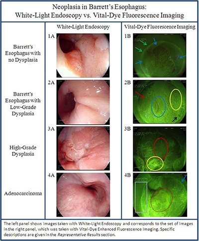
Figure 1. Neoplasia in Barrett’s Esophagus: White-light endoscopy vs. vital dye fluorescence imaging. (Left) Images taken with white-light endoscopy. (Right) Corresponding images taken with vital-dye enhanced fluorescence imaging. Please click here to view a larger version of this figure.
With standard endoscopic surveillance, neoplasia in BE is often missed8 because benign metaplasia can be indistinguishable from high-grade dysplasia and adenocarcinoma. As a tool that would better enable gastroenterologists to remedy this currently unavoidable error, vital-dye enhanced fluorescence imaging highlights a tissue’s glandular morphology thereby providing a distinct feature to differentiate the tissue types. Moreover, by providing a field of view up to 2.5 cm, VFI enables endoscopists to pan over the entire distal esophagus efficiently and systematically, making suspicious lesions more prominent.
To utilize this novel technique, first spray a topical contrast agent, proflavine, over the tissue of interest. Then, after removing the endoscope a 495-nm long-pass filter needs to be securely capped to the MDM tip. Finally, after reinserting the endoscope near the tissue of interest and then switching from the white light to the laser diode, a green fluorescence provides in vivo imagery of the tissue’s glandular framework.
In cases of continuous suboptimal fluorescence, using fresh proflavine can improve results as well as turning on the laser diode a few minutes before actual usage and adjusting its voltage. Separately, for patient safety, make certain the cap has been pushed completely over the filter, such that the filter is flush with the tip of the endoscope. This, in turn, will enhance image quality by preventing mucus buildup behind the filter itself.
While VFI is able to characterize glandular morphology, a tangible improvement to white-light endoscopy, it cannot resolve cellular dysplasia. That is, the spatial resolution of 50-100 μm makes VFI incapable of differentiating benign metaplasia from low-grade dysplasia. To distinguish these diagnoses, higher resolution devices that visualize cellular traits, such as nuclear polarity and cellular crowding, are needed 9,10. As an example, high-resolution microendoscopy is a more novel technique that can differentiate neoplastic tissue on a cellular level as nuclei are illuminated bright white and nuclear to cytoplasmic ratio can be appreciated. Narrow band imaging is another option, but it requires use of two different scopes and processors and follow-up administration of either proflavine or fluorescein for microendoscopy; in contrast VFI allows immediate confocal imaging with one scope and one processor, lengthening anesthesia time by approximately 6-8 min. However, because these latter devices focus on minute tissue areas and consequently miss neoplasias, VFI, in addition to diagnosing neoplasia, may also act as a widefield, red-flagging device for the placement of higher-resolution probes. Thus, the future of endoscopic screening for Barrett’s dysplasia will likely involve a combination approach of widefield surveillance technology, like VFI, along with a cellular classification technology like microendoscopy.
The authors have nothing to disclose.
This work is supported by the National Cancer Institute at the National Institute of Health grant R01 CA140257-01.
Filter* | Schott North America, Inc., Duryea, Pennsylvania | Not Applicable | 495-nm long-pass filter |
Halo Cap – Medium* | Barrx Medical | CP-002A | |
Processor* | Pentax | EPK-i | |
Multispectral Digital Microscope** | Not Applicable | Not Applicable | |
Kimwipes | Kimberly-Clark | KimTech Science | S-8115 |
Cidex | Advanced Sterilization Products | CIDEX OPA Solution | |
Proflavine hemisulfate (0.01% w/v) | FDA (IND 102,217) | ||
Laser Diode* | Nichia Corporation | Not Applicable | 455-nm |
Image Capture* | Labview | Not Applicable | |
Spray Catheter | Olympus | Not Applicable | |
*Equipment specifics within Reference 6. **Equipment specifics within Reference 7 |