With the intent of characterizing changes in miRNAs on differentiated human neural stem cells (hNSCs) we describe hNSC differentiation on a three dimensional system,the evaluation of changes in microRNA expression by miRNA PCR array, and computational analysis for miRNA target prediction and its validation by dual luciferase assay.
Method Article
Differentiation of a Human Neural Stem Cell Line on Three Dimensional Cultures, Analysis of MicroRNA and Putative Target Genes
In This Article
Summary
Abstract
Neural stem cells (NSCs) are capable of self-renewal and differentiation into neurons, astrocytes and oligodendrocytes under specific local microenvironments. In here, we present a set of methods used for three dimensional (3D) differentiation and miRNA analysis of a clonal human neural stem cell (hNSC) line, currently in clinical trials for stroke disability (NCT01151124 and NCT02117635, Clinicaltrials.gov). HNSCs were derived from an ethical approved first trimester human fetal cortex and conditionally immortalized using retroviral integration of a single copy of the c-mycERTAMconstruct. We describe how to measure axon process outgrowth of hNSCs differentiated on 3D scaffolds and how to quantify associated changes in miRNA expression using PCR array. Furthermore we exemplify computational analysis with the aim of selecting miRNA putative targets. SOX5 and NR4A3 were identified as suitable miRNA putative target of selected significantly down-regulated miRNAs in differentiated hNSC. MiRNA target validation was performed on SOX5 and NR4A3 3’UTRs by dual reporter plasmid transfection and dual luciferase assay.
Introduction
Stem cell research plays a central role in regenerative medicine, which also spans the disciplines of tissue engineering, developmental cell biology, cellular therapeutics, gene therapy, and biomaterials (scaffolds and matrices). Stem cells are important in both organ developmentand tissue repair; understanding how stem cells work is pivotal for developing new stem cell based treatments1. CTX0E03 is a clonal, conditionally immortalized human neural stem cell (hNSC) line2 isolated from first trimester human fetal cortex. CTX0E03 has defined quality characteristics that are essential for clinical cell banking under good manufacturing practice (GMP)3. HNSCs can spontaneously differentiate into neurons, glia, and oligodendrocytes and have been proven to ameliorate neurological deficits when transplanted in a rodent model of focal ischemia2,4,5. The CTX0E03 hNSCs are currently in clinical trials for stroke disability (NCT011511246 and NCT02117635, Clinicaltrials.gov).
MiRNAs have an average 22 nucleotides in length and are proven regulatory molecules7, they do not encode proteins, but rather bind 3 prime untranslated region (3’UTR) of mRNAs, regulating their stability and translation into proteins. MiRNAs are reported to participate in the regulation of a number of cellular processes, including development, proliferation, and differentiation8. Several miRNAs have been implicated in regulating the fate and specification of neural stem cells9.
In two dimensional (2D) standard culture environment the complexity of the in vivo environment is not matched, and consequently the induction and regulation of hNSC differentiation is not optimal. In vivo, cells are surrounded by a three dimensional (3D) microenvironment composed by other cells and extracellular ligands, including many types of collagens, laminin, and other matrix proteins (extracellular matrix, ECM). Previous studies have reported that the architectural topography of the materials mimicking ECMs and their geometry influence cell phenotype and fate10-15. A number of commercially available 3D scaffolds are available; we used a highly porous polystyrene scaffold engineered with a well-defined and uniform architecture into a 200 µm thick membrane which provides a 3D space into which cells can invade and differentiate16-18.
Herein 2D and 3D differentiation assays are used to assess axonprocess outgrowth. Furthermore, we describe a method to profile the miRNA expression of a clinical grade hNSC line to further investigate miRNA effects on its differentiation. The methods in here illustrated are based on the ones originally described in19.
Protocol
1. HNSC differentiation on Three Dimensional (3D) Culture
NOTE: Isolate and derive hNSCs,from an ethical approved tissue, described in a previous publication2. Please follow the ethical and institutional guidelines while following this procedure.
1.1) 3D Culture Using 12-well Plate
- Use aseptic technique throughout the procedure. In a biological safety cabinet, re-hydrate scaffolds by adding 2 ml/well of 70% ethanol prepared using water for irrigation (WFIr). Avoid touching the scaffolds by dispensing the 70% ethanol down the wall of each well.
- Carefully aspirate the 70% ethanol and immediately wash the scaffolds with 2 ml/well DMEM: F12 for 1 min. Repeat the wash procedure twice. Carefully aspirate the DMEM: F12 after the final wash.
- Coat scaffolds by adding 2 ml/well of laminin (10 µg/ml) in DMEM: F12. Incubate plate at 37 °C in a 5% CO2 humidified incubator for a minimum of 2 hr. Wash plate with warm DMEM: F12.
- Seed each scaffold with approximately 500,000 hNSCs in a volume of 150 µl of RMM+GFs.
- Incubate the plate for 3 hr at 37 °C in a 5% CO2 humidified incubator to allow cells to settle into the scaffolds.
- Add 3.5 ml of RMM to each well taking care not to dislodge cells from scaffolds.
- Incubate the plate for 7 days; replace RMM medium (3.5 ml/well) after 1 day (first feeding) and again after 2-3 days from the first feeding.
- After 7 days, remove medium and fix cells with 4% paraformaldehyde (PFA) for immunocytochemistry (ICC) or add 500 µl/well of lysis reagent for total RNA extraction (store plate at ≤ -80 °C or proceed to total RNA extraction).
1.2) Measurement of Axon Process Outgrowth
- Stain 4% PFA fixed differentiated hNSCs according to a standard ICC protocol20 using β3-tubulin (TUBB3) primary antibody detected with a fluorochrome conjugated secondary antibody. Counterstain cell nuclei with Hoechst (1 µM).
- Capture representative images of β3-tubulin stained cells using a fluorescent microscope; save images as tiff or jpeg. Measure of axon process outgrowth using a software measurement tool (e.g. Image-Pro Plus).
- Open the image, select measurements option in the tool bar, and select trace feature.
- To start the trace, select the start point on the axon outgrowth by clicking the mouse button. Trace along the entire length of the axon outgrowth; right click to finish each trace. Repeat to measure all axon outgrowthswith in the field of view.
- Express measurements as pixels or µm (pending calibration set up). Export length measurements to a spreadsheet program for sample comparative and statistical analysis.
2. MiRNA Expression Using a Stem Cells and Developmental Pathways Focused miRNA PCR Array
2.1) miRNA total RNA extraction
- Perform miRNA extraction on scaffolds and 2D grown samples (differentiated and undifferentiated, hNSC control).
- Thaw scaffolds, inserted in a 12 well plate, if required. Combine lysis reagent contents (500 µl) of 2 wells in the same 1.5 ml Eppendorf tube, final volume 1 ml of Lysis reagent. Take care to leave the scaffold in the well.
- For previously collected as dry cell pelleted 2D grown samples (differentiated hNSC 2D cultures and undifferentiated control) add 1 ml of lysis reagent and transfer contents into 1.5 ml microcentrifuge tube.
- Homogenize each test sample using a 1 ml syringe and a short 20 to 25 gauge needle. Pass the lysate through the needle using a 1 ml syringe up to 5 times until a homogeneous lysate is achieved.
- Add 200 µl of chloroform to each Eppendorf tube and cap securely. Invert and vortex tubes to mix contents.
- Centrifuge each 1.5 ml microcentrifuge tube containing homogenate for 15 min at ≥12,000 x g.
- Transfer the upper aqueous phase of each sample (avoid transferring any interphase, pink coloured liquid) to a fresh 1.5 ml microcentrifuge tube containing 750 µl of 100% ethanol. Mix thoroughly by inversion.
- Pipette up to 700 μl of each test sample, including any precipitate, into a mini column in a 2 ml collection tube. Close the lid and centrifuge at ≥8,000 x g for approximately 30 sec at room temperature. Discard the flow-through. Repeat this step using the remainder of the sample.
- Perform a DNA digest for 15 min.
- Add 700 μl buffer RWT to the mini column and centrifuge for approximately 30 sec at ≥8,000 x g. Discard the flow-through.
- Wash mini column by adding 500 μl buffer RPE and centrifuging for approximately 30 sec at ≥8,000 x g. Discard the flow-through. Repeat wash.Centrifuge at ≥12,000 x g for 1 min to dry the membrane further.
- Elute total RNA in a 1.5 ml Eppendorf tube by adding 40 μl of RNase-free water to the mini column and centrifuging for 1 min at ≥8,000 x g. Measure total RNA concentration with the aid of an appropriate spectrophotometer.
2.2) miRNA cDNA Preparation
- Prepare the reverse-transcription master mix. Each sample requires 4 µl of 5x miScript HiSpec buffer, 2 µl of 10x nucleics mix, 2 µl miScript reverse transcriptase mix.
- Aliquot 8 µl of master mix in a PCR tube, add required volume of total RNA (250 ng), and add RNase-free water to a final volume of 20 µl.
- Incubate for 60 min at 37 °C.Incubate for 5 min at 95 °C to inactivate miScript Reverse Transcriptase Mix.
- Add 200 μl RNase-free water to each 20 μl reverse-transcription reaction, store at –20 °C, or proceed with real-time PCR immediately.
2.3) Stem Cells and Developmental Pathways Focused miRNA PCR Array
- Prepare the PCR components mix in a 15 ml tube by adding 1375 µl of 2x SYBR green master mix, 100 µl miRNA cDNA preparation, 275 µl of 10x miScript Universal Primer, and 1000 µl of RNase-free water (total volume 2,750 µl).
- Transfer PCR components mix on a PCR loading reservoir. Carefully remove the 96-well plate PCR array from its sealed bag.
- Add 25 μl PCR components mix to each well of the PCR Array using an 8-channel pipettor, or a 12-channel pipettor using only 8 tips. Change pipet tips following each pipetting step to avoid cross-contamination between the wells.
- Seal plate with optical adhesive film, and centrifuge for 1 min at 1,000 x g at room temperature (15–25 °C) to remove bubbles. Visually inspect the plate from underneath to ensure no bubbles are present in the wells.
- Place the 96 well plate PCR array in the real-time cycler.
- Program the real-time cycler accordingly: 1 cycle at 95 °C for 10 min, and 45 cycles for 15 sec at 95 °C, and 1 min at 60 °C set a single (fluorescence data collection).
- Export the Ct values for all wells to a blank Excel spreadsheet for use with PCR Array Data Analysis Template Excel and web-based software.
NOTE: Software is available at www.SABiosciences.com/pcrarraydataanalysis.php.
3. Computational Analysis for miRNA Target Prediction and Validation of Target mRNAs by Reporter Plasmid Transfection and Ddual Luciferase Assay.
3.1) Computational Analysis for miRNA Target Prediction
- Browse PicTar (http://pictar.mdc-berlin.de/)21, online available algorithm for the identification of miRNA target prediction mRNAs.
- Click on "click here for PicTar prediction on vertebrates". A new page will open.
- In the new page, PicTar web interface, choose species in the drop down menu and insert miRNA of interest in the miRNA ID box. Click search for targets of a miRNA.
- Observe a list of target mRNAs ranked by aPicTar score.
NOTE: PicTar computes a score reflecting the likelihood that a given UTR will be targeted by the selected miRNA, and based on seed complementarity, thermodynamics and a combinatorial prediction for common targets in sets of co-expressed miRNAs22. - Similarly retrieve list of target mRNAs using DIANA-LAB (http://diana.cslab.ece.ntua.gr/microT/)23, and TargetScan (http://www.targetscan.org/)24, alternative on line available algorithms.
- Compile a comparative table of miRNAs based on ranking obtained using different software tools. PicTar ranks by score, DianaLab by precision (based on signal to noise ratio and a precision score for the evaluation of the significance of the predicted results), and TargetScan by aggregate PCT (based on seed complementarity, thermodynamic free energy of binding, conservation over different species), see Table 1.
3.2) Validation of Target mRNAs by Reporter Plasmid Transfection and Dual Luciferase Assay.
NOTE: 3’ UTR luciferase reporters for the validation of target mRNA has been previously described25.
- Transfection
- Seed HeLa cells at a density of 105 cells/well in 24-well plate.
- After 24 hr, co-transfect HeLa cells with 1.5 µl of transfection reagent and either 100 ng of NR4A3 3’ UTRor SOX5 3’ UTR plasmid, as well as 20 nM miRNA mimics (hsa-miR-96-5p for SOX5 3’ UTR, and hsa-miR-7-5p, and hsa-miR-17-5p for NR4A3 3’ UTR respectively) per well.
- Co-transfect control wells with NR4A3 3’ UTRor SOX5 3’ UTRplasmid and fluorochrome labelled negative control siRNAs. The transfection of control wells with fluorochrome labelled negative control siRNAs allows evaluation of transfection efficiency and accounts for mimic miRNA 3’UTR bind specificity.
- Measurement of Dual Luciferase Activities
- Measure Firefly and Renilla luciferase activities 24 hr post transfection. Remove cell growth medium.
- Prepare working solution I by adding 50 µl of substrate I into 10 ml of Solution I; mix thoroughly. Add 300 µl of working solution I into each 24 well.
- Transfer the contents of each 24 well into 3 wells of a 96 white plate, 100 µl each. Wait 10 min and measure Firefly luciferase in luminometer set for luminescence acquisition.
- Prepare working solution II by adding 50 µl of substrate II into 10 ml of solution II; mix thoroughly. Add 100 µl of working solution I into each 96 well already containing 100 µl of working solution I.
- Wait 10 min and measure Renilla luciferase in luminometer set for luminescence acquisition. Calculate the ratio of luminescence from the Firefly luciferase to the Renilla luciferase.
Results
In Figure 1 is visualized the investigation and quantification of hNSC differentiation on 3D scaffolds compared with traditional flat surface cultures (2D) and expressedas axon process outgrowth measurements. In Figure 2 is representedthe workflow and the results for the miRNA quantification using stem cells and developmental pathways focused miRNA PCR array to investigate differential expression of miRNAs in 2D and 3D differentiated hNSCs compared with undifferentiated control. Following miRNA comparative analysis between differentiated and undifferentiated hNSCs, and computational analysis for target prediction, SOX5 and NR4A3 (NOR1) were identified as putative target mRNAs. To study the direct interaction between the miRNAs and their putative site on the 3’UTR mRNA we used 2 commercially available plasmids containing either SOX5 or NR4A3 3′UTR sequence inserted downstream of the firefly luciferase reporter gene, and containing in the same plasmid renilla luciferase gene for normalization. The representative results of 3’ UTR luciferase activity down-regulation is presented in Figure 3.
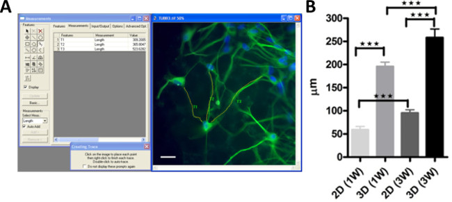
Figure 1: Differentiation of hNSCs on 3D scaffolds, and measurement of axon processes outgrowth. (A) Following standard ICC staining for β3-tubulin (green) and nuclei counterstained with Hoechst (blue) representative microphotographs were acquired and measurement of axon processes were performed with the aid of an image analysis software. (B) Diagrams reporting the measurement of axon process outgrowth performed in 2D and 3D differentiated hNSCs for 1 (1W) or 3 weeks (3W). Please click here to view a larger version of this figure.
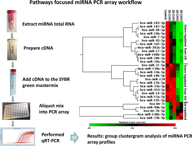
Figure 2: Schematic workflow of miRNA profiling in differentiated hNSCs. Total RNA,including miRNAs, was extracted from either differentiated hNSCs on 3D scaffolds and standard 2D cultures, or undifferentiated control. 250 ng of total RNA were retro-transcribed with a method that facilitates the selective conversion of mature miRNAs into cDNA suitable for miRNA quantification with human cell differentiation and development miScript miRNA PCR array. The geo-mean of SNORD61, SNORD68, SNORD72, SNORD95, SNORD96A, and RNU6-2 was used for data analysis based on the 2-ΔΔct method. Significant changes were defined as +/- 1.5 fold up and down regulation to a statistically significant extent. Data were analyzed using web-based software available at Results are expressed as a clustergram. Modified from Figure 319. Please click here to view a larger version of this figure.
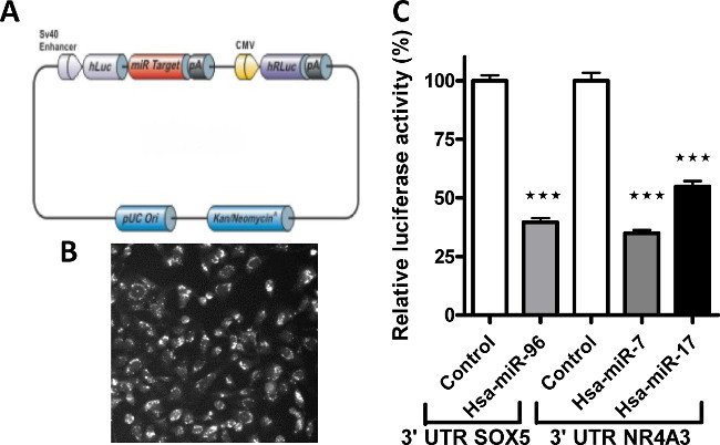
Figure 3: Validation of miRNA target predicted mRNA using a dual luciferase report assay. (A) Representative diagram of a dual luciferase reporter plasmid used as a tool to validate 3’UTR sequence as a direct target of a miRNA. (B) Representative micrograph showing transfection efficiency. (C) Signals, expressed as relative luciferase activity, from human 3'UTR reporters ofSOX5 and NR4A3 genes were significantly knocked down in the presence of hsa-miR-96, and hsa-miR-7 and 17 miRNA mimics respectively. Modified from Figure 519. Please click here to view a larger version of this figure.
| DIANALAB | PicTar | TargetScan | |||
| miRNA | Target gene | Target sites/genes found | Precision | Score | Aggregate PCT |
| hsa-miR-96 | SOX5 | 1214/763 | 1 | 6.74 | 0.94 |
| hsa-miR-183 | DUSP10 | 302/190 | 0.72 | 4.75 | 0.85 |
| hsa-miR-302a | NR4A3 | 863/473 | 0.84 | 3.05 | NF |
| hsa-miR-182 | FOXN3 | 1358/794 | 0.94 | NF | 0.96 |
| hsa-miR-7 | NR4A3 | NF | NF | 1.61 | 0.17 |
| hsa-miR-7 | FOXN3 | 399/136 | 0.17 | NF | 0.88 |
| hsa-miR-20a | NR4A3 | 1650/841 | 0.85 | 5.87 | 0.63 |
| hsa-miR-20b | NR4A3 | 1955/973 | 0.9 | 5.87 | 0.63 |
| hsa-miR-17 | NR4A3 | 1928/961 | 0.9 | 5.38 | 0.63+0.37 |
| hsa-miR-20a | EIF4G3 | 1650/841 | 0.97 | NF | NF |
| hsa-miR-20b | EIF4G3 | 1955/973 | 0.94 | NF | NF |
| hsa-miR-17 | EIF4G3 | 1928/961 | 0.94 | NF | NF |
Table 1: List of miRNA target prediction mRNAs. Representative table of miRNAs, target predictive genes,and algorithms analysis (prediction/score/aggregate provided using Diana Lab, PicTar, and TargetScan respectively). Modified from Table 319.
Discussion
The development of new in vitro assays to assess and improve the differentiation of stem cells has always presented a challenge for the investigation of their functionalities and therapeutic capabilities. Long term and stable attachment of hNSCs, required for the development of a robust 3D in vitro differentiation assay, was addressed by testing several 3D substrates with different characteristics in term of materials and structures. As part of the assay development we also assessed optimal cell seeding concentration and identified the requirement for the scaffolds to be laminin coated. Although our hNSCs can spontaneously differentiate upon 4-OHT and growth factor removal, the implementation of a 3D culture system showed a significant improvement in their differentiation capability as measured by axon process outgrowth when compared to standard 2D differentiated hNSCs.
To investigate the effect of 3D induced differentiation on hNSC miRNA expression we used a PCR array. Compared with standard chip microarray PCR array offers the advantages of a direct quantification and validation of miRNA of interest. Although the PCR array is limited in term of total number of miRNA per array, for example in here less than 96, it can be tailored to specifically include miRNAs of interest, in our case previously reported in stem cell development. The expression of pathway focused miRNAs was profiled in 3D and 2D differentiated hNSCs compared with undifferentiated hNSCs. The most significantly down-regulated miRNAs were selected and analyzed to assess their putative target mRNAs with the intent of determining their functionality and biological interpretation using online available algorithms (DIANA Lab, PicTar, and TargetScans).
A single miRNA can recognize hundreds of targets and for this reason we validated miRNA interactions with 3’ UTR mRNAsthat may be suitable candidates in axonal regulation. NR4A3, a target of the hsa-miR-7, and miR-17 family, is a member of the NR4A family nuclear receptors which, depending on their level of expression, are involved in the differentiation, survival, apoptosis and regulation of hippocampal axon guidance26. Similarly SOX5, a target of hsa-miR-96, is reported to control cell cycle progression in neural progenitors27axon length28, migration, post-migratory differentiation, and projections of neurons29. Both SOX5 and NR4A3 were identified as a direct target mRNA of hsa-miR-96, and hsa-miR-7 and 17 respectively by dual luciferase reporter assays. Dual luciferase (Firefly and Renilla) relies on two different reporter genes in the same plasmid and offers an improved system allowing normalization for effects caused by suboptimal transfection and cytotoxic effects compared with a single luciferase plasmid system. As part of our assay development we also optimized our transfection conditions in order to obtain high efficiency.
The protocolsdescribed herein may be exploited to further optimize and characterize in vitro hNSC differentiation which may beimplemented intransplantation protocols for pre-clinical studies and future clinical applications.
Disclosures
All authors are employees, stock and/or stock option holders in ReNeuron Ltd or its parent company. This study was supported by ReNeuron (RENE.L).
Acknowledgements
We acknowledge Julie Heward for helping in the preparation of hNSCs, and Lavaniya Thanabalasundaram for her technical support with HeLa culturing and transfection.
Materials
| Name | Company | Catalog Number | Comments |
|---|---|---|---|
| DMEM:F12 | Invitrogen | 21331-020 | For RMM medium preparation |
| Human Albumin Solution | Baxter health care limited | 1501052 | For RMM medium used at a final concentration of 0.03% |
| Transferrin, Human recombinant | Sigma | T8158 | For RMM medium used at a final concentration of 5 µg/ml |
| Putrescine DiHCl | Sigma | P5780 | For RMM medium used at a final concentration of 16.2 µg/ml |
| Insulin, Human recombinant | Sigma | I9278 | For RMM medium used at a final concentration of 5 µg/ml |
| Progesterone | Sigma | P6149 | For RMM medium used at a final concentration of 60 ng/ml |
| L-Glutamine | Invitrogen | 25030-24 | For RMM medium used at a final concentration of 2 mM |
| Sodium Selenite | Sigma | S9133 | For RMM medium used at a final concentration of 40 ng/ml |
| bFGF | Peprotech | AF-100-15 | To be added to RMM medium, used at a final concentration of 10 ng/ml |
| EGF | Peprotech | AF-100-188 | To be added to RMM medium, used at a final concentration of 20 ng/ml |
| 4-OHT | Sigma | H7904 | To be added to RMM medium, used at a final concentration of 100 nM |
| Laminin | AMS Biotech | 3400-010-10 | |
| TrypZean/EDTA | Lonza | BE02-034E | |
| Water for irrigation | Baxter Healthcare | UKF7114 | |
| Defined Trypsin Inhibitor (DTI) (store -20°C) | Invitrogen | R007-100 | |
| Alvetex 12 well plate | Reinnervate Ltd | AVP002 | |
| Ethanol | Merck | 1.00986.1000 | |
| βIII tubulin antibody | Sigma | T8660 | |
| Hoechst | Sigma | B2261 | |
| miRNeasy mini kit | Qiagen | 217004 | |
| RNase free DNase | Qiagen | 79254 | |
| Chloroform | Sigma | C7559-5VL | |
| miScript II RT Kit | Qiagen | 218160 | |
| SYBR green PCR kit | Qiagen | 218073 | |
| Cell Development & Differentiation miScript miRNA PCR Array | Qiagen/ SABiosciences | MIHS-103Z | |
| Lightcycler 480 white 96 plate | Roche | 4729692001 | |
| Lightcycler 480 sealing foil | Roche | 4729757001 | |
| Lipofectamine 2000 | Invitrogen | 11668-027 | |
| Vector NR4A3 miRNA 3' UTR target clones | GeneCopeia | HmiT019538-MT0 | |
| Vector SOX5 miRNA 3' UTR target clones | GeneCopeia | HmiT017632-MT01 | |
| Allstars negative control siRNA AF 488 (Qiagen). | Qiagen | 1027284 | MiRNA transfection control |
| Luc-Pair miR Luciferase Assay | GeneCopeia | LPFR-M010 | |
| Lightcycler 480 | Roche | ||
| GloMax 96 microplate luminometer | Promega |
References
- Falanga, V. Stem cells in tissue repair and regeneration. J Invest Dermatol. 132 (6), 1538-1541 (2012).
- Pollock, K., et al. A conditionally immortal clonal stem cell line from human cortical neuroepithelium for the treatment of ischemic stroke. Exp Neurol. 199 (1), 143-155 (2006).
- Hodges, H., et al. Making stem cell lines suitable for transplantation. Cell Transplant. 16 (2), 101-115 (2007).
- Smith, E. J., et al. Implantation site and lesion topology determine efficacy of a human neural stem cell line in a rat model of chronic stroke. Stem Cells. 30 (4), 785-796 (2012).
- Stroemer, P., et al. The neural stem cell line CTX0E03 promotes behavioral recovery and endogenous neurogenesis after experimental stroke in a dose-dependent fashion. Neurorehabil Neural Repair. 23 (9), 895-909 (2009).
- Kalladka, D., Muir, K. W. Brain repair: cell therapy in stroke. Stem Cells Cloning. 7, 31-44 (2014).
- Fire, A., et al. Potent and specific genetic interference by double-stranded RNA in Caenorhabditis elegans. Nature. 391 (6669), 806-811 (1998).
- Anokye-Danso, F., et al. Highly efficient miRNA-mediated reprogramming of mouse and human somatic cells to pluripotency. Cell Stem Cell. 8 (4), 376-388 (2011).
- Li, X., Jin, P. Roles of small regulatory RNAs in determining neuronal identity. Nat Rev Neurosci. 11 (5), 329-338 (2010).
- Badylak, S. F. The extracellular matrix as a scaffold for tissue reconstruction. Semin Cell Dev Biol. 13 (5), 377-383 (2002).
- Buxboim, A., Discher, D. E. Stem cells feel the difference. Nat Methods. 7 (9), 695-697 (2010).
- Li, W. J., et al. A three-dimensional nanofibrous scaffold for cartilage tissue engineering using human mesenchymal stem cells. Biomaterials. 26 (6), 599-609 (2005).
- Lutolf, M. P., Gilbert, P. M., Blau, H. M. Designing materials to direct stem-cell fate. Nature. 462 (7272), 433-441 (2009).
- Ortinau, S., et al. Effect of 3D-scaffold formation on differentiation and survival in human neural progenitor cells. Biomed Eng Online. 9 (1), 70(2010).
- Saha, K., Pollock, J. F., Schaffer, D. V., Healy, K. E. Designing synthetic materials to control stem cell phenotype. Curr Opin Chem Biol. 11 (4), 381-387 (1016).
- Knight, E., Murray, B., Carnachan, R., Przyborski, S. Alvetex(R): polystyrene scaffold technology for routine three dimensional cell culture. Methods Mol Biol. 695, 323-340 (2011).
- Bokhari, M., Carnachan, R. J., Przyborski, S. A., Cameron, N. R. Emulsion- templated porous polymers as scaffolds for three dimensional cell culture: effect of synthesis parameters on scaffold formation and homogenity. J. Mater. Chem. 17, 4088-4094 (2007).
- Bokhari, M., Carnachan, R. J., Cameron, N. R., Przyborski, S. A. Novel cell culture device enabling three-dimensional cell growth and improved cell function. Biochem Biophys Res Commun. 354 (4), 1095-1100 (2007).
- Stevanato, L., Sinden, J. D. The effects of microRNAs on human neural stem cell differentiation in two- and three-dimensional cultures. Stem Cell Res Ther. 5 (2), 49(2014).
- Ippolito, D. M., Eroglu, C. Quantifying synapses: an immunocytochemistry-based assay to quantify synapse number. J Vis Exp. (45), (2010).
- Chen, K., Rajewsky, N. Natural selection on human microRNA binding sites inferred from SNP data. Nat Genet. 38 (12), 1452-1456 (2006).
- Krek, A., et al. Combinatorial microRNA target predictions. Nat Genet. 37 (5), 495-500 (2005).
- Maragkakis, M., et al. Accurate microRNA target prediction correlates with protein repression levels. BMC Bioinformatics. 10, 295(2009).
- Lewis, B. P., Burge, C. B., Bartel, D. P. Conserved seed pairing, often flanked by adenosines, indicates that thousands of human genes are microRNA targets. Cell. 120 (1), 15-20 (2005).
- Aldred, S. F., Collins, P., Trinklein, N. Identifying targets of human micrornas with the LightSwitch Luciferase Assay System using 3'UTR-reporter constructs and a microRNA mimic in adherent cells. J Vis Exp. (55), (2011).
- Ponnio, T., Conneely, O. M. nor-1 regulates hippocampal axon guidance, pyramidal cell survival, and seizure susceptibility. Mol Cell Biol. 24 (20), 9070-9078 (2004).
- Martinez-Morales, P. L., Quiroga, A. C., Barbas, J. A., Morales, A. V. SOX5 controls cell cycle progression in neural progenitors by interfering with the WNT-beta-catenin pathway. EMBO Rep. 11 (6), 466-472 (2010).
- Kwan, K. Y., et al. SOX5 postmitotically regulates migration, postmigratory differentiation, and projections of subplate and deep-layer neocortical neurons. Proc Natl Acad Sci U S A. 105 (41), 16021-16026 (2008).
- Lai, T., et al. SOX5 controls the sequential generation of distinct corticofugal neuron subtypes. Neuron. 57 (2), 232-247 (2008).
Reprints and Permissions
