Liver macrophages, named Kupffer cells, are responsible for the capture of circulating nanoparticles. We describe here a method, of high cell purity and yield, for Kupffer cell isolation. The modified LDH assay is used here to measure the toxicity induced by carbon nanotubes in Kupffer cells.
A subscription to JoVE is required to view this content. Sign in or start your free trial.
Method Article
Kupffer Cell Isolation for Nanoparticle Toxicity Testing
In This Article
Summary
Abstract
The large majority of in vitro nanotoxicological studies have used immortalized cell lines for their practicality. However, results from nanoparticle toxicity testing in immortalized cell lines or primary cells have shown discrepancies, highlighting the need to extend the use of primary cells for in vitro assays. This protocol describes the isolation of mouse liver macrophages, named Kupffer cells, and their use to study nanoparticle toxicity. Kupffer cells are the most abundant macrophage population in the body and constitute part of the reticulo-endothelial system (RES), responsible for the capture of circulating nanoparticles. The Kupffer cell isolation method reported here is based on a 2-step perfusion method followed by purification on density gradient. The method, based on collagenase digestion and density centrifugation, is adapted from the original protocol developed by Smedsrød et al. designed for rat liver cell isolation and provides high yield (up to 14 x 106 cells per mouse) and high purity (>95%) of Kupffer cells. This isolation method does not require sophisticated or expensive equipment and therefore represents an ideal compromise between complexity and cell yield. The use of heavier mice (35-45 g) improves the yield of the isolation method but also facilitates remarkably the procedure of portal vein cannulation. The toxicity of functionalized carbon nanotubes f-CNTs was measured in this model by the modified LDH assay. This method assesses cell viability by measuring the lack of structural integrity of Kupffer cell membrane after incubation with f-CNTs. Toxicity induced by f-CNTs can be measured consistently using this assay, highlighting that isolated Kupffer cells are useful for nanoparticle toxicity testing. The overall understanding of nanotoxicology could benefit from such models, making the nanoparticle selection for clinical translation more efficient.
Introduction
The field of nanotoxicology research aims to characterize the biological effect of nanoparticles. Toxicological studies based on in vivo investigations remain the most accurate methods. However, their use is limited by their cost, labor and time requirements1. As alternatives, in vitro assays have been used because of their simplicity and the possibility of developing high-throughput in vitro testing platforms2 - so extending the number of conditions tested. Most nanotoxicological studies are performed using in vitro assays with immortalized cell lines. However, there are concerns regarding the extrapolation of these experimental findings to in vivo toxicological effects3. Indeed, the properties of immortalized cell lines can be significantly different from tissues they were derived from, e.g. genetic transformation4, deterioration of key morphological features5, loss of cellular polarity6 and functional alterations such as the regulation of inflammatory mediators7.
Kupffer cells are the most abundant macrophage population in the body and are directly in contact with blood by lining the wall of liver sinusoids. As part of the reticulo-endothelial system (RES), these macrophages are responsible for the capture of circulating nanoparticles and therefore, constitute a highly suitable model to study nanoparticle toxicity. In vivo8 and in vitro9 studies of inflammatory responses associated with Kupffer cells exposed to nanoparticles have been published elsewhere. Kupffer cells have also been involved in the pathogenesis of liver diseases such as alcoholic liver disease10, liver fibrosis11 or viral hepatitis12. It was reported that isolated Kupffer cells provide useful insight to describe cellular mechanisms involved in liver disruption13,14.
Several methods have been reported to isolate and purify Kupffer cells. Cell isolation can result from mechanical or enzymatic dissociation15. Collagenase digestion shows the advantage of conserving the functional integrity of Kupffer cells, while leading to high Kupffer cell yield16. Many experimental approaches, varying in complexity and costs, have been used to separate Kupffer cells from other liver cell populations. For example, Kupffer cell purity can be achieved by immunoaffinity17, flow electrophoresis18, selective adhesion16 or by centrifugal techniques16, which select cells according to their size and density. A combination of these methods can be chosen to increase the purity of the population16. There is no consensus about the ideal method for Kupffer cell isolation, as it mostly depends on the application and available equipment. However, in the case of nanoparticle toxicity testing, the simplicity and high yield of the technique associated with the functional preservation of Kupffer cells appeared to be most suitable for this application.
The Kupffer cell isolation method reported here is based on a 2-step perfusion method followed by purification on density gradient. The method was modified from the original protocol developed by Smedsrød et al.16 designed for rat liver cell isolation. Most studies reported and described the isolation of Kupffer cells from rat livers. Herein, we describe a method to isolate Kupffer cells from mouse liver, at a high yield and purity. The use of mice reduces the experiment cost and allows the processing of several livers to obtain large amounts of Kupffer cells for nanoparticle toxicity testing.
In the following protocol, Kupffer cells were incubated with functionalized carbon nanotubes (f-CNTs). The unique physico-chemical properties of CNTs –i.e. high length to diameter ratio and large surface area, have made CNTs interesting candidates as vectors for therapy and diagnosis purposes. However, concerns have been raised regarding the toxicity of CNTs19, and the development of new in vitro tests aims at increasing the understanding of CNT biological effect. Toxicity in Kupffer cell is associated with a lack of structural integrity of the cell membrane. This is measured by the loss of the cytoplasmic enzyme LDH from the cell into the supernatant. The principle of this method, therefore, is to remove any released LDH and measure what is left in the cells20. This is done in preference to measuring the released LDH in the supernatant because the presence of CNTs in the supernatant interferes with the assay21.
We propose the use of this simple and cost effective Kupffer cell isolation method to isolate high number of functional Kupffer cells. This allows the screening of toxicity of a range of nanoparticles, in a relevant primary macrophage model.
Access restricted. Please log in or start a trial to view this content.
Protocol
All animal experiments were executed in compliance with all relevant guidelines, regulations and regulatory agencies. The protocol being demonstrated was performed under the guidance and approval of the UK Home office regulation
1. Perfusion and Cell Collection (Figure 1)
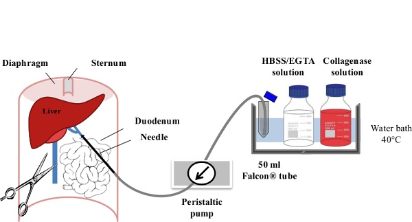
Figure 1: Liver Perfusion. After anaesthesia of the mouse, the digestive tract is laterally moved to the left of the abdomen in order to make the portal vein (PV) accessible. The PV is cannulated using a slow flow rate (1-3 ml/min) of EGTA/HBSS Solution and the inferior veina cava (IVC) is immediately ruptured to avoid any excess pressure within the liver. Within the first minute of perfusion, the flow rate is gradually increased to 7 ml/min. The Collagenase Solution is then perfused at 10 ml/min until its full digestion is achieved.
- Prepare freshly all reagents described in the material table.
- Warm the EGTA (Ethylene Glycol Tetra-acetic Acid)/HBSS (Hank's Balanced Salt Solution) Solution (50 ml per mouse) and the Collagenase Solution (100 ml per mouse) for 30 min at 40 °C.
- Rinse the pump flexible tubing first with 70% ethanol. Pour 40 ml of EGTA/HBSS Solution into a centrifuge tube immersed in the water bath and rinse the pump flexible tubing with pre-warmed EGTA/HBSS Solution.
- Perform terminal anesthesia using a barbiturate to reliably produce unconsciousness before respiratory depression and death. Inject phenobarbitone at 1 mg/kg, i.p. into a female or male CD1 mouse (35-45 g). Confirm the anesthesia by toe pinching.
- Shave abdominal hairs and sterilize the abdominal surface using 70% ethanol solution.
- Cut through the abdominal cavity and expose the portal vein and inferior vena cava by moving the intestine laterally to the left of the abdomen.
- Start the pump at a speed of 1-3 ml/min with EGTA/HBSS Solution and cannulate the portal vein using a 23 g butterfly needle (wings cut). Clamp the section of the portal vein cannulated with the 23 G needle and then flip the serrefine forceps at the surface of the opened mouse abdomen. The liver should quickly pale within the first 30 sec of EGTA/HBSS Solution perfusion.
- Rapidly incise the lower part of the inferior vena cava to avoid excess pressure building in the liver, and then increase the flow rate gradually to 7 ml/min, over the first minute of perfusion. The animal dies due to bleeding secondary to vena caval venipuncture.
- When less than 5 ml of EGTA/HBSS Solution is remaining into the centrifuge tube, fill it up with Collagenase Solution (40-50 ml). Increase the flow rate gradually to 10 ml/min, over 30 sec.
- Make the liver swell by applying pressure to the inferior vena cava, using tweezers, for 5-10 sec intervals. This can be done periodically (5-10 times during digestion). This step will improve the liver cell dissociation and reduce the perfusion time with collagenase.
- When less than 10 ml of Collagenase solution remains within the centrifuge tube, pour 40-50 ml of pre-warmed Collagenase solution into the centrifuge tube.
- After 10-15 min of perfusion and about 70-80 ml of Collagenase Solution perfused, apply a small pressure on the surface of the liver with a forceps. A print of the pressure indicates that liver cells are dissociated.
- Remove the liver from the abdominal cavity as one piece and place it in a centrifuge tube containing 20-30 ml of Kupffer Cells Isolation Medium. Keep liver cells on ice or at 4 °C for a maximum period of 3 hr to avoid affecting liver cell viability.
- Repeat the perfusion procedure on several animals if needed, while keeping the perfused liver on ice. Pool one to three livers for purification and proceed with stage 3.
2. Preparation of the Density Gradient for Centrifugation (Figure 2)
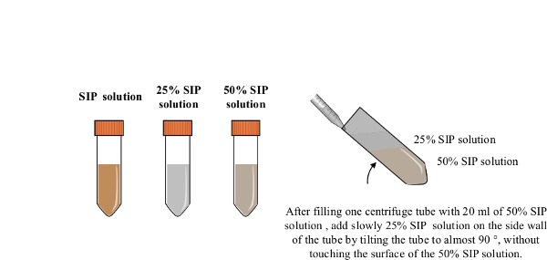
Figure 2: Preparation of the density gradient. All procedures are carried out under sterile condition. The SIP is prepared by mixing 15.3 ml of coated silica particle solution with 1.7 ml of 10 x PBS. A 5 ml of SIP are mixed with 15 ml of PBS to make 20 ml of 25% SIP solution. 10 ml of SIP are mixed with 10 ml of PBS to make 20 ml of 50% SIP solution. A centrifuge tube containing the 50% SIP solution (20 ml) is tilted and the 25% SIP solution (20 ml) is slowly added using a serological 25 ml pipette.
- Proceed with the following steps (section 2., 3. & 5.) under sterile condition and keep cells on ice or at 4 °C. Prepare the SIP (Solution of Isotonic coated silica Particles) by mixing 15.3 ml of coated silica particle solution with 1.7 ml of 10 x PBS.
- To make 20 ml of 25% SIP solution, mix 5 ml of SIP with 15 ml of PBS. To make 20 ml of 50% SIP solution, mix 10 ml of SIP with 10 ml of PBS.
- Fill one centrifuge tube with 20 ml of the 50% SIP solution. Tilt the centrifuge tube at an angle close to 90° and add slowly the 25% SIP solution (20 ml) on the side wall of the tube using a 25 ml serological pipette, without touching the surface of the 50% SIP layer. When adding the 25% SIP solution, progressively reduce the angle of the tube. Keep the density gradient on ice until use.
3. Kupffer Cell Purification (Figure 3)
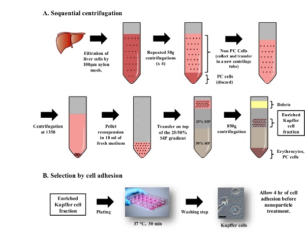
Figure 3: Purification of Kupffer Cells by density gradient centrifugation and cell adhesion. All procedures are carried out under sterile condition. (A) After rupture of the Glisson’s capsule, liver cells are filtered through a 100 µm strainer. The suspension is centrifuged at x 50 g to discard hepatocytes (pellets) and collect the non-parenchymal cell fraction (supernatant). This step is repeated 3 times. Non-parenchymal cells are added on top of a discontinuous isotonic gradient and centrifuged at 800 x g for 15 min. Kupffer cells collected from the 25% SIP cushion are purified further by cell adhesion selection. (B) Cells are plated in 24-well plate and incubated at 37 °C and 5% CO2 for 30 min. Non-adherent cells are washed once with 500 µl of HBSS. Kupffer cells display their adherent morphology 4 hr after plating, when f-CNTs can be added to the cells. The scale bar represents 25 µm. Please click here to view a larger version of this figure.
- Put one perfused liver in a Petri dish with 15 ml of Kupffer Cell Isolation Medium. Rupture the Glisson’s capsule (membrane of the liver) using scissors and release all liver cells into the Kupffer cell isolation medium. Filter the solution though a 100 µm cell strainer to be collected in a centrifuge tube. Repeat the perfusion procedure on additional perfused livers and pool the cells in the same centrifuge tube (15 ml from one mouse, up to 45 ml from three mice).
- Centrifuge the cell suspension at 50 x g for 2 min at 4 °C. Parenchymal cells (hepatocytes) will be in the pellet and non-parenchymal cells (including Kupffer cells) will be in the supernatant.
- Collect the supernatant in a clean centrifuge tube and centrifuge at 50 x g for 2 min each. Repeat this step three more times.
- Centrifuge at 1,350 x g for 15 min to pellet non-parenchymal cells. Discard the supernatant and resuspend the pellet in 10 ml of Kupffer Cell Isolation Medium.
- Add the non-parenchymal cell solution on the discontinuous isotonic gradient 25/50% as described in 2.3. Centrifuge at 850 x g for 15 min without acceleration or break.
- Localize the enriched Kupffer cell fraction appearing turbid within the 25% SIP fraction close to the 25/50% SIP interface (Figure 3). Using a 10 ml serological pipette, aspirate about 12 ml of the enriched Kupffer cell fraction. Transfer cells into a centrifuge tube containing 35-40 ml of Kupffer Cell Isolation Medium. Mix gently and centrifuge at 1,350 x g for 15 min at 4 °C to pellet cells.
- Discard the supernatant and resuspend cells in 5-10 ml of pre-warmed Kupffer Cell Culture Medium. Count cells (haemocytometer) and measure the viability using trypan blue staining.
- Plate the purified non-parenchymal cells in 24-well plates at a density of 5 x 10⁵ cells/well. Incubate the cells at 37 °C and 5% CO2 (Figure 3). After 30 min, gently remove the medium, wash with pre-warmed Hank's Balanced Salt Solution (HBSS) once then replace it with 500 µl of fresh and pre-warmed Kupffer Cell Culture Medium (per well).
- Leave Kupffer cells for at least 4 hr at 37 °C and 5% CO2 before treatment with nanoparticles, to allow them to acquire their adherent morphology.
4. Characterization
- Kupffer Cells Purity
- Prepare fresh PBS/BSA solution used to maintain Kupffer cell viability. Wash cells once with 500 µl of PBS/BSA solution. Remove the PBS/BSA solution and detach Kupffer cells in 500 µl of fresh PBS/BSA solution using gentle scraping. When cells are plated in 24-well plates, shorten the extremities of scraper blades with scissors to make the detachment easier to proceed. Transfer cells into flow cytometry tubes.
- Count Kupffer cells using a haemocytometer and centrifuge 1 x 10⁵ cells at 1,350 x g. Resuspend Kupffer cells in 30 µl of F4/80 antibody (neat). Mix well and incubate at room temperature for 30 min. Keep the unstained cells (not incubated with the antibody) on ice.
- Add 2 ml of PBS/BSA solution in stained cells, centrifuge at 1,350 g for 5 min and discard the resulting supernatant. Resuspend stained cells in 200 µl of PBS/BSA.
- For flow cytometry analysis, gate 10,000 unstained Kupffer cells according to their size (forward scatter) and granularity (side scatter). Analyze fluorescence of gated cells in FL-1 channel.
- Repeat the procedure for stained Kupffer cells keeping the same settings used for the flow cytometry analysis of unstained Kupffer cells. Select and quantify the fluorescence of stained cells to assess the purity of Kupffer cell.
- Kupffer Cell Phagocytic Activity
- Replace media with 0.5% (v/v) fluorescent beads in Kupffer Cell Culture Medium and incubate for 4 hr at 37 °C and 5% CO2.
- In order to remove non-internalized beads, centrifuge Kupffer cells at 1,350 x g, discard the supernatant and resuspend cells in 500 µl PBS/BSA Solution. Repeat this step 2 more times.
- Scrape Kupffer cells in 500 µl of PBS/BSA Solution and transfer cells into a flow cytometry tube.
- Centrifuge Kupffer cells at 1,350 x g, discard the supernatant and resuspend cells in 200 µl of PBS/BSA Solution.
- For flow cytometry analysis, gate 10,000 unstained Kupffer cells according to their size (forward scatter) and granularity (side scatter). Analyze fluorescence of gated cells in FL-2 channel.
5. Incubation of Chemically Functionalized Carbon Nanotubes (f -CNTs)
- Prepare a dispersion of f-CNTs (1 mg/ml) in water by sonication for 15 min before use. f-CNTs are prepared in-house22.
- Prepare 2x concentrated f-CNTs dispersion in Kupffer Cell Culture Medium. Sonicate f-CNTs dispersed in medium for 2 min before proceeding with step 5.3).
- For each well, replace 250 µl of old media with 250 µl of 2x f-CNTs dispersion. Untreated wells (250 µl of conditioned medium + 250 µl of fresh Kupffer Cell Culture Medium) and wells treated with 10% DMSO are used as negative and positive control, respectively.
- Incubate Kupffer cells at 37 °C and 5% CO2 for 24 or 72 hr.
6. Toxicity Assessment of f -CNTs in Kupffer Cells by the Modified Lactate Dehydrogenase (LDH) Assay
- Discard the supernatant.
- Add 200 µl (per well of 24-well plate) of Lysis Buffer and incubate at 37 °C for 30 min-1 hr.
- After carrying out vigorous pipetting, collect lysed Kupffer cells into microcentrifuge tubes and centrifuge at 40,000 x g for 10 min to pellet f-CNTs that were taken up by cells and the cell debris.
- Collect the supernatants (ƒ-CNT free) into microcentrifuge tubes and store at -20 °C or proceed to step 6.5).
- Transfer 50 µl into a 96-well plate and add an equal volume of the substrate mix solution (LDH kit) to each well. Cover the plate and incubate at room temperature for 15 min before adding 50 µl of the stop solution (LDH kit). Add triplicates of blank wells containing 50 µl of lysis buffer, 50 µl of substrate mix solution and 50 µl of stop solution.
- Read the absorbance at 490 nm in a microplate reader and use the following formula to calculate the (%) cell viability.

Access restricted. Please log in or start a trial to view this content.
Results
The number of non-purified parenchymal cells was consistent and ranged between 8 and 14 x 106 cells per mouse (17 isolations were performed). Each mouse isolation was sufficient to plate 16 to 28 wells. Cell viability by trypan blue staining showed cell viability ~95%. Kupffer cells showed a round shape within 30 min of incubation at 37 °C, related to their incomplete adherent morphology (Figure 4A). At 4 hr of incubation and afterwards, cells were spread and cell clusters started to form.
...Access restricted. Please log in or start a trial to view this content.
Discussion
The following steps are critical to achieve high yield and high viability of Kupffer cells. Aseptic conditions should be used to limit the risk of bacterial and fungal contamination. All instruments need to be sterilized before use. Reagents should be freshly prepared before carrying out the isolation procedure.
The choice of collagenase IV, with low tryptic activity, is crucial. Different lots from the same supplier have different enzymatic activity and they may need to be compared initially ...
Access restricted. Please log in or start a trial to view this content.
Disclosures
The authors declare that they have no competing financial interests.
Acknowledgements
This work was supported by EU FP7-ITN Marie-Curie Network program RADDEL. MB and KAJ would like to acknowledge Joe Varguese and Rui Serra Maia for their help and suggestions along the optimization of the Kupffer cell isolation.
Access restricted. Please log in or start a trial to view this content.
Materials
| Name | Company | Catalog Number | Comments |
|---|---|---|---|
| Euthatal (pentobarbital sodium) | Merial | ||
| CD1 mice | Charles River | - | Mouse weight should vary from 35 to 45 g. We advise the use of male CD1 mice as their weight increase rapidly (e.g. a male CD1 mouse of 7 weeks old exceeds 35 g in weight). It is also advised to contact the animal supplier in advance to arrange for the delivery of older animals. Alternatively animals can be in-house to reach the desired weight. |
| HBSS (Ca2+ and Mg2+ free, with bicarbonate) | Life Technologies | 14175-053 | HBSS must be Ca2+ free. |
| Ethylene Glycol Tetraacetic Acid (EGTA) tetrasodium salt | Sigma-Aldrich | E8145 | EDTA can also be used but EGTA has the advantage of chelating Ca2+ selectively. |
| HEPES (1 M) | Life Technologies | 15630-056 | 100 ml |
| Collagenase type IV | Worthington | CSL-4 | It is advised to test different batches of collagenase or at least mention to the supplier the product has to be suitable for liver cell isolation |
| Low glucose DMEM | Sigma-Aldrich | D5523 | 500 ml |
| RPMI (with sodium pyruvate and Glutamax) | Life Technologies | 12633-012 | 500 ml |
| Penicillin/Steptomycin | Life Technologies | 15140-122 | 100 ml |
| Fetal Bovine Serum | First-Link | 60-00-850 | 500 ml |
| Trypan blue solution | Sigma-Aldrich | T8154 | 100 ml |
| Coated silica particle solution (Percoll) | GE Healtcare | 17-0891-02 | Percoll® is very stable and can be kept for several years |
| 1x Phosphate Buffered Saline | Life Technologies | 10010-023 | 500 ml |
| 10x Phosphate Buffered Saline | Life Technologies | 70011-036 | 500 ml |
| Dimethyl sulfoxide | Fisher | D/4120/PB08 | 500 ml |
| LDH kit (CytoTox 96) | Promega | G1781 | Keep protected from light |
| DMEM phenol red free | Life Technologies | 31053-028 | 500 ml |
| F4/80 antibody (Alexa 488) | AbD Serotec | MCA497A488 | Do not dilute, used neat for flow cytometry |
| Fluorescent beads | Sigma | L2778 | Latex beads, amine-modified polystyrene, fluorescent red. 1 ml |
| Name of the Material | Company | Catalog number | Comments/Description |
| Butterfly blood collection set (23 G/305 mm long tubing) | BD | 367288 | |
| Syringe Filters (0.22 μm Blue Rim) | Minisart | 16534-K | |
| Centrifuge Tubes (50 ml Blue Cap) | BD Biosciences, Falcon | 35 2070 | |
| Petri dish (90 x 15 mm) | Thermo Fisher Scientific | BSN 101VR20 | |
| 100 μm cell strainer | BD | 352360 | |
| Peristaltic pump | Watson Marlow | SciQ 300 | Rinse tubing before and after each usage with sterile PBS and 70% ethanol. |
| 24-well plates | Corning | 3526 | |
| 96-well plates | Corning | 3595 | |
| Centrifuge | Eppendorf | 5810R | |
| Plate reader | BMG Labtech | FLUOstar Omega | |
| Serrefine forceps | Hammacher GmbH | Art. Nr. HSE 004-35 / Cat. Nr. 221-0051 | The serrefine forceps allow to clamp the vessel cannulated with the 23 G needle without the need of holding the forceps during the perfusion procedure. URL: (http://www.hammacher.de/Laboratory-Products/Clamps-forceps/Serrefines/HSE-004-35-Serrefine::25126.html) |
| Flow cytometry tubes | BD Biosciences, Falcon | 352052 | |
| Microcentrifuge tubes (1.5 ml) | Elkay | 000-MICR-150 | |
| Cell scraper | BD Biosciences, Falcon | 353086 | Cut blade extremities with a pair of scissors to scrape cells in 24-well plates. |
| Name of the Reagent | Company | Catalog number | Comments/Description |
| EGTA (Ethylene Glycol Tetraacetic Acid)/HBSS (Hank's Balanced Salt Solution) Solution | HBSS containing 0.5 mM EGTA and 25 mM HEPES. Adjust pH to 7.4. Prepare 50 ml for each liver to perfuse. | ||
| Collagenase Solution | DMEM low glucose containing collagenase type IV at 100 UI/ml, 15 mM HEPES and 1% Penicllin/Streptamycin (v/v). Adjust pH to 7.4. Prepare 100 ml for each liver to perfuse. After adding the collagenase, it is advised to warm up the solution for 30 min before use. This allows the collagenase activity to be optimum. | ||
| Kupffer Cell Isolation Medium | RPMI containing, 1% Non-Essential Amino-Acids (v/v),1% glutamax® (v/v) and 1% Penicllin/Streptomycin (v/v). Prepare at least 100 ml for 1-3 livers. | ||
| Kupffer Cell Culture Medium | RPMI containing 10% Fetal Bovine Serum, 1% Non-Essential Amino-Acids (v/v),1% Glutamax® (v/v) and 1% Penicllin/Streptamycin (v/v). Prepare at least 100 ml for 1-3 livers. | ||
| SIP (solution of isotonic coated silica particles) | Mix 1.7 ml of 10x Phosphate Buffered Saline with 15.3 ml of Percoll® to obtain 17mL of SIP. | ||
| 25% SIP solution | Mix 5 ml of SIP with 15 ml of 1x Phosphate Buffered Saline | ||
| 50% SIP solution | Mix 10 ml of SIP with 10 ml of 1x Phosphate Buffered Saline | ||
| Lysis Buffer | DMEM media with 0.9% Triton X-100 | ||
| PBS/BSA Solution | Prepare fresh Phosphate Buffered Saline pH 7.4 with 1% Bovine Serum Albumin. |
References
- Hoffmann, D., et al. Performance of novel kidney biomarkers in preclinical toxicity studies. Toxicol Sci. 116 (1), 8-22 (2010).
- Nel, A., et al. Nanomaterial Toxicity Testing in the 21st Century: Use of a Predictive Toxicological Approach and High-Throughput Screening. Accounts of Chemical Research. 46 (3), 607-621 (2013).
- Bregoli, L., et al. Toxicity of antimony trioxide nanoparticles on human hematopoietic progenitor cells and comparison to cell lines. Toxicology. 262 (2), 121-129 (2009).
- Yamasaki, K., et al. Genomic Aberrations and Cellular Heterogeneity in SV40-Immortalized Human Corneal Epithelial Cells. Investigative Ophthalmology, & Visual Science. 50 (2), 604-613 (2009).
- Motoyoshi, Y., et al. Megalin contributes to the early injury of proximal tubule cells during nonselective proteinuria. Kidney International. 74 (10), 1262-1269 (1038).
- Prozialeck, W. C., Edwards, J. R., Lamar, P. C., Smith, C. S. Epithelial barrier characteristics and expression of cell adhesion molecules in proximal tubule-derived cell lines commonly used for in vitro toxicity studies. Toxicology in Vitro. 20 (6), 942-953 (2006).
- Chamberlain, L. M., Godek, M. L., Gonzalez-Juarrero, M., Grainger, D. W. Phenotypic non-equivalence of murine (monocyte-) macrophage cells in biomaterial and inflammatory models. J Biomed Mater Res A. 88 (4), 858-871 (2009).
- Kermanizadeh, A., et al. The role of Kupffer cells in the hepatic response to silver nanoparticles. Nanotoxicology. , (2013).
- Fischer, H. C., Hauck, T. S., Gomez-Aristizabal, A., Chan, W. C. W. Exploring Primary Liver Macrophages for Studying Quantum Dot Interactions with Biological Systems. Advanced Materials. 22 (23), 2520-2524 (2010).
- Wan, J. H., et al. M2 Kupffer Cells Promote M1 Kupffer Cell Apoptosis: A Protective Mechanism Against Alcoholic and Nonalcoholic Fatty Liver Disease. Hepatology. 59 (1), 130-142 (2014).
- Matsuoka, M., Tsukamoto, H. Stimulation of hepatic lipocyte collagen production by Kupffer cell-derived transforming growth factor β: Implication for a pathogenetic role in alcoholic liver fibrogenesis. Hepatology. 11 (4), 599-605 (1990).
- Polakos, N. K., et al. Kupffer Cell-Dependent Hepatitis Occurs during Influenza Infection. The American Journal of Pathology. 168 (4), 1169-1178 (2006).
- Tanner, A., Keyhani, A., Reiner, R., Holdstock, G., Wright, R. Proteolytic enzymes released by liver macrophages may promote hepatic injury in a rat model of hepatic damage. Gastroenterology. 80 (4), 647-654 (1981).
- Knittel, T., et al. Expression patterns of matrix metalloproteinases and their inhibitors in parenchymal and non-parenchymal cells of rat liver: regulation by TNF-α and TGF-β1. Journal of Hepatology. 30 (1), 48-60 (1999).
- Branster, M. V., Morton, R. K. Isolation of Intact Liver Cells. Nature. 180 (4597), 1283-1284 (1038).
- Smedsrød, B., Pertoft, H. Preparation of pure hepatocytes and reticuloendothelial cells in high yield from a single rat liver by means of Percoll centrifugation and selective adherence. Journal of Leukocyte Biology. 38 (2), 213-230 (1985).
- Morin, O., Patry, P., Lafleur, L. Heterogeneity of endothelial cells of adult rat liver as resolved by sedimentation velocity and flow cytometry. J Cell Physiol. 119 (3), 327-334 (1984).
- Miller, S. B., et al. Purification of cells from livers of carcinogen-treated rats by free-flow electrophoresis. Cancer Res. 43 (9), 4176-4179 (1983).
- Firme, C. P., Bandaru, P. R. Toxicity issues in the application of carbon nanotubes to biological systems. Nanomedicine. 6 (2), 245-256 (2010).
- Ali-Boucetta, H., Al-Jamal, K. T., Kostarelos, K. Cytotoxic assessment of carbon nanotube interaction with cell cultures. Methods in Molecular Biology. 726, 299-312 (2011).
- Wang, G., et al. Understanding and correcting for carbon nanotube interferences with a commercial LDH cytotoxicity assay. Toxicology. 299 (2-3), 99-111 (2012).
- Wang, J. T. -W., et al. Magnetically Decorated Multiwalled Carbon Nanotubes as Dual MRI and SPECT Contrast Agents. Advanced Functional Materials. 24 (13), 1880-1894 (2014).
- Li, P. Z., Li, J. Z., Li, M., Gong, J. P., He, K. An efficient method to isolate and culture mouse Kupffer cells. Immunol Lett. 158 (1-2), 52-56 (2014).
- Froh, M., Konno, A., Thurman, R. G. Isolation of liver Kupffer cells. Curr Protoc Toxicol. Chapter 14, Unit14 14(2003).
Access restricted. Please log in or start a trial to view this content.
Reprints and Permissions
