Lymphocytes are the major players in adaptive immune responses. Here, we present a lymphocyte purification protocol to determine the physiological functions of the desired molecules in lymphocyte activation in vitro and in vivo. The described experimental procedures are suitable for comparing functional capacities between control and genetically modified lymphocytes.
Method Article
Isolation and Activation of Murine Lymphocytes
In This Article
Summary
Abstract
B and T cells, with their extremely diverse antigen-receptor repertoires, have the ability to mount specific immune responses against almost any invading pathogen1,2. Understandably, such intricate abilities are controlled by a large number of molecules involved in various cellular processes to ensure timely and spatially regulated immune responses3. Here, we describe experimental procedures that allow rapid isolation of highly purified murine lymphocytes using magnetic cell sorting technology. The resulting purified lymphocytes can then be subjected to various in vitro or in vivo functional assays, such as the determination of lymphocyte signaling capacity upon stimulation by immunoblotting4 and the investigation of proliferative abilities by 3H-thymidine incorporation or carboxyfluorescein diacetate succinimidyl ester (CFSE) labeling5-7. In addition to comparing the functional capacities of control and genetically modified lymphocytes, we can also determine the T cell stimulatory capacity of antigen-presenting cells (APCs) in vivo, as shown in our representative results using transplanted CFSE-labeled OT-I T cells.
Introduction
Mature lymphocytes generally exist in the resting state if there is no pre-existing infection or inflammation in the individual. Therefore, it is important to retain the naïve status of lymphocytes during the isolation process before performing in vitro or in vivo functional assays. The key to ensuring consistent and reproducible results is to limit any unnecessary manipulation of the cells.
Magnetic cell sorting utilizes antibodies and microbeads to label cells so as to enrich the cell population of interest. With this approach, there are two purification strategies: positive enrichment and negative depletion. Positive enrichment enriches the cell population of interest using an antibody that binds to the target cells. Negative depletion, on the other hand, depletes non-target cells, leaving the cell population of interest. In our lab, we prefer negative depletion to positive enrichment because the binding of antibodies to the target cells could potentially alter cell features and behavior. In fact, many established cell surface markers suitable for the isolation of a particular cell population are also functional receptors.
Magnetic cell sorting not only yields highly pure populations of viable target cells, it is also less time-consuming and avoids the cellular stress induced by high-pressure flow used in fluorescence-activated cell sorting (FACS). By labeling the unwanted cell populations and depleting them using a magnetic separation column, we are able to perform rapid cell isolation without compromising the viability of the target cell population. In this protocol, we demonstrate the use of negative depletion strategies to purify naïve B cells or T cells.
Protocol
All mice are bred and maintained under specific pathogen-free conditions and all mouse protocols are conducted in accordance with the guidelines of the Institutional Animal Care and Use Committee.
1. Preparation of Buffers and Reagents
- Prepare complete Roswell Park Memorial Institute (RPMI) medium (10% heat-inactivated fetal bovine serum (FBS), 2 mM L-glutamine, penicillin (100 IU/ml)/streptomycin (100 µg/ml), 55 µM 2-mercaptoethanol).
- Prepare 20x Balanced Salt Solution (BSS) Stock 1 and Stock 2, Separately.
- Prepare 20x BSS stock 1 (111 mM dextrose, 8.8 mM potassium phosphate, 26.7 mM sodium phosphate dibasic in 1 L sterile water) and add 40 ml 0.5% Phenol Red to 20x BSS stock 1 before final volume adjustment. Sterile filter using 0.2 µm filter before storing at 4 °C.
- Prepare 20x BSS stock 2 (25.8 mM calcium chloride dihydrate, 107 mM potassium chloride, 2.73 M sodium chloride, 19.6 mM magnesium chloride hexahydrate, 16.6 mM magnesium sulfate in 1 L sterile water). Sterile filter using 0.2 µm filter before storing at 4 °C.
- To prepare 1x BSS for experimental use, dilute 50 ml BSS stock 1 and 50 ml BSS stock 2, separately, in 400 ml sterile water each. Combine both diluted solutions, adjust to pH 7, and add 20 ml FBS (2%). Top up to 1 L using sterile water and sterile filter using 0.2 µm filter.
NOTE: BSS stock solutions should be prepared separately because the mixing of concentrated BSS stocks directly could result in precipitation.
- Red Blood Cell (RBC) Lysis Buffer
- To prepare RBC lysis buffer, mix 9 parts stock ammonium chloride (155 mM ammonium chloride in sterile water) to 1 part stock Tris-base (130 mM Tris(hydroxymethyl)aminomethane in sterile water, pH 7.65) before use.
NOTE: Store sterile stock solutions of ammonium chloride and Tris-base at 4 °C. Prepare lysis buffer fresh to ensure efficient lysis of RBCs.
- To prepare RBC lysis buffer, mix 9 parts stock ammonium chloride (155 mM ammonium chloride in sterile water) to 1 part stock Tris-base (130 mM Tris(hydroxymethyl)aminomethane in sterile water, pH 7.65) before use.
2. Generation of Lymphocyte Suspension from Spleen or Lymph Nodes
NOTE: It is important to prepare all reagents and equipment required for the experiment before mouse euthanasia and to generate single cell suspensions of lymphocytes as soon as possible to maintain high cell viabilities.
- Euthanize experimental mouse by cervical dislocation or CO2 asphyxiation.
NOTE: From this step onwards, all experimental procedures should be performed aseptically. - Dip the entire mouse into 70% ethanol before making any incisions. Remove the spleen and lymph nodes aseptically8, and place them in separate 15 ml tubes containing 5 ml ice-cold RPMI/FBS (RPMI with 2% FBS) or BSS/FBS (from step 1.2.3).
NOTE: Since BSS prevents efficient RBC lysis, use RPMI for the preparation of the splenic cell suspension and switch to BSS after RBC lysis. - To generate a single cell suspension from spleen or lymph nodes, place the organ(s) in between two pieces of sterile 100 µm cell strainer mesh in a petri dish containing 2 ml ice-cold RPMI/FBS or BSS/FBS. Using the plunger of a 1 ml syringe, mash the organ(s) until it has been torn into very fine parts.
- Transfer the cell suspension to a 15 ml tube and wash the cell strainer mesh with ice-cold RPMI/FBS or BSS/FBS. Collect the remaining cell suspension, add it to the same 15-ml tube, and spin down at 453 x g for 5 min at 4 °C. Remove the supernatant.
NOTE: Re-suspend the pelleted cells by flicking the tube with fingers before adding RBC lysis buffer or medium in the subsequent steps. - Prepare room temperature (RT) RBC lysis buffer during centrifugation of the cell suspension (see step 1.3.). After pelleting the cells and removing the supernatant, re-suspend the cells with 1 ml RBC lysis buffer for every 108 cells. Incubate the lysis reaction at RT for 3-4 min.
- Stop RBC lysis with 14 ml ice-cold BSS/FBS and spin down at 453 x g for 5 min at 4 °C.
3. Purification of B and T Cells
- Purification of B Cells
- Count the cells using a hemocytometer. Re-suspend up to 108 splenic cells in 300 µl BSS/FBS and add 50 µl anti-CD43 magnetic microbeads9,10. To remove dead cells, add 30 µl Annexin V magnetic beads. Incubate the cell suspension in a 4 °C refrigerator for 30 min.
- Purification of T Cells
- Count the cells using a hemocytometer. Re-suspend up to 108 cells in non-T cell depletion antibody cocktail (biotinylated antibodies against CD19, B220, Gr-1, TCR-γδ, CD49b, CD11c, CD11b, Ter119 and CD4 or CD8 depending on the target cell population to be purified), diluted 1:200 in 200 µl BSS/FBS4,5. Incubate the cell suspension in a 4 °C refrigerator for 15 min.
- After incubation, add 10 ml BSS/FBS to wash the cells and spin down at 453 x g for 5 min at 4 °C. Remove the supernatant and re-suspend the cells in 165 µl BSS/FBS with 30 µl streptavidin microbeads and 15 µl Annexin V magnetic beads. Incubate the cell suspension in a 4 °C refrigerator for 30 min.
NOTE: To ensure even labeling with the magnetic microbeads, incubate the cells with microbeads for 15 min, then mix the cell suspension gently by tapping the 15 ml tube and incubate another 15 min during step 3.1.1. or 3.2.2.
- Preparation of Separation Column for Cell Purification
- Prepare an unused separation column during microbead labeling of the cells (step 3.1.1. or 3.2.2.). Pre-warm BSS (without FBS) to RT and use 2 ml to wash and equilibrate the column aseptically. After equilibration with BSS, the washed column should not be allowed to dry out.
NOTE: We use the LS column instead of the recommended LD column due to its re-usability (see step 3.4.). - After labeling with the magnetic beads, add 14 ml BSS/FBS to wash the cells and spin down at 453 x g for 5 min at 4 °C. Remove the supernatant and re-suspend the cells in 1-3 ml RT BSS.
- Attach a sterile 21 G needle to the tip of the column to reduce the flow rate during the process of purification. Load the cell suspension onto the equilibrated column and collect the flow through containing the purified target cells.
NOTE: Avoid introducing bubbles into the column while loading. - Wash the column once with 1 ml BSS/FBS and collect the flow through containing the purified target cells. Reload the column with the flow through once again. Collect the flow through after the second loading in the same 15 ml tube.
- Wash the column 3 times with 1 ml BSS/FBS and collect the flow through containing the purified target cells. Thereafter, add 5 ml BSS/FBS to the column and, with a plunger, flush the magnetically labeled cells out of the column into a new 15 ml tube.
- Check the purity of the cells collected by flow cytometry using antibodies that bind to surface antigens of purified B or T cells4,5.
- Prepare an unused separation column during microbead labeling of the cells (step 3.1.1. or 3.2.2.). Pre-warm BSS (without FBS) to RT and use 2 ml to wash and equilibrate the column aseptically. After equilibration with BSS, the washed column should not be allowed to dry out.
- Re-using the Separation Column
NOTE: The LS column can be reused up to 4 times without affecting purification efficiency.- Wash the column 3 times with 5 ml phosphate buffered saline (PBS) and 3 times with 5 ml distilled water from the top using the plunger.
- Wash the column with 5 ml 70% ethanol and dry the column extensively using an air tap to prevent the buildup of rust in the column.
- To prepare a used LS column for a separate purification experiment, wash the column from the bottom up with 5 ml 70% ethanol using the syringe adaptor. Next, wash the column from the bottom up twice with 5 ml sterile PBS, followed by 5 ml PBS once from the top of the column. Add 2 ml RT BSS to equilibrate the column and then proceed to loading the column with labeled cells.
4. CFSE Labeling and Stimulation
NOTE: Purified cells can be subjected to a variety of in vitro and in vivo functional assays. Here, we use purified T cells to determine the T cell stimulation capability of APCs5.
- Pre-warm labeling solution (0.1% FBS in PBS) to 37 °C prior to CFSE loading.
NOTE: Using a low percentage of FBS in PBS reduces cell death during CFSE loading and minimizes cell loss during centrifugation. However, too much FBS can interfere with CFSE loading. - Wash purified cells twice with labeling solution, then re-suspended at 2 x 10⁷ cells/ml in pre-warmed labeling solution in a 15 ml tube.
- Prepare 10 µM CFSE solution (1:500 dilution of 5 mM CFSE stock solution) in pre-warmed labeling solution. CFSE solution should be freshly prepared each time to achieve optimal labeling.
- To load cells with CFSE, add 1 part cell suspension to 1 part 10 µM CFSE solution in a 15-ml tube and incubate in the dark for 10 min at 37 °C. A final concentration of 5 µM CFSE is used to label 1 x 10⁷ cells/ml.
- Invert the tube every 2 min to ensure a homogenous mixture of cells during CFSE loading.
- To stop the reaction, add several volumes of ice-cold complete RPMI medium and spin down at 453 x g for 5 min at 4 °C. Upon successful CFSE loading, the cell pellet will appear yellowish.
- Wash CFSE loaded cells one more time with ice-cold complete RPMI medium and spin down at 453 x g for 5 min at 4 °C before using for in vitro culturing or in vivo stimulation.
5. In Vitro Stimulation
- Prepare a 2x stock solution of stimuli (2x stimuli stock solution) immediately before use so that 100 µl of 2x stimuli stock solution can be added to 100 µl of cells to a final volume of 200 µl per well in a 96-well plate.
- If a plate coated with stimuli (IgM or CD40 for B cells or CD3 and CD28 for T cells) is required, dilute the stimuli in PBS and pre-coat the culture plate at 4 °C overnight. Alternatively, the culture plate can be coated at 37 °C for 1 hr on the day of the experiment. Wash the coated plate twice with PBS (Do not allow the plate to dry out at any time).
| For B cells | |
| Stimuli | Final concentration |
| F(ab’)2 goat anti-mouse IgM | 0.6-2.4 µg/ml |
| Anti-mouse CD40 mAb | 0.5-2 µg/ml |
| Recombinant mouse IL-4 | 25 U/ml |
| Lipopolysaccharide | 0.1-10 µg/ml |
| (LPS) from E. coli Serotype 055:B5 | |
| For T cells | |
| Stimuli | Final concentration |
| Anti-CD3 (plate coated) | 2-10 µg /ml |
| (50 µl/well for coating) | |
| Anti-CD28 (plate coated) | 2 µg/ml |
| Recombinant IL-2 | 40 U/ml |
| PDBu (Phorbol ester) | 5-50 ng/ml |
| A23187 (Calcium ionophore) | 250 ng/ml |
Table 1: Concentrations of stimuli used to stimulate lymphocytes in in vitro culture.
- For CFSE-labeled B cells, re-suspend to 3 x 10⁶ cells/ml in complete RPMI medium and culture in triplicate with 3 x 10⁵ cells/well in 96-well flat bottom plates for 72 hr.
- For CFSE-labeled T cells, re-suspend to 0.5-3 x 10⁶ cells/ml in complete RPMI medium and culture in triplicate with 0.5-3 x 10⁵ cells/well in 96-well round bottom plates for 48 or 72 hr.
6. In Vivo Stimulation
- For in vivo stimulation, adoptively transfer 4 x 10⁶ CFSE-labeled T cells per mouse (intravenously (i.v.) in 200 µl PBS) into each MHC-matched recipient mouse.
NOTE: In this protocol, CFSE-labeled T cells can be adoptively transferred using tail vein or retro-orbital injection as these cells will home to lymphoid organs such as the spleen and lymph nodes. - Challenge the recipient mice one day later with the antigen.
NOTE: In this example, we use ovalbumin (OVA protein, 50 µg/mouse) as the antigen because OVA-specific, T cell receptor (TCR)-transgenic T cells were adoptively transferred into recipient mice. Prepare OVA protein in sterile PBS and inject 100 µl of OVA protein/PBS or PBS control, via subcutaneous injection (s.i.), into each recipient mouse5. - Harvest and generate single cell suspensions from lymphoid organs (lymph nodes and spleens) of recipient mice 3 days after immunization with OVA protein or PBS. Separate lymph nodes into proximal lymph nodes (pLN), which includes axillary, brachial and superficial cervical lymph nodes) and distal lymph nodes (dLN), which includes mesenteric, popliteal, inguinal, lumbar, and caudal lymph nodes. Stain cells using the appropriate FACS antibodies to check for T cell proliferation.
NOTE: In this CFSE cell tracking experiment, CFSE-labeled T cells from PBS-injected control mice establish a baseline fluorescence for non-dividing cells. Cell divisions of proliferating, antigen-stimulated, CFSE-labeled cells are visualized by measuring fluorescence peaks12,13. With the PBS control, the number of cell divisions of proliferating, CFSE-labeled T cells can be determined12,13. - Analyze the CFSE-labeled cell proliferation data by comparing the number of cell divisions or peaks between samples (Figures 1 and 2).
NOTE: For example, CFSE-labeled, OVA-specific T cells adoptively transferred into recipient mice receiving the OVA antigen will undergo active proliferation compared to the PBS-injected control mice5,12. Furthermore, it is noteworthy to point out that there are many ways to analyze CFSE cell proliferation data, as demonstrated by Hawkins and colleagues14.
Results
Magnetic cell purification of lymphocytes allows users to purify a target cell population in a relatively short amount of time. Using our depletion protocol, we were able to increase the percentage of CD8 T cells (OT-I in recombination-activating gene-1 (RAG-1)-deficient mice) from 72.8% (before purification) to 94.2% (after purification; Figure 1A)4,5. These purified lymphocytes can then be used for downstream functional assays to determine lymphocyte proliferation and signal transduction4,5. For example, we can study the in vivo T cell stimulation capacity of APCs by transferring CFSE-labeled, antigen-specific T cells into wildtype (WT) and mutant (MT) mice immunized with suitable antigen5.
In our representative experiment, we transferred purified, CFSE-labeled, ovalbumin (OVA) specific OT-I CD8 T cells into control (WT) and mutant (MT) mice and immunized these mice one day later with OVA protein. Three days after immunization, we harvested lymph nodes (proximal and distal) and spleens and analyzed the T cells by flow cytometry. CD45 allelic forms can be utilized to better separate the dividing, CFSE-labeled, donor OT-I CD8 cells from non-labeled, recipient CD8 T cells. In this example, donor and recipient cells are from CD45.1 and CD45.2 mice, respectively (Figure 1B). The CFSE levels of surviving, unstimulated OT-I T cells at this time point are used to define the peak for non-proliferating cells (Figure 1C, dotted line). Upon stimulation, the intensity of the CFSE levels in OT-I T cells will reduce by half with each division. T cell proliferation can thereby be determined by counting the number of CFSE peaks6,12. In our representative results, we do not see differences in the cross-presentation capacities of WT and MT APCs (Figure 1C, solid lines), since the T cells proliferate at similar rates in both mice.
In a separate experiment, we performed a [3H]-thymidine incorporation assay to study the cell proliferation of activated control and MT T cells. Purified WT and MT T cells were stimulated with various stimuli for 48 hr and pulsed with [3H]-thymidine for the final 8 hr. Proliferating cells in the S-phase of the cell cycle will incorporate the radiolabeled nucleotide, [3H]-thymidine, into newly synthesized deoxyribonucleic acid (DNA), therefore, using liquid scintillation counting, cell proliferation can be measured by [3H]-thymidine uptake. The number of counts per minute (c.p.m.) directly correlates with the amount of [3H]-thymidine uptake in proliferating T cells. In our representative results, the reduced number of c.p.m. obtained from MT T cells upon stimulation compared to counts from WT T cells indicates a compromised proliferative capacity of MT T cells (Figure 1D).
To determine B cell proliferative capacity, we purified splenic B cells using the described depletion strategy. Upon purification, we were able to increase the percentage of B220 B cells (WT mice) from 63.9% (before purification) to 98.4% (after purification; Figure 2A)4. Similar to purified T cells, purified splenic B cells can also be used for downstream functional assays to assess lymphocyte proliferation and signal transduction4. Subsequently, we labeled purified WT splenic B cells with CFSE and stimulated these cells in vitro using plates coated with anti-CD40 supplemented with the cytokine IL-4. Three days after stimulation, we analyzed viable, activated B cells by flow cytometry. The CFSE levels of surviving, unstimulated B cells at this time point are used to define the peak for non-proliferating cells (Figure 2B, dotted line). Similar to T cells, the intensity of the CFSE levels in B cells will reduce by half with each division6,12.
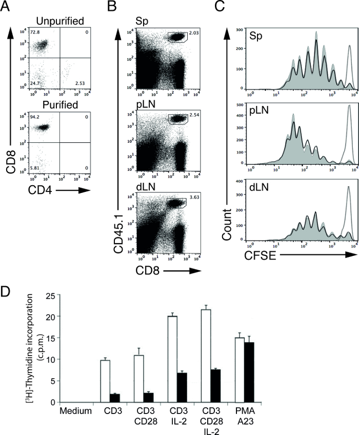
Figure 1: CFSE profiles of adoptively transferred purified OT-I T cells after immunization. (A) CD4 and CD8 staining of cells isolated from OT-I; RAG-1-deficient mice before (upper panel) and after (lower panel) non-T cell depletion. (B) Representative FACS plots of T cells from spleen (Sp), proximal lymph nodes (pLN) and distal lymph nodes (dLN). Donor OT-I CD8 T cells are CD45.1 positive. (C) Representative CFSE profiles of transferred, CFSE-labeled OT-I donor T cells from spleen (Sp), proximal lymph nodes (pLN) and distal lymph nodes (dLN) of WT (shaded curves) and MT (solid lines) recipient mice 3 days after immunization with OVA protein and LPS. Dotted line indicates OT-I T cells from WT recipient mice without immunization (PBS injected). (D) Cell proliferation of activated control or MT T cells as measured by [3H]-thymidine uptake assay. Results are presented in counts per minute (c.p.m.). Purified control (open bar) or MT (closed bar) T cells were incubated for 48 hr in medium only (medium) or in the presence of plate-bound anti-CD3 or anti-CD3 plus anti-CD28 with or without recombinant IL-2. Polyclonal cell activation was triggered by PMA (phorbol ester) and A23 (calcium ionophore). Please click here to view a larger version of this figure.
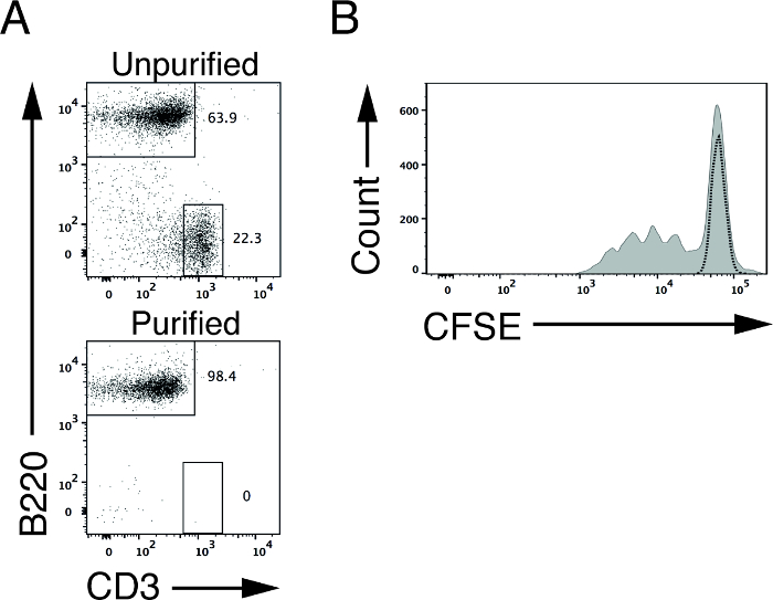
Figure 2: CFSE profile of B cells upon in vitro stimulation. (A) B220 and CD3 staining of splenic cells isolated from WT mice before (upper panel) and after (lower panel) non-B cell depletion. (B) Representative CFSE profile of CFSE-labeled WT (shaded curve) splenic B cells after 3 days of in vitro stimulation with plates coated with anti-CD40 and IL4. Dotted line indicates CFSE-labeled WT B cells without stimulation. Please click here to view a larger version of this figure.
Discussion
In this protocol, we demonstrate a procedure for purifying lymphocytes from lymphoid organs. Cell purification using magnetic bead sorting is a fast and simple method that yields viable, highly purified target cells.
Critical Steps within the Protocol
Cell viability and cell yield
Maintaining viability of hematopoietic lineage cells in vitro is critical to ensuring successful and reproducible experiments. Chemical and biological reagents, sub-optimal experimental conditions or improper storage conditions of excised organs can all affect cell viability. Upon excision from mice, lymphoid organs need to be stored on ice and single cell suspensions prepared as soon as possible. High-speed centrifugation of cell suspensions should also be avoided. Furthermore, cell pellets should be loosened by flicking tubes with fingers after removal of supernatant. It is not recommended to re-suspend pellets directly with large amounts of medium by pipetting.
Approximately 2-4 x 10⁷ splenic B cells and 2 x 10⁷ T cells from all major lymph nodes can be isolated from one 8-10 week-old WT mouse. Reduced numbers of purified B and T cells (below the expected value) indicates sub-optimal conditions, as mentioned earlier.
Cell purity
The average purity of isolated cells using this protocol is within the range of 90 to 95%, which is sufficiently pure for subsequent in vitro or in vivo experiments (Figure 1A). It is advisable not to overload the separation column with excessive numbers of cells during the process of separation because doing so can compromise cell purity due to the column's limited binding capacity.
Quality of FBS
The quality of FBS used to supplement the culture medium is critical for the in vitro survival of lymphocytes. FBS from different sources and batches may vary in their ability to support in vitro lymphocyte responses. Therefore, it is important to test different types of FBS to find one that gives high-specific response with low background. A substantial number of bottles should then be reserved as a stock.
Modifications and Troubleshooting
CFSE labeling conditions
Even though CFSE labeling works in plain RPMI, we have found that using a low percentage of FBS in PBS reduces cell death during the loading process, without compromising the efficiency of labeling. Moreover, the presence of FBS minimizes cell loss during centrifugation.
An important aspect of CFSE labeling is to ensure even labeling of the cells in order to visualize the distinct peaks that represent cell division. Overloading of CFSE could result in increased cell death in vitro or poor recovery of labeled cells in vivo. On the other hand, insufficient CFSE labeling or heterogeneity in the target cell suspension during the process of labeling can result in poorly resolved CFSE peaks12. Therefore, it is important to optimize the CFSE labeling conditions. The amount of CFSE used for labeling should be kept as low as possible to reduce potential toxic effects from overloading, but still achieve sufficient labeling to detect nicely resolved peaks upon stimulation within the experimental time frame. In addition, to avoid poorly resolved peaks, cell clumps should be removed from single cell suspensions prior to CFSE loading.
Cell clumps and cell loss
It is crucial to ensure that cells are fully re-suspended before proceeding to the next step because the perpetual removal of cell clumps during the experiment drastically reduces cell numbers. Cell clumps are usually associated with reduced cell viability or inadequate re-suspension of the cell pellet.
CFSE and emission spectra overlap
CFSE-labeled cells can be further defined with fluorophore-conjugated antibodies after one day in culture, however, these CFSE-labeled cells still remain brightly fluorescent after a day in culture. Using combinations of antibodies conjugated to bright fluorophores with minimal amounts of emission spectra overlap with CFSE, such as phycoerythrin (PE) and phycoerythrin-cyanine 7 (PeCy7), provides optimal staining results. However, compensation to reduce emission spectra overlap is still required. Additionally, due to the high intensity of CFSE signal (FL-1 channel) that spills into the FL-2 channel, the FL-2 channel has to be compensated by deducting the high percentage of FL-2 value to achieve an optimal CFSE profile. One can refer to the paper published by Quah et al., which provides solutions for troubleshooting CFSE labeling and analysis of results12.
Alternatives to CFSE
The emission spectrum of CFSE restricts the use of combinations of CFSE with fluorescein derivatives such as green fluorescent protein (GFP) and fluorescein isothiocyanate (FITC)-conjugated antibodies. There are, however, other commercially available cell dyes with equally high fluorescence intensity and low cell toxicity in violet (CTV, excitation/emission: 405/450 nm), yellow (CTY, 555/580 nm), far red (CTFR, 630/661 nm or CPD670, 647/670 nm) and red (PKH26, 551/567 nm). Using cell labeling dyes with different emission spectra provides flexibility in experimental design. On the other hand, not all of the above-described cell labeling dyes can generate distinct cell division peaks. A comparative study performed using various cell labeling dyes concluded that CTV is a better replacement for CFSE because CTV enables the detection of a higher number of clearly defined cell division peaks13.
Limitations
The major limitation of magnetic cell sorting is that it is only suitable for simple cell sorting. In our example, we could use CD43 to deplete all non-B cells from spleen; however, we would not be able to separate follicular B cells from marginal zone B cells. These sub-populations can only be defined with multiple surface markers. While it is possible to positively select cells using magnetic cell sorting based on multiple surface markers, it is dependent on specially prepared, commercially available reagents that enable the release of microbeads from target cells after the first round of sorting before proceeding to the second marker. Thus, the user would be completely dependent on the commercially available kits. In such a case, FACS is much more advantageous in separating refined cell populations.
Significance of Technique
Antibody-based cell sorting approaches, such as magnetic cell sorting and FACS, are the most reliable cell sorting techniques to date15. Other methods of cell separation exist, including density-based and adherence-based techniques, however, lymphocytes are poorly adherent cells and their subpopulations are relatively similar in density, thus adherence- and density-based techniques are either not applicable or are very inefficient15.
As mentioned in the introduction, rapid, simple, high cell viability, and independent of any sophisticated equipment are the winning features of magnetic cell sorting over FACS. In particular, negative depletion using magnetic cell sorting labels and depletes undesirable cells using a magnetic separation column, while isolating target cells with minimal modifications to the cell surface and maintaining the naïve state.
Future Applications
The highly enriched, viable, and naïve lymphocytes purified using this magnetic-based purification technique can be subjected to various functional assays of lymphocyte behavior and signaling mechanisms in vitro and in vivo. In this protocol, we demonstrated using two different cell proliferation assays — [3H]-thymidine incorporation assay and CFSE cell proliferation assay — to investigate the cell proliferative capacities of activated lymphocytes.
The choice between [3H]-thymidine incorporation assay and CFSE cell proliferation assay depends largely on sample size and experimental conditions. Utilizing the [3H]-thymidine incorporation assay in experiments with large sample sizes generates high-throughput cell proliferation data. In order to ensure optimal [3H]-thymidine uptake and reproducibility of results, [3H]-thymidine should be added to the culture when a majority of the cells are actively dividing. Optimization of the labeling protocol is required for different cell types and stimuli. Furthermore, due to the radioactive nature of [3H]-thymidine, this incorporation assay can only be performed in vitro, unlike CFSE labeling of cells, which can be activated in vitro (Figure 2B) or adoptively transferred into recipient mice (Figure 1B and 1C).
The CFSE cell proliferation assay, on the other hand, offers more information on cell proliferation than [3H]-thymidine incorporation. Since the intensity of CFSE in CFSE-labeled cells reduces by half with each cell division, one can determine the number of cell divisions and the proportion of cells at each division by counting the number and size of peaks12,13. Furthermore, CFSE-labeled cells can be further defined into different subsets according to their surface molecule expression by flow cytometry using the corresponding fluorophore-conjugated antibodies. Most importantly, unlike the [3H]-thymidine incorporation assay that measures cell proliferation at the final time point, the CFSE cell proliferation assay allows for the tracking of cell division, providing more information on the kinetics of cell proliferation capacity. However, the CFSE cell proliferation assay may not be suitable for experiments with large sample sizes.
Disclosures
The authors have nothing to disclose.
Acknowledgements
The study is supported by the Ministry of Education, Singapore (AcRF Tier1-RG40/13 and Tier2-MOE2013-T2-2-038). The manuscript was edited by Amy Sullivan from Obrizus Communications.
Materials
| Name | Company | Catalog Number | Comments |
|---|---|---|---|
| Materials | |||
| RPMI 1640 (without L-Glutamine) | Gibco | 31870025 | |
| Fetal Bovine Serum | Heat inactivated | ||
| L-glutamine | Gibco | 25030024 | |
| Penicillin/Streptomycin | Gibco | 15140114 | |
| 2-mercaptoethanol | Gibco | 21985023 | |
| Anti-CD43 magnetic microbeads | Miltenyi Biotec | 130-049-801 | Mix well prior use |
| Streptavidin microbeads | Miltenyi Biotec | 130-048-101 | Mix well prior use |
| Anti-Annexin V magnetic beads | Miltenyi Biotec | 130-090-201 | Mix well prior use |
| MACS LD | Miltenyi Biotec | 130-042-901 | |
| 96-well U-bottom sterile culture plate | Greiner Bio-one | 650180 | |
| 96-well F-bottom sterile culture plate | Greiner Bio-one | 655180 | |
| 100 μm cell strainer mesh | To sterilize using UV radiation prior use | ||
| 0.2 μm sterile disposable filter units | Nalgene | 567-0020 | Can be substituted with any sterile filter device |
| CellTrace Violet | Invitrogen | C34557 | CTV for short; alternative to CFSE |
| CellTrace Yellow | Invitrogen | C34567 | CTY for short; alternative to CFSE |
| CellTrace Far Red | Invitrogen | C34564 | CTFR for short; alternative to CFSE |
| Cell Proliferation Dye eFluor 670 | eBioscience | 65-0840 | CPD670 for short; alternative to CFSE |
| PKH26 | Sigma Aldrich | PKH26GL | PKH26, alternative to CFSE |
| Name | Company | Catalog Number | Comments |
| Chemicals | |||
| Dextrose | Sigma Aldrich | G7021 | |
| Potassium phosphate monobasic | Sigma Aldrich | P5655 | |
| Sodium phosphate dibasic | Sigma Aldrich | S5136 | |
| Phenol Red | Sigma Aldrich | P0290 | |
| Calcium chloride dihydrate | Sigma Aldrich | C7902 | |
| Potassium chloride | Sigma Aldrich | P5405 | |
| Sodium chloride | Merck Millipore | S7653 | Can use from other sources |
| Magnesium chloride hexahydrate | Sigma Aldrich | M2393 | |
| Magnesium sulfate | Sigma Aldrich | M2643 | |
| Ammonium chloride | Sigma Aldrich | A9434 | |
| Tris-base | |||
| Dimethyl Sulfoxide | Sigma Aldrich | D8418 | |
| (5-(and 6-) carboxyfluorescein diacetate succinimidyl ester (CFSE) | Molecular Probes | C-1157 | Reconstitute in DMSO |
| Phorbol 12,13-dibutyrate (PBDU, Phorbol ester) | Sigma Aldrich | P1269 | |
| A23187 (Calcium ionophore) | Sigma Aldrich | C7522 | |
| Name | Company | Catalog Number | Comments |
| Antibodies and recombinant protein | |||
| CD11b biotin (clone m1/70) | Biolegend | 101204 | T cell depletion cocktail |
| CD11c biotin (clone N418) | Biolegend | 117304 | T cell depletion cocktail |
| Gr-1 biotin (clone RB6-8C5) | Biolegend | 108404 | T cell depletion cocktail |
| Ter119 biotin (clone Ter119) | Biolegend | 116204 | T cell depletion cocktail |
| TCR-γδ biotin (clone GL-3) | Biolegend | 118103 | T cell depletion cocktail |
| CD19 biotin (clone 6D5) | Biolegend | 115504 | T cell depletion cocktail |
| B220 biotin (clone RA3-6B2) | Biolegend | 103204 | T cell depletion cocktail |
| CD49b biotin (clone DX5) | Biolegend | 108904 | T cell depletion cocktail |
| CD4 biotin (clone GK1.5) | Biolegend | 100404 | T cell depletion cocktail |
| CD8 biotin (clone 53-6.7) | Biolegend | 100704 | T cell depletion cocktail |
| F(ab’)2 goat anti-mouse IgM (plate coated) | Jackson ImmunoResearch | 115-006-075 | 50 µl/well for coating (96-well) |
| Anti-mouse CD40 mAb (plate coated) | Pharmingen | 553722 | 50 µl/well for coating (96-well) |
| Recombinant IL-4 | ProSpec | Cyt-282 | |
| LPS from E. coli Serotype 055:B5 | Sigma Aldrich | L-4005 | |
| Anti-CD3 (clone clone OKT3) (plate coated) | eBioscience | 16-0037-85 | 50 µl/well for coating (96-well) |
| Anti-CD28 (clone clone 37.51) (plate coated) | eBioscience | 16-0281-85 | 50 µl/well for coating (96-well) |
| Recombinant IL-2 | ProSpec | Cyt-370 | |
| Albumin from chicken egg white, Ovalbumin | Sigma Aldrich | A7641 |
References
- Nikolich-Žugich, J., Slifka, M. K., Messaoudi, I. The many important facets of T-cell repertoire diversity. Nat. Rev. Immunol. 4 (2), 123-132 (2004).
- LeBien, T. W., Tedder, T. F. B lymphocytes: how they develop and function. Blood. 112 (5), 1570-1580 (2008).
- Brownlie, R., Zamoyska, R. T cell receptor signaling networks: branched, diversified and bound. Nat. Rev. Immunol. 13 (4), 257-269 (2013).
- Neo, W. H., Lim, J. F., Grumont, R., Gerondakis, S., Su, I. C-rel regulates ezh2 expression in activated lymphocytes and malignant lymphoid cells. J. Biol. Chem. 289 (46), 31693-31707 (2014).
- Gunawan, M., et al. The methyltransferase Ezh2 controls cell adhesion and migration through direct methylation of the extranuclear regulatory protein talin. Nat Immunol. 16 (5), 505-516 (2015).
- Lyons, A. B., Parish, C. R. Determination of lymphocyte division by flow cytometry. J. Immunol. Methods. 171 (1), 131-137 (1994).
- Cabatingan, M. S., Schmidt, M. R., Sen, R., Woodland, R. T. Naïve B lymphocytes undergo homeostatic proliferation in response to B cell deficit. J. Immunol. 169 (12), 6795-6805 (2002).
- Bedoya, S. K., Wilson, T. D., Collins, E. L., Lau, K., Larkin, J. Isolation and Th17 differentiation of naïve CD4 lymphocytes. J. Vis. Exp. (79), e50765(2013).
- Su, I., et al. Ezh2 controls B cell development through histone h3 methylation and Igh rearrangement. Nat. Immunol. 4 (2), 124-131 (2003).
- Mecklenbräuker, I., Saijo, K., Zheng, N., Leitges, M., Tarakhovsky, A. Protein kinase Cδ controls self-antigen-induced B-cell tolerance. Nature. 416 (6883), 860-865 (2002).
- Rush, J. S., Hodgkin, P. D. B cells activated via CD40 and IL-4 undergo a division burst but require continued stimulation to maintain division, survival and differentiation. Eur. J. Immunol. 31 (4), 1150-1159 (2001).
- Quah, B. J. C., Warren, H. S., Parish, C. R. Monitoring lymphocyte proliferation in vitro and in vivo with the intracellular fluorescent dye carboxyfluorescein diacetate succinimidyl ester. Nat. Protoc. 2 (9), 2049-2056 (2007).
- Quah, B. J. C., Parish, C. R. New and improved methods for measuring lymphocyte proliferation in vitro and in vivo using CFSE-like fluorescent dyes. J. Immunol. Methods. 379 (1-2), 1-14 (2012).
- Hawkins, E. D., Hommel, M., Turner, M. L., Battye, F. L., Markham, J. F., Hodgkin, P. D. Measuring lymphocyte proliferation, survival and differentiation using CFSE time-series data. Nat. Protoc. 2 (9), 2057-2067 (2007).
- Tomlinson, M. J., Tomlinson, S., Yang, X. B., Kirkham, J. Cell separation: Terminology and practical considerations. J. Tissue Eng. 4 (1), 1-14 (2013).
Reprints and Permissions
