In view of the 3Rs principle, respiratory models as alternatives to animal studies are evolving. Especially for risk assessment of respiratory substances, there is a lack of appropriate assays. Here, we describe the use of human precision-cut lung slices for the assessment of airborne substances.
Method Article
Assessment of the Cytotoxic and Immunomodulatory Effects of Substances in Human Precision-cut Lung Slices
* These authors contributed equally
In This Article
Summary
Abstract
Respiratory diseases in their broad diversity need appropriate model systems to understand the underlying mechanisms and enable development of new therapeutics. Additionally, registration of new substances requires appropriate risk assessment with adequate testing systems to avoid the risk of individuals being harmed, for example, in the working environment. Such risk assessments are usually conducted in animal studies. In view of the 3Rs principle and public skepticism against animal experiments, human alternative methods, such as precision-cut lung slices (PCLS), have been evolving. The present paper describes the ex vivo technique of human PCLS to study the immunomodulatory potential of low-molecular-weight substances, such as ammonium hexachloroplatinate (HClPt). Measured endpoints include viability and local respiratory inflammation, marked by altered secretion of cytokines and chemokines. Pro-inflammatory cytokines, tumor necrosis factor alpha (TNF-α), and interleukin 1 alpha (IL-1α) were significantly increased in human PCLS after exposure to a sub-toxic concentration of HClPt. Even though the technique of PCLS has been substantially optimized over the past decades, its applicability for the testing of immunomodulation is still in development. Therefore, the results presented here are preliminary, even though they show the potential of human PCLS as a valuable tool in respiratory research.
Introduction
Respiratory diseases such as allergic asthma, occupational asthma, chronic obstructive lung disease (COPD), emphysema, and infections of the upper and lower airways are on the rise and represent a worldwide health burden1,2. Suitable test systems are required, in order to identify some of the basic mechanisms underlying these diseases, in addition to the development and testing of appropriate substances. Basic research as well as preclinical drug development are focused on results gained in in vitro or in vivo assays. These assays, however, have their limitations3. Firstly, in vitro assays utilize human cells that were isolated and removed from adjacent tissue or organs and are thus no longer able to interact with or be protected by other cells3. Secondly, animal models are often not translatable to humans due to differences in physiology and biochemical divergences4. In order to minimize these limitations, and in the context of the 3Rs (replacement, refinement, reduction) principle5, new alternative models are constantly evolving.
Alternative human 3D tissue models, such as PCLS, are a link between human-based in vitro and complex animal in vivo models. PCLS reflect the functional heterogeneity with all relevant cell types present in the respiratory tract6. Additionally, the PCLS technique has the advantage of reproducible preparation of several thin slices of precise thickness from a single animal or human tissue donor. This allows an internal control as well as different concentrations or drugs to be tested.
Since the first introduction of agar-filled human lung slices in 1994 by Fisher et al.7 the technique for slicing and culturing of lung tissue has been substantially improved. Christian Martin et al. have improved this technique for further chemical and pharmacological applications8. Our group was introduced to this technique by Christian Martin in 2007. Since then, the application of PCLS in research has broadened from testing of functional responses, such as airway9,10 and vasoconstriction11, to immunological, pharmacological12,13, and toxicological7 testing in various species in several laboratories. For instance, Schlepütz et al.14 investigated airway responses for species differences and compared peripheral neuron activation either by electrical field stimulation (EFS) or by capsaicin in mice, rats, guinea pigs, sheep, marmosets, and humans. They found the various species to have distinct but different patterns of nerve-mediated bronchoconstriction and concluded that the commonly used laboratory animals (mice and rats) do not always reflect the human response. For lung toxicity testing and to reduce the number of animals in this context, Hess et al.15 pre-validated rat PCLS as an ex vivo alternative for inhalation toxicity studies. This multicentric pre-validation study resulted in the development of two prediction models using PCLS with promising results.
Moreover, in basic research, PCLS have been used to elucidate calcium signaling16, early allergic responses17, and viral infection responses18,19. Technical progress is ongoing and further advances are being explored. For example, the field is increasing the benefit of human tissue by the storage and reuse of frozen tissue. Rosner et al. described a technique of freezing and thawing murine PCLS that preserves the ability of the airways to contract upon stimulation: therefore, the technique prolongs the limited time window within which tissues remain viable, and so further assays can be applied over time to the same donor20. In addition to these research advances, Lauenstein et al.21 recently investigated risk assessment of various chemicals that might act as potential sensitizers for occupational asthma in human PCLS.
Occupational asthma, whose symptoms are airflow obstruction and airway hyperresponsiveness, similar to allergic asthma, is induced by exposure to high-molecular-weight (HMW)22 or low-molecular-weight (LMW) substances23 (e.g., platinum compounds) in the workplace environment. LMW agents have a high sensitization potential when forming haptens and bind to carrier proteins21. Registration of new HMW or LMW chemicals requires in vitro and in vivo risk assessment regarding their putative sensitization potential (e.g., OECD Guideline 429)24. The tests used to determine the putative sensitization potential, however, were originally not designed for risk assessment of respiratory sensitizers, but for contact sensitizers, though there seemed to be some congruence for a small subset of substances25. The work by Lauenstein et al. was designed as part of the European Union project Sens-it-iv, to develop alternative testing strategies for risk assessment of putative contact or lung sensitizers21. For this project, we focused on testing the usability of human PCLS as an alternative testing tool. Therefore, a set of immunomodulatory endpoints (e.g., viability and cytokine secretion) was selected to determine the irritant or inflammatory potential of chemicals, such as LMW platinum compounds. Lauenstein et al. found no general pattern that could be applied to all respiratory sensitizers; however, their work provided the foundation for recently published protocols26.
In summary, the protocol presented here for preparation and subsequent exposure of human PCLS provides a helpful method for evaluation of potentially lung-toxic and/or immunomodulatory substances that might be involved in the development of respiratory diseases, such as occupational asthma.
Protocol
Experiments with human PCLS must be performed according to The Code of Ethics of the World Medical Association and approved by the local ethics committee (the exemplary PCLS experiments shown here were approved by the local ethics committee of the Hannover Medical School; number 2701-2015). Written informed consent to the use of human lung material is required from all donor patients or their next of kin. The results described in this manuscript were obtained with tissue slices from donors of different ages, genders, medical history, and cause of resection, though most of the tissue received was from patients suffering from lung cancer. Only tumor-free tissue was used for experiments.
1. General Preparation of Human PCLS and Subsequent Exposure to Chemicals
NOTE: Two persons are required to fill a lung. An up-to-date vaccination record for hepatitis A and B is recommended. Patients are routinely screened for HCV and HIV prior to lung transplantation. If an active infection with Mycobacterium tuberculosis is diagnosed or suspected, the lung should be rejected. Nevertheless, all fresh human lung tissue and samples derived from it must be treated as potentially infectious and corresponding protective measures must be taken (particle filter masks (FFP2), protective eyewear, gloves) to ensure occupational safety of the staff. The procedure takes 60 - 90 min.
- Confirm the intactness of the human lung lobe.
NOTE: A tear in the pleura prevents homogeneous filling of the tissue. Human lung material must be from the day of resection. Storage periods of about 2 h at room temperature (RT) prior to filling it with agarose on ice are tolerated. The tissue that we use in this protocol is obtained from patients undergoing resection. It is not from deceased patients. If the tissue has been stored on ice, pre-warm it to RT before filling it, otherwise the agarose will polymerize immediately, and no homogeneous filling will be possible.
Caution: Ensure that all persons in contact with native human material put on protective clothing consisting of a lab coat, two pairs of gloves, cap, face mask, and a pair of safety glasses. Human material is potentially infectious. - Weigh 7.5 g of low-gelling agarose and add it to 250 mL of bi-distilled water. Boil the agarose in a microwave until the agarose is dissolved. Cool it to approximately 40 °C, depending on the melting and gelling point of the agarose.
NOTE: Several flasks are required, depending on the lobe size. - Pre-warm and keep the culture medium (Table of Materials) at 37 °C.
NOTE: If the agarose is too hot, vitamins in the culture medium will lose effectiveness and cells will be damaged. If the temperature of the medium/agarose solution is below 37 °C, the gelling process will start and impair homogeneous filling of the lung. Only a temperature range of 37 °C to 39 °C is recommended. - Place all the required materials within reach before starting: 5 - 10 clamps, flexible catheter of approximately 1 m length, a suitable syringe (e.g., 50 mL) fitting the connection to the catheter, and an ice box filled with ice.
- Cannulate the trachea/main bronchus by inserting the silicone tube and fixating it with a clamp oriented parallel to the tube, so that the clamp squeezes the tissue together alongside the silicone tube without pinching it off. Verify that the silicone tube is fixated inside and cannot slip out during the filling procedure. Close all other bronchi, blood vessels and injuries with clamps, so that no agarose can leak out during the filling procedure.
- Mix an equivalent volume of 3% low-gelling agarose with culture medium in a beaker. Instill the mixture into the lung using a 50-mL syringe. Prior to refilling the syringe with medium, clamp shut the catheter with fingers or a clamp to avoid air bubbles and agarose reflux. Fill the lung lobe until it is fully inflated. Carefully touch the lung pleura on the side; the pleura should be even and hard.
NOTE: Depending on the size of the lung lobe, up to 2 or 3 L of agarose/medium solution can be required. - Repeat steps 1.5 - 1.6 for each bronchus in the case of several bronchi within a single specimen.
- Put the lung on ice for polymerization of the agarose to gel for 20 - 40 min, depending on the amount of instilled agarose. Carefully touch the pleura on several sides to check whether it is hard and cool, indicating that polymerization is complete, otherwise continue to wait. Let the pathologists will cut the lung into slices, take their samples and store the lung material on ice for transportation.
- Cut the human lung tissue into 3 - 5 cm slabs with a sharp knife.
NOTE: Use a new blade for every lung to ensure sharpness. - Fill the tissue slicer with 400 mL ice-cold Earle's balanced salt solution (EBSS). Immediately cut cylindrical tissue cores out of the lung slabs using a semi-automated screwdriver with a coring tool of the preferred diameter (e.g., 8 mm; 10 mm).
NOTE: This diameter must be equivalent to the diameter of the tissue holder inside the machine. - Adjust the thickness of the lung slices to the desired thickness value.
NOTE: A thickness of approximately 250 µm is commonly used in PCLS experiments. The manufacturer of the tissue slicer recommends their Tissue Slice Thickness Gauge for verification of the slice thickness. Alternatively, whole-mount staining combined with confocal microscopy can be used to determine the slice thickness as described by Brismar et al.27 - Transfer the tissue cores into the tissue holder of the tissue slicer. Put the weight (part of the tissue holder) on top of the tissue core. Set the arm speed and blade speed to 6 on the tissue slicer. Start slicing the tissue core into PCLS.
- Supplement 500 mL of commercially available Dulbecco's Modified Eagle Medium (DMEM): Nutrient Mixture F-12 DMEM/F12 (1:1) with 5 mL of penicillin (10,000 units/mL) and streptomycin (10,000 µg/mL). Store the culture medium for several days under sterile conditions in a refrigerator. Pre-warm only the required volume of medium.
NOTE: Penicillin will be inactivated if kept at 37 °C for longer periods. Use serum-free and dye-free culture medium, as serum composition varies from batch to batch and dyes, such as phenol-red, can interfere with assays. - Fill a Petri dish (100 x 15 mm) with 25 mL ice-cold culture medium. Medium should be drained out of the tissue slicer into a beaker by opening the clamp of the glass cylinder. Transfer the slices from a beaker into the Petri dish with culture medium by using an applicator (e.g., inoculation loop). Put the Petri dish into an incubator (37 °C, 5% CO2, 100% humidity). Allow the medium to warm up prior to washing steps.
NOTE: All further steps are performed under sterile conditions. - Place a cell strainer into the Petri dish for washing the PCLS. Completely remove the culture medium with a 10-mL serological pipette through the cell strainer and add 25 mL of 37 °C pre-warmed fresh medium. Repeat this step 3 - 4 times every 30 min.
NOTE: The cell strainer prevents the slices from being sucked into the pipette, to avoid any damage to the slices. - Transfer the lung slices carefully into a 24-well culture plate with a minimum of 500 µL culture medium for two slices per well. Expose the lung tissue to substances (see steps 3.1 - 3.4), e.g., for 24 h at 37 °C, 5% CO2.
NOTE: For the transfer, preferably use an inoculation loop and let the slices float onto the loop in order to prevent tissue damage. No further separation of the slices is needed, as only shortage of sufficient fresh medium will induce cell death in tissue slices. Slices can overlap or touch each other in the well: this has no influence on tissue viability. - Put all residual human material into a plastic flask with a fixative (e.g., 10% buffered formaldehyde) for at least 24 h and burn this in the disposal process.
Caution: Formaldehyde is toxic, perform this step under a hood.
2. Histological Analysis of PCLS
- Prepare biopsy cassettes with two foam pads soaked in fixative (e.g., 4% buffered formaldehyde). Place the PCLS between the foam pads in the biopsy cassette and immediately transfer the tissue into fixative solution. Fix the tissue overnight in 4% buffered formaldehyde.
- Process the tissue for paraffin embedding by dehydrating samples in ethanol (70%, 90%, 100%, 100%, 100%, 100%) for 1 h each, followed by washing in xylene (3 x 1 h) and liquid paraffin (3 x 1 h) as per standard histopathology protocols.
- Transfer the PCLS into an embedding mold. Use an inoculation loop. Embed the PCLS tissue in liquid paraffin. Make sure to place the tissue evenly in the mold when casting the paraffin block.
- Cut the tissue block containing the PCLS with a rotary microtome until it has a flat and even surface. Take serial sections of the desired thickness (e.g., 4 µm), placing them individually on consecutively numbered coated glass slides (usually 25 - 30 sections can be taken) until the whole PCLS has been processed.
- Stain single glass slides (every 8 - 10 sections) with hematoxylin and eosin stain (H&E) for orientation. Select sections for the experiment based on the orientation slides (the first and last 8 sections are usually incomplete).
NOTE: (Optional) For prolonged storage, slides can be dipped in liquid paraffin for preservation.
3. Preparation of Solutions for Substances
NOTE: Prepare working solutions and controls immediately before use.
Caution: Handle substances according to safety instructions or, if unknown, as potentially harmful and follow routine safety precautions.
- Dissolve water-soluble substances directly in culture medium. For insoluble or poorly soluble chemicals, first dissolve the substance in the appropriate solvents depending on substance solubility. Substances with limited water solubility (<0.1 mg/mL) can be dissolved, for example, in DMSO or ethanol. Non-toxic solvent concentrations should be determined by titration beforehand. Make sure the substances do not precipitate out of solution when diluted into medium.
NOTE: HClPt and sodium laureth sulfate (SLS) solutions are prepared. - Prepare substance stock solutions at 100-fold of the desired highest concentration in the culture medium or solvent. Weigh 12.5 mg HClPt and 34.4 mg SLS and prepare stock solutions by dissolving the chemicals in 1 mL culture medium.
NOTE: No solvent is required for these chemicals. - Prepare a final dilution of 1:100 in pre-warmed medium. In case of prior use of solvent, this approach results in the same final solvent concentration for all substance concentrations.
- Use the final solvent concentration (e.g., 1% as described in step 3.3) for reference treatment of PCLS. No solvent control was required for the chemicals mentioned in the results section.
4. Positive and Negative References for Cytotoxicity Assays
- For all viability assays, prepare the following positive and negative controls:
- Tissue control: incubate PCLS in culture medium only as a reference for untreated PCLS for 24 h at cell culture conditions (37 °C, 5% CO2, 100% humidity).
- Vehicle control (if necessary): incubate PCLS with the final solvent concentration as a reference for PCLS treated with vehicle only (see step 3.4) for 24 h at cell culture conditions (37 °C, 5% CO2, 100% humidity).
- Positive control: incubate PCLS with 1% detergent in buffer solution for 1 h at 4 °C.
NOTE: If PCLS become colorless, the tissue is dead. Total L-lactate dehydrogenase (LDH) is determined in the supernatant, with an absorption of approximately 1.9 - 2.3 (see steps 6.1 - 6.4). The tissue is used for the WST-1 assay (see steps 5.1 - 5.5), with an absorption of approximately 0.
5. Measurement of Chemically-induced Cytotoxicity in Human PCLS by WST-1 Assay
NOTE: The WST-1 assay is performed in a 24-well plate with two PCLS per well. Preferably, use duplicates for each parameter and pool the results of these duplicates after measurement.
- Prepare the working solution by diluting the WST-1 reagent in medium immediately before starting. The required amount of working solution is 250 µL/well of a 24-well plate. Therefore, mix 25 µL of the reagent with 225 µL of the culture medium for one well.
- After incubation of PCLS from step 1.16, discard or use the supernatant for cytokine measurements or other tests such as the LDH assay. The remaining tissue is used for the next step.
- Pipet 250 µL of the working concentration of the WST-1 reagent per well and incubate the plate at 37 °C and 5% CO2 for 1 h. Ensure that the PCLS are fully covered by the WST-1 reagent during incubation.
- Place the plate on an orbital shaker (200 rpm) and shake carefully for 30 s to ensure thorough mixing of the WST-1 reagent. Take a new flat-bottom 96-well plate and pipet 100 µL in duplicates from the supernatant of each well of the 24-well plate.
- Measure the absorption of each well at 450 nm (reference: 630 nm) using a microplate reader. Subtract the absorption at 630 nm from 450 nm. These values will be used for the calculation in step 5.6).
NOTE: Reliable data for cytotoxicity assessment can be obtained only from viable tissue. Therefore, these acceptance criteria published by Hess et al.15 should be met in each experiment. If these criteria are not met, the experiment should be repeated. - Absorption of the untreated medium control should be above 0.6, otherwise the experiment should be repeated. If the data meet this requirement, set the absorption value of the tissue control to 100% and calculate the WST-1 reduction of treated samples in relation to the tissue control.
6. LDH Assay
Note: The LDH assay is performed in a 96-well plate with 100 µL culture supernatant in total after the post-incubation period.
- After incubation of the PCLS with or without the test agents, take 50 µL of supernatant from step 1.16 and transfer it in duplicates into a new 96-well plate. This generates duplicates from each treated well of a 24-well plate.
- Prepare the working solution of LDH reagent immediately before the assay. The working solution consists of the catalyst solution (lyophilisate dissolved in 1 mL bidistilled water) and dye solution supplied by the manufacturer. For one 96-well plate, thoroughly mix 125 µL of catalyst solution with 6.25 mL of dye solution.
Caution: Do not expose the solution to direct light. - Pipet 50 µL of the working solution into each well already containing 50 µL of supernatant and incubate the plate for 20 min at RT in the dark. No further mixing is required.
- Measure the absorption of each well at 492 nm (reference: 630 nm) using a microplate reader. Subtract the absorption at 630 nm from 492 nm. These values will be used for the calculation in step 6.5.
NOTE: Reliable data for cytotoxicity assessment can be obtained only from viable tissue. Absorption of the detergent-treated control should be over 1. - For analysis, set the absorption value of the positive control from step 4.1 as 100% and calculate the LDH release in treated samples in relation to the positive control (i.e., maximum LDH release).
7. Microscopic Viability Assay with Confocal Laser Scanning Microscope (cLSM)
Note: For the microscopic viability assay, two of the normal 250-µm-thick PCLS are stained in 250 µL/well of a 24-well plate. As each slice is imaged separately, no further duplicates are required.
- After incubation of PCLS with or without test items, discard the supernatant or use it for cytokine measurements or other tests such as the LDH assay. Use the remaining tissue for the procedure described here.
- Prepare the working dilution of calcein-AM and ethidium homodimer 1 (EthD-1) with a working concentration of both reagents of 4 µM in culture medium. For this purpose, add 1 µL of calcein-AM (4 mM) and 2 µL of EthD-1 (2 mM) to 997 µL culture medium. This volume is required to stain 4 wells with two PCLS each.
NOTE: Avoid exposure of the reagents to direct light. - Pipet 250 µL of the working dilution in each well and incubate the 24-well plate for 45 min at RT in the dark. Place the plate on an orbital shaker at 150 rpm during the incubation to ensure better exposure of the tissue to the staining solution.
- After 45 min, discard the staining solution and wash the PCLS with 1 mL buffer solution through shaking at 150 rpm for 3 - 5 min at RT in the dark. Repeat this step twice.
- Apply 500 µL of buffer solution to each well containing the stained PCLS to avoid autofluorescence from culture medium. For microscopic assessment, perform at least two randomly distributed z-stacks with a cLSM.
- Click on the Ocular tab to choose a 10X/0.3 objective and click on Online to use the cLSM as a standard light microscope to find the surface of PCLS. Click on Offline to exit the ocular setting.
- Click on the Acquisition tab and turn on the appropriate lasers for the fluorophores.
NOTE: We used an argon laser with a wavelength of 488 nm for the polyanionic dye calcein (excitation wavelength of 495 nm) and a HeNe laser with a wavelength of 543 nm for detection of the EthD-1/DNA complex (excitation wavelength of 533 nm). The argon laser should generally run at 30 - 50% of the maximum current to enable a tube current of around 5.7 A, as this prolongs laser lifetime significantly. - Click on Light Path under the Acquisition tab and set up the necessary filters and mirrors for the experiment.
NOTE: In the present study, a filter-based system was used with a main dichroic beam splitter (e.g., HFT 488/543) separating the excitation and emission light. Through a reflecting mirror this light is directed to the secondary dichroic beam splitter (e.g., NFT 545) to further separate the light emitted from the sample into two channels. - Set up band pass filters (e.g., BP 505-530 for the calcein signal and BP 560-615 for the EthD-1/DNA complex signal) that transmit only a certain range of wavelengths and assign the calcein signal to channel 2 and the EthD-1/DNA complex signal to channel 3.
- Check the Z-Stacks box to set the upper and lower limits for the microscopic volume. Press the Live button to see a live view of the corresponding layer on the screen. Move the focus up or down to find the surface of the PCLS with a sharp signal.
- Click on the Channels subsection and check the box Show all. Lock up the table by pressing on the button of the corresponding channel underneath the live image and activate the channel color.
NOTE: A blue color in the live image will indicate that there is no signal, whereas a high signal will appear in white and an overexposed signal will appear in red. - Set the pinhole for the red EthD-1 channel to 1 Airy unit for the best trade-off between efficiency of light collection and optical sectioning and adjust the calcein channel accordingly. Increase the detected signal of the gain such that it appears white but not red in the live image with activated channel color lockup table.
NOTE: The master gain should not exceed 600 units. - Increase or decrease the digital offset to adjust the background such that it appears in blue in the live image with activated channel color lockup table.
- Click on the Z-Stack tab under Multidimensional Acquisition and use the focus to shift to a z-layer of interest. Press Set First to save this position for later acquisition. Slowly shift the focus up or down until a range of 30 µm has been reached and press Set Last to save.
- Click on Live once again to deactivate the live image and press on Start Experiment to start the imaging.
NOTE: Viable tissue is detected through fluorescence of the polyanionic dye calcein. Dead cells are discriminated by complex formation of EthD-1 and nucleic acids.
8. Image Rendering
Note: The computer recommendation of the image rendering software must be taken into account, as this program requires the appropriate hardware.
- Load the LSM file acquired from microscopic viability staining of PCLS (see Section 7) into the image rendering software. Save it as .ims file before proceeding. Set all parameters on the tissue control and apply these to all images. No further changes are permitted.
- Use the Volume button for surface reconstruction of viable tissue (calcein staining) (Supplementary Figure 1A) and the Spots button for dead cell nuclei (Supplementary Figure 1H). When rendering one channel, switch off the other channel in the display adjustment window.
- Start by rendering the volume of vital tissue: set the channel of the surface, process the entire image, set the smooth factor to 0.5 µm, and use absolute intensity for threshold (Supplementary Figure 1B, C). Press the blue forward arrow.
- Adjust the absolute intensity threshold of the reconstructed volume; noise and background intensity should be subtracted. Surface overlay is shown in grey and the accuracy of surface coverage should be checked (Supplementary Figure 1D). After completing the adjustment, press the blue forward arrow. Most of the time this step is required for calculation of the settings. If the software is not able to calculate the volume, increase the smooth factor.
- In the last step, choose a filter. Use the sphericity filter (with approximately 10 µm) for surface rendering to filter out macrophages in the alveolar space (Supplementary Figure 1E). Press the green forward button.
- Optically adjust the output image and export statistics as .xls files. The total volume (sum) will be used for further analysis (Supplementary Figure 1F, G). Save the settings and use them for all further analyses of z-stacks taken on the same day with the same cLSM settings (Supplementary Figure 1I).
- Reconstruct the red channel (spots) for dead cell nuclei. Randomly measure the dot size in µm scaling in the surpass window; dot size is approximately 5 µm. Mark Enable and set the dot size. Check whether all dots are marked and each dot is counted only once. Correct manually if required (Supplementary Figure 1B, H, J). Press the blue forward arrow.
- Press the green forward button to let the program calculate/count the total number of spots according to the parameters set and export the result as .xls file (Supplementary Figure 1K, L, M). For graphical or statistical analysis of the image set the number of calculated dead cell nuclei in ratio to the volume of vital tissue.
9. Measurement of Cytokines and Chemokines in Human PCLS
Note: The cytokine and chemokine immune response can be measured extracellularly in the culture supernatant and intracellularly in tissue lysates after the incubation time. ELISA is measured in duplicates in a 96-well plate with 100 µL/well.
- Prepare a 1% detergent solution by mixing 5 mL of detergent with 495 mL of buffer solution. The solution can be stored at RT for several months.
- Mix the 1% detergent solution with protease inhibitor (PI) at a ratio of 1:500. The required amount depends on the culture plate; for example, use 500 µL/well for a 24-well plate. For one well, mix 1 µL of PI with 499 µL of detergent solution.
- Prepare tubes with 1 µL of PI solution and collect 500 µL of culture supernatant in these tubes. Immediately replace the medium in the 24-well plate by 500 µL detergent solution containing PI as prepared in step 9.2. Lyse tissue for 1 h by placing the 24-well plate in at 4 °C. Pipet the tissue lysate in a new tube and store these tubes at -80 °C until further analysis.
NOTE: The ELISA protocol highly depends on the manufacturer and the manufacturer's instructions should be followed. A standard ELISA protocol is described in the following. - Perform the ELISA to measure cytokine levels in supernatant or lysate according to the manufacturer's instructions. To measure IL-1α and TNF-α, coat a96-well plate by pipetting 100 µL of the capture antibody (for dilution follow the manufacturer's instructions) and incubate overnight at RT.
- Prepare washing buffer by dissolving a washing buffer tablet in 1 L bidistilled water. Remove the capture antibody and pipet 400 µL washing buffer into each well. Replace this volume three times. Block the plate by adding the blocking solution (e.g., 1% BSA in buffer solution, according to the manufacturer's instructions). Incubate at RT for 1 h.
NOTE: Standard commercially available ELISAs require 2 x 100 µL of tissue supernatant or lysate. Samples should be thawed once and just prior to use. Depending on the sensitivity of the assay and on the released cytokine, samples must be diluted prior to measurements. Therefore, dilute the sample in the reagent provided by the assay. - Remove the blocking solution and add diluted samples for the standard curve (for dilution follow the manufacturer's instructions) and 100 µL of tissue supernatant or 100 µL of tissue lysate in duplicates collected in step 9.3. Incubate for 2 h at RT.
- Remove the samples and pipet 400 µL washing buffer into each well. Replace this volume three times. Pipet 100 µL biotinylated detection antibody (for dilution follow the manufacturer's instructions) and incubate for 2 h at RT.
- Remove the antibody and pipet 400 µL washing buffer into each well. Replace this volume three times. Add 100 µL/well of HRP-labeled streptavidin (for dilution follow the manufacturer's instructions) and incubate for 20 min at RT (check the manufacturer's instructions).
- Remove streptavidin and pipet 400 µL washing buffer into each well. Replace this volume three times.
- Add 100 µL of the substrate (recommended by the manufacturer) and incubate for 20 min in the dark (check the manufacturer's instructions).
NOTE: This step is light-sensitive. Avoid light exposure of the substrate and plate. - Stop the reaction by adding 50 µL stop solution (1 M H2SO4). Measure the absorption of each well at 450 nm (reference: 570 nm) using a microplate reader. Subtract the absorption at 570 nm from 450 nm.
NOTE: Untreated and/or vehicle tissue controls serve as negative controls. To induce an immune response in lung tissue, use appropriate stimulation (e.g., 100 ng/mL lipopolysaccharide (LPS)) as a positive control. Incubate two wells with two PLCS per well with 100 ng/mL LPS in culture medium, for example, for 24 h, and collect the supernatant and tissue lysate as described in step 9.3.
NOTE: The measured results are accepted, if the absorption of the highest standard is equal to or above 1.0; otherwise the measurement needs to be repeated. The standard curve should follow the sigmoidal dose-response curve fitting. - Cytokine concentrations (pg) determined by ELISA in step 9.11 are normalized to the total protein content determined in tissue lysate of each sample (see Section 10).
10. Measurement of Total Protein Content in Human PCLS
Note: The PCLS need to be lysed to measure total protein concentration. The tissue lysates from step 9.3 are used for this step. The assay is performed in a flat 96-well plate with 25 µL/well of tissue lysate, pipetted in duplicates.
- Prepare a 7-point BSA standard with BSA concentrations of 2,000 µg/mL, 1,500 µg/mL, 1,000 µg/mL, 750 µg/mL, 500 µg/mL, 250 µg/mL, and 125 µg/mL. Apply 25 µL in duplicates for each standard and use 25 µL buffer solution as a blank on a 96-well plate (use a plate with a plate cover).
- Pipet 25 µL of the lysates (see step 9.3) from each well in duplicates into a 96-well plate. In total, 50 µL of each well is required. Prepare the working reagent of BCA solution by diluting BCA reagent B 1:50 in BCA reagent A. For a 96-well plate use 400 µL of reagent B and 20,000 µL of reagent A.
- Carefully pipet 200 µL of the working reagent into each well, avoiding air bubbles. Place the plate on an orbital shaker (150 rpm) for 30 s to ensure proper mixing of lysates and working reagent, and put a plate cover on the plate. Incubate the plate at 37 °C for 30 min in the dark. Measure the absorption of each well at 540 - 590 nm using a microplate reader.
NOTE: The measurement results are accepted, if the absorption of the highest BSA standard is equal to or above 0.8; otherwise the measurement needs to be repeated. The standard curve should follow the sigmoidal dose-response curve fitting. - For analysis, calculate the standard curve using a 4-parameter Marquardt equation after subtraction of the blank. Calculate the protein concentrations of unknown samples by using the standard curve fit.
Results
With the present research method, human PCLS were prepared and exposed to five concentrations of industrial substances to assess chemically induced immunomodulatory effects in human lung material. Lung tissue was exposed under submerged conditions as permanent culture for 24 h. Toxicity was assessed by measurement of released LDH, mitochondrial activity, and microscopic viability staining. In addition, total protein content for normalization and pro-inflammatory cytokines were measured. Whenever possible, inhibitory concentration (IC) 75 values were calculated by non-linear sigmoidal curve fitting. The positive reference substances, detergent for cytotoxicity and LPS for the innate cytokine response, were used for validation of each run. The logarithmically transformed IC75 [µM] values (log IC50) were used as "irritant" concentrations for subsequent cytokine release measurements.
Figure 1 and Figure 2 show the procedure of filling human lung material and exemplary H&E staining of human PCLS. Routine hygienic procedures have to be followed to avoid adverse effects on health and ensure safety of the staff handling the human lung material. The technique requires personnel training and practice. Experienced pathologists who distinguish different regions (upper/lower lobes and subpleural versus more central) and diseased versus healthy areas are needed to assess the pathological status of the human lung material. The generated human PCLS should be used for preparation of H&E-stained thin sections, which can be re-evaluated by a pathologist. This procedure helps users gain practice in discriminating different diseased areas and locations within the lungs.
Figure 3 presents the measurement results for LDH and WST-1 activity in human PCLS after 24 h incubation with SLS. The untreated medium control without SLS, shown at 0 µg/mL SLS, is an important quality control. Values should be below 20% for LDH compared with detergent-lysed tissue and >0.7 absorbance units for the WST-1 assay. These quality controls also serve as indicators of insufficient tissue viability. Responsiveness of the human tissue towards the detergent solution, as an effective toxic substance, must be included as positive control. It should result in an increase by 300 - 500% (>2.0 absorbance units) of LDH compared with untreated tissue and in values <0.1 absorbance units for the WST-1 assay. SLS resulted in a dose-dependent reduction of cell viability as measured by an increase in LDH and a decrease of metabolic activity in the WST-1 assay at concentrations >87 µg/mL. A sigmoid concentration-response model was fitted to the data, so that it is possible to calculate IC75 or IC50 values. These values can subsequently be used to select concentrations for cytokine and chemokine levels: at highly toxic concentrations, usually no or only diminished cytokines and chemokines can be detected. Therefore, non-toxic or only slightly toxic concentrations (IC75) are recommended. It is important to note here that the expected increase in LDH activity in the cell culture supernatant is often influenced by the applied substance21. Frequently, interferences occur between the substance and enzyme activity, leading to misinterpretation of results if the LDH assay was the only cytotoxicity assay used. Thus, it is important and necessary to perform several cytotoxicity assays to obtain reliable and valid results. Moreover, it should be mentioned that sensitivity of the LDH and WST-1 assays is highly comparable.
In Figure 4, microscopic viability images of PCLS demonstrate responsiveness of the human tissue to detergent as an effective toxic substance. The results are very useful to confirm other viability data, but this requires additional equipment. Toxic effects are visually assessed by evaluation of calcein-positive tissue compared to EthD-1-positive cell nuclei. Randomly chosen segments within parenchyma generally result in comparable data, whereas airways or blood vessels should be avoided, as bigger cavities may shift the results. From our experience and with the microscopic settings described above, an average volume between 1.5 x 10⁶ and 5 x 106 µm3 of the control tissue is measured. The values depend on lung material quality, PCLS quality, and also on the disease of the lung tissue donor. Even though the viability of human lung tissue varies among donors, the number of dead cell nuclei compared with viable tissue should not exceed 15% of the tissue control. If dead cell nuclei are predominantly present in the tissue control, however, the experiment should be abandoned.
Figure 5 displays representative data for the immunomodulatory effect of HClPt and SLS on human lung tissue. The pro-inflammatory cytokines IL-1α and TNF-α are biomarkers of inflammation, indicating the beginning of the inflammatory processes after exposure to stimulating substances. Both cytokines were measured by ELISA. Extracellular IL-1α is usually low in human PCLS, whereas intracellular IL-1α reaches levels of 1,000 pg/mL and above. A decrease in intracellular IL-1α indicates ongoing cytotoxic processes - and is mostly observed after exposure of human PCLS to chemicals. If tissue is stimulated with LPS, an activator of the innate immune system and as such the positive treatment control, then most of the induced IL-1α can only be detected intracellularly after 24 h. In contrast, TNF-α secretion induced by LPS stimulation can mainly be measured extracellularly. The use of a suitable positive control, such as LPS, demonstrates the validity of a method. With the present research method, human PCLS were exposed to three concentrations of HClPt (32, 64, and 125 µg/mL) or two concentrations of SLS (1 and 10 µg/mL) for 24 h. Cytokine secretion was determined as a sum of the extracellular and intracellular cytokines normalized to total protein concentrations as depicted in Figure 5. It is worth mentioning here that the BCA assay is generally used for the determination of total protein content, however, it also reflects substance-induced cytotoxicity. Hence, at highly toxic concentrations the total protein content cannot be used for normalization of cytokine data. Secretion of IL-1α increased significantly from 263 ± 38 pg/mg to 887 ± 216 pg/mg and for TNF-α from 263 ± 38 pg/mg to 1,160 ± 286 pg/mg (Figure 5A, B). Moreover, LPS-induced IL-1α and TNF-α secretion is presented in comparison to an unstimulated tissue control. Induction of pro-inflammatory cytokines by pathogen-associated molecular patterns, such as LPS, demonstrates the validity of a method and is highly recommended to be used when working with human PCLS to investigate immunomodulatory effects. For more details on HClPt and SLS data, please refer to the study by Lauenstein et al.21
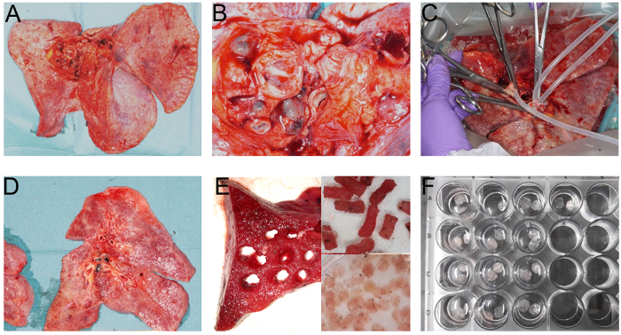
Figure 1: Procedure for filling human lung material. The human lung is prepared immediately after resection. The lung should be stored at room temperature for consistent filling with agarose and to avoid sudden agarose polymerization during filling. The lung is positioned horizontally, with the lobes spread out for an optimal view on the main bronchus or bronchi (A & B for closeup on bronchus). The bronchi are cannulated with a flexible silicone tube and fixed with clamps (C). After the filling procedure and agarose polymerization in the tissue, trained pathologists cut the lung into slices of about 3 - 5 cm thickness (D). From these slices cylindrical tissue cores (Ø e.g., 8 mm) are prepared with a coring tool (E). These tissue cores are cut with a tissue slicer into PCLS, which are immediately transferred into medium-filled Petri dishes for incubation under normal cell culture conditions (E). After several washing steps the PCLS are transferred into cell culture plates (e.g., 24-well plate with two PCLS per well and 500 µL medium) to remove residual cell debris and released cell mediators (F). Please click here to view a larger version of this figure.
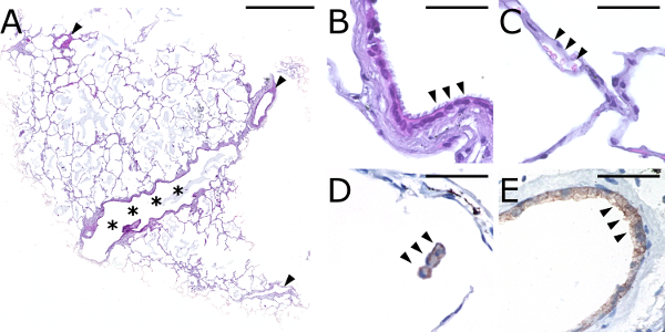
Figure 2: Determination of the health status of human PCLS by H&E staining. The PCLS in this image, prepared from a donor with a lung tumor, shows intact lung tissue with alveolar parenchyma, peripheral airways (***), and blood vessels (arrowheads) in the overview (A). At higher magnification, conducting airways with intact ciliated respiratory epithelium (arrowheads) (B), an intact alveolar structure lined with viable pneumocytes, including a capillary with numerous erythrocytes (arrowheads) (C), alveolar spaces containing alveolar macrophages (CD68 immunohistochemistry) (D), as well as blood vessels with intact endothelium (CD31 immunohistochemistry) (E) can be observed. Only tumor-free tissue was used for the PCLS, which is why no macroscopically diseased areas are visible. In contrast to PCLS prepared from donors with other lung diseases, such as emphysema or fibrosis, the alveolar structure is not disrupted and the PCLS is only slightly frayed at the outer boarder, most likely due to preparation steps. Scale bars indicate 1,000 µm (A) and 50 µm (B-E), respectively. Please click here to view a larger version of this figure.
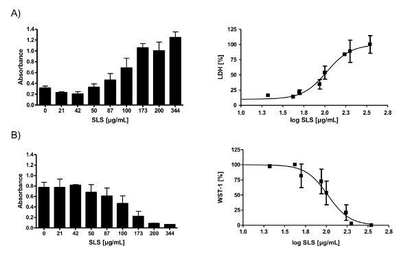
Figure 3: Determination of cytotoxicity induced by SLS in human PCLS, measured by leakage of the enzyme LDH into culture medium (A) and loss of metabolic enzyme activity assessed with the dye WST-1 (B). Human PCLS were exposed to increasing concentrations of SLS. A sigmoid concentration-response model was subsequently fitted to the data, so that IC75 or IC50 values (inhibitory concentration at 25% and 50% reduction of cell viability) can be calculated (right figures). Data are presented as mean ± SEM, n = 3 - 5. Absorbance of LDH assay was measured at 492 nm (reference at 630 nm) and of WST-1 assay at 450 nm (reference at 630 nm). Data were adapted and modified from Lauenstein et al.21 Please click here to view a larger version of this figure.
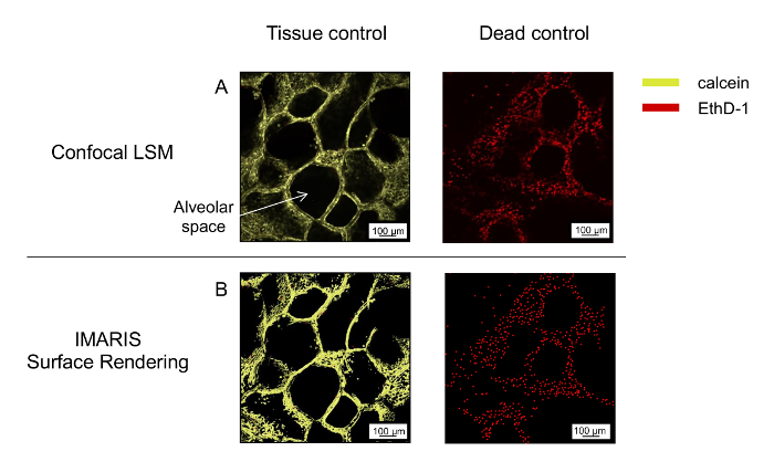
Figure 4: Representative images of the three-dimensional microscopic staining and image rendering of human PCLS. Live tissue control and responsiveness of tissue to detergent, as an effective toxic substance, are shown after staining of human PCLS with calcein AM (yellow) and EthD-1 (red). Stacks of 30 µm were taken with a 10X objective (A) and rendered (B). The scale bar indicates 100 µm. Excitation wavelengths 488 nm and 543 nm, emission filters BP 505-550 nm and LP 560 nm. Please click here to view a larger version of this figure.
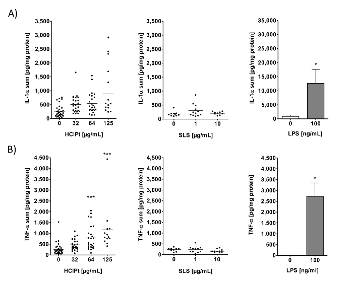
Figure 5: Ammonium hexachloroplatinate (HClPt)-induced IL-1α and TNF-α secretion in human PCLS after 24 h exposure. PCLS were exposed to three concentrations (32 µg/mL, 64 µg/mL, and 125 µg/mL) of HClPt and two different concentrations (1 µg/mL and 10 µg/mL) of SLS. Extracellular and intracellular IL-1α (A) and TNF-α (B) were determined by ELISA and normalized to the total protein content. To show the validity of the method a positive activator of the innate immune system, LPS, is shown as a reference for intracellular IL-1α (A) and for extracellular TNF-α (B) release, normalized to total protein content. Data are presented as mean ± SEM, *p <0.05 and ***p <0.001; HClPt and SLS: n = 9, IL-1αLPS: n = 5, TNF-αLPS: n = 4 in technical duplicates (Mann-Whitney test). This figure has been modified from Lauenstein et al.21 Please click here to view a larger version of this figure.
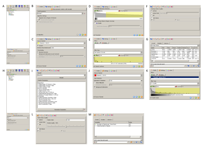
Supplementary Figure 1: Detailed computational processing for images of microscopic viability staining using rendering software. Step-by-step operating procedure for image analysis of uploaded LSM images for surface rendering of calcein-stained viable tissue (A-G, I) and spot detection of ethidium homodimer-stained cell nuclei (H, J-M). Please click here to view a larger version of this figure.
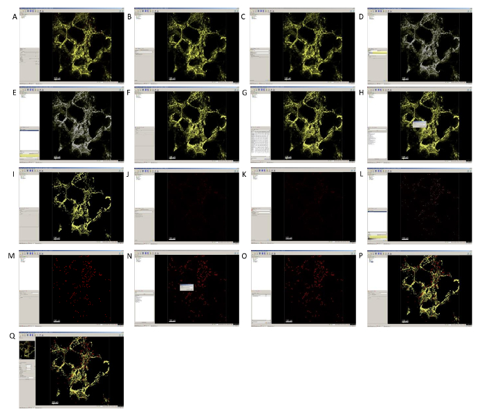
Supplementary Figure 2: Overview of computational image analysis of microscopic viability staining of human lung tissue. Viable tissue (yellow) was analyzed by surface rendering (B-I) and dead cell nuclei (red) were detected by spot detection (J-P). The snapshot mode was used to export the image (Q). Please click here to view a larger version of this figure.
Discussion
The human PCLS technique is well established in our laboratory. The present paper gives a description of this technique and its use for toxicity testing of substances in lung tissue ex vivo. In general, any laboratory using this technique should seek to set up an assay related definition of quantifiable ranges, estimation of variabilities, and quality controls guaranteeing validity of the experiment. Possible standard procedures could be, for example, to repeat each endpoint, e.g., the cytotoxicity assay, in a minimum of three biological donors (individual runs) with a minimum of two to three technical replicates per sample including positive and negative references. Laboratory personnel should be trained to increase assay consistency and minimize assay variability.
Endpoints frequently used to assess immunomodulatory effects of substances on lung tissue include cytotoxicity measurements using different assays (e.g., LDH assay, WST-1 assay, microscopic staining assay), cytokine release assays, as well as changes in expression profile and characterization of changes in cellular populations by immunohistopathological methods6. Furthermore, there are techniques describing the visualization of cells, such as pulmonary dendritic cells, in murine PCLS28 that can also be transferred to human PCLS and might thus provide more detailed insight into the cellular composition before and after treatment with putative cytotoxic substances.
This basic protocol and technique for preparation of human lung tissue sections is comparable to techniques that have been well described in publications29. In short, the organ material is obtained from patients suffering from live-threatening chronic diseases such as lung cancer, who have to undergo surgery for resection or transplantation. The studies must be approved by the local ethics committee. Patients' informed written consent is required. Setting up a workflow between clinic and laboratory is a critical issue and requires communication, and definition of interfaces and infrastructure between both sites. Human lung material has to be processed directly after resection to preserve viability of the tissue. It is worth mentioning that the technique described here for human PCLS can be applied to both young and old lungs and to healthy and diseased lungs. In the US, for example, it is possible to obtain lungs from healthy organ donors who died in accidents or whose organs have been rejected for transplantation.
The first critical step in the protocol is inflation of the airways and surrounding parenchyma with agarose solution. This step is necessary to solidify the very soft tissue for the subsequent slicing procedure. The quality of human lung material, based on the disease background, is critical here. Only lobes with intact pleura can be filled. End-stage tumors near the bronchus sometimes prevent the filling process. Before inflating the human material, the temperature of the agarose solution has to be thoroughly checked. Too much blood (or other fluids, exudates) inside the human lung tissue will result in undesired dilution of agarose and will influence the polymerization process. After inflation of the lung tissue and gelling of agarose on ice, lungs are cut into sections of 200 to 300 µm thickness. Consistency of the tissue is a very critical issue. If the tissue is too soft, slicing of equal sections is difficult. Even if the same microtome parameters are set for each donor, the thickness of the slices between donors may vary, due to individual conditions and changes during the inflating process for each lung. Inhomogeneous filling of the tissue will result in different slice thicknesses. Instead of measuring and standardizing the thickness of slices, measurement of total protein content can be used to indirectly monitor lung slice thicknesses. Several end-stage diseases hamper the slicing process; e.g., blood vessels are extremely thickened in pulmonary hypertension, and fibrotic tissue can be so stiff that slicing of tissue cylinders is hardly possible and the microtome blade needs to be replaced very often.
After preparation of human PCLS and intensive washing steps, which are necessary to remove cell debris and released enzymes, tissue sections can be used for experiments29. Human PCLS are cultured under normal cell culture conditions and exposed, for example, to chemicals, drugs, or lipopolysaccharides. Occasional contamination of PCLS due to (unknown) infections is a special issue in the culture of human lung material. Tissue cultures showing infections must be discarded and the equipment must be thoroughly disinfected. Spatial separation between laboratory places used for preparation on the one hand and culturing on the other hand may help to avoid cross-infections. With regard to the equipment, the microtome may leak, and loose hex screws may lead to motor damage and stop of blade movement. Not every part of the slicer is made of stainless steel, and so it will oxidize if not dried immediately alter cleaning. To overcome equipment issues, it may be necessary to have at least one backup device.
In previous publications, agarose has been reported to be washed out and removed during intensive washing steps after preparation. In fact, this is not possible, the agarose cannot be removed. For complete removal of the agarose, it needs to be re-melted at high temperatures, which would destroy the tissue. The agarose in alveoli and airways does not interfere with the described endpoints. Other endpoints might be influenced by the presence of agarose (see also limitations). The need to prepare very fresh tissue sections has to be emphasized, as tissue viability is a critical issue in culture. Bronchoconstriction is not a valid parameter for viability. We recommend using at least two or three independent cytotoxicity assays to check viability of the surrounding parenchyma; This has to be checked in every experiment30. Quality controls in cytotoxicity assays serve as indicators of insufficient tissue viability. Therefore, it is recommended to assess the responsiveness of the tissue, for example, to an effective toxic substance such as a detergent in all cytotoxicity assays. Based on dose-response curves, minimum and maximum values of absorption need to be defined for cytotoxicity assays and met for subsequent experiments. Further modifications to the protocol mostly depend on the applied chemicals and the endpoints of interest. The applicability of insoluble or highly reactive chemicals is limited. The highest solvent concentration for DMSO is limited to 1%. Higher concentrations can be used but may result in pronounced release of pro-inflammatory cytokines, such as IL-8. On the other hand, the stimulus used might be relatively weak. In this case, the amount of tissue can be increased from two to four slices per well. This approach limits the viability to 24 h.
A major limitation of human PCLS is that in Germany they can only be prepared from diseased human lung material. Patients who undergo surgery are normally older than 50 years and 80% of patients suffering from lung cancer are or used to be smokers. Medication of patients, such as glucocorticoids, can also influence the outcome of experiments using human tissue. Therefore, it is essential to: i) validate each experiment by positive references verifying viability, functionality, and sensitivity of the individual tissue, and ii) cross-validate the results using healthy, non-diseased, middle-age lung tissue from laboratory animals (non-human primates such as cynomolgus and, if possible, mouse, rat, guinea pig). The better the pathology score of the diseased tissue, the better the experimental outcomes. Heavily diseased tissue can hardly be used and very often shows limited viability, inadequately low or extremely high cytokine levels, bacterial or fungal infections, and less bronchoconstriction. Donor-to-donor variation is higher compared with results obtained from laboratory animals, reflecting the individual variability of humans. This is, however, not a limitation in general; as mentioned above, in other countries (e.g., the U.S.) it is possible to obtain healthy lungs from deceased organ donors rejected for transplantation.Responsiveness of the tissue has been well described for the first up to 48 h in acute exposure experiments. Viability and functionality of the tissue is reduced after many days of culture or after storage at -80 °C. It is possible to culture human lung tissue for up to about 14 days. Viability continues during this time; however, an increase in variability, as well as a loss of functionality of several cell populations, such as macrophages, in the tissue is observed, resulting in limited cytokine release in response to mitogens. Another limitation for some endpoints is the presence of agarose in the tissue, hindering, for example, the isolation of high-quality and sufficient amounts of RNA31 or the preparation of single-cell suspensions for subsequent flow cytometry and phenotyping of cells. The possibility to gain mechanistic insights into single-cell responses and functionality is thus limited.
Organotypic tissue models, such as human PCLS, are considered to have a high impact on basic and non-clinical research. Human lung material has a biological composition which closely reflects the normal organ architecture. It contains, for example, residential alveolar and bronchial epithelial cells, smooth muscle cells, fibroblasts, endothelial cells, nerve fibers, and macrophages. The tissue is viable and cells respond to several stimuli. Nerve fibers, although cut, can be locally activated, leading to terminal reflex responses14. Consequently, this ex vivo model offers the possibility to study cellular innate immune responses, defense responses, cytokine signaling, and induction of cell surface markers. Several improvements in technique, culturing, and validation of endpoints allow the use of human PCLS in translational science. Examples of future approaches are: i) validation of new targets in human lung tissue, ii) assessment of immune responses after exposure, for example, to chemicals, drugs, nanoparticles, etc., iii) supplementation of lung tissue with immune cells, such as T-lymphocytes, iv) identification and modification of molecular patterns, for example, after exposure to respiratory sensitizers, disease-inducing substances, or active compounds inhibiting pathways; furthermore, v) airway remodeling and vi) neuronal regulation32. The scientific field is interested in these present and future approaches with PCLS. In addition, there are a variety of different developments that will help to improve the PCLS technique, such as cryo-preservation20, and tissue stretching33 to mimic the natural movement of the tissue during breathing or mechanical ventilation.
The major advantage of human PCLS compared with other 3D models is the presence of immune cells and nerve fibers. Experiments can also be performed in mouse, rat, and non-human primates, which are the animal species that are still used most often in pharmacology and toxicology. The complexity of human lung tissue supports the translation of results from animal to human and from in vitro to in vivo. In the context of existing alternative assays for the identification of respiratory sensitizers, human PCLS are very complex and do not allow insights into single cell responses. Yet, microscopy and flow cytometry might give information about cellular responses, if the right cellular marker is used in combination, for example, with apoptosis, necrosis, or intracellular markers. There are published assays which have been validated and reported to have been used for the identification of respiratory sensitizers. However, the advances in the use of PCLS with all their advantages over single-cell assays are making a valuable contribution in the sense that the technique can be used for high-throughput screening, as described by Watson et al.34 They developed a high-throughput screening assay to predict airway toxicity in murine cryo-preserved PCLS. With their miniaturized 96-well PCLS format, they detected similar readouts in murine lavage fluid, making PCLS a feasible high-throughput assay.
Disclosures
The authors have nothing to disclose. Fraunhofer ITEM is an independent public non-profit research organization for applied research, performing contract research on behalf of the biotech, pharmaceutical, chemical, and cosmetics industries. Funding of the institute comes from national and international public projects.
Acknowledgements
We would like to thank Lan Lauenstein for her pre-work regarding alternative testing strategies for risk assessment with human PCLS. Some of the research was supported by a grant from the European Commission within the 6th framework program SENS-IT-IV "Novel Testing Strategies for In Vitro Assessment of Allergens".
Materials
| Name | Company | Catalog Number | Comments |
|---|---|---|---|
| Silicon hose 3.0 x 5.0 mm | A. Hartenstein (Leipzig, Germany) | SS04 | |
| Syringe | Faust Lab Science (Klettgau, Germany) | 9.410 050 | |
| Coring tools | custom-made | custom-made | |
| Trimming Blade Handle (FEATHER) | pfm medical ag (Cologne, Germany) | 205530001 | |
| Trimming Blades (FEATHER) | pfm medical ag (Cologne, Germany) | 205500000 | |
| Microtome: Tissue Slicer | Alabama R&D (Bad Homburg, Germany) | 303400-ADPT | Krumdieck Tissue Slicer (MD6000) |
| Microtome blade | Wilkinson Sword (Solingen, Germany) | ENR-4027800011506 | |
| Tissue culture dishes | Sigma (München, Germany) | Z666246-420EA | |
| Cell strainer filter (100 µm Nylon) | Becton Dickinson (Heidelberg, Germany) | BD352360 | |
| Inoculation loop | Copan Diagnostics (Murrieta, USA) | CD176S01 | |
| TPP Tissue culture plates 24wells | Sigma (München, Germany) | Z707791-126EA | |
| Cassette | Thermo Scientific (Schwerte, Germany) | 1000957 | |
| Nunc MaxiSorp flat-bottom | Fisher Scientific GmbH (Hannover, Germany) | 44-2404-21 | |
| Nunc MicroWell 96-Well | Thermo Scientific (Schwerte, Germany) | 260836 | |
| Confocal Microscope | Zeiss (Jena, Germany) | Confocal LSM Meta 510 | |
| Image rendering software | Bitplane AG (Zürich, Switzerland) | IMARIS 7.6 | |
| LSM Image Browser | Zeiss (Jena, Germany) | ||
| Multiwell-Reader | Tecan Group Ltd. (Männedorf, Switzerland) | Tecan infinite F200Pro | Plate Reader |
| Plate shaker | Edmund Buehler GmbH (Hechingen, Germany) | KM-2 Akku | |
| BenchMark ULTRA | Ventana Medical Systems (Tucson, USA) | Automated IHC/ISH slide staining system | |
| Assays | |||
| Cell Proliferation Reagent WST-1 | Roche (Basel, Switzerland) | 11644807001 | |
| Cytotoxicity Detection Kit (LDH) | Roche (Basel, Switzerland) | 11644793001 | |
| Microscopical vitality staining | Invitrogen (Carlsbad, USA) | L-3224 | LIVE/DEAD Viability/Cytotoxicity Kit |
| BCA Protein Assay | Thermo Scientific (Schwerte, Germany) | 23225 | |
| ELISA assay kit | R & D systems | various, e.g. DY210 (TNF-α) or DY200 (IL-1α) | DuoSet |
| Reagents | |||
| Agarose, low gelling temperature | Sigma (München, Germany) | A9414-100G | |
| Balanced Salt Mixtures and Solutions, Cell Culture, Classic Media and Salts, Earle’s Balanced Salts (EBSS) | Sigma (München, Germany) | E2888-500ML | |
| Penicillin and streptomycin | Lonza (Verviers, Belgium) | 17-602E | |
| Dulbecco´s Modified Eagle´s Medium Nutrient Mixture F-12 Ham (DMEM F-12) | Gibco (Darmstadt, Germany) | 11039-047 | Culture medium |
| Dulbecco´s Phosphate with Ca and Mg (DPBS) | Lonza (Verviers, Belgium) | BE17-513F | Buffer solution |
| Albumin from bovine serum (BSA) | Sigma (München, Germany) | A9647-500G | |
| Detergent | Sigma (Saint Louis, USA) | X100-100ML | Triton X-100 |
| Washing buffer | Merck (Darmstadt Germany) | 524653 | 0.05% Tween 20 in PBS |
References
- D'Amato, G., et al. Climate change, air pollution and extreme events leading to increasing prevalence of allergic respiratory diseases. Multidiscip Respir Med. 8 (1), 12(2013).
- Pawankar, R., et al. State of world allergy report 2008: allergy and chronic respiratory diseases. World Allergy Organ J. 1 (6), Suppl 4-17 (2008).
- Hartung, T., Daston, G. Are in vitro tests suitable for regulatory use. Toxicol Sci. 111 (2), 233-237 (2009).
- Hartung, T. Toxicology for the twenty-first century. Nature. 460 (7252), 208-212 (2009).
- Balls, M., Straughan, D. W. The three Rs of Russell & Burch and the testing of biological products. Dev Biol Stand. 86, 11-18 (1996).
- Sewald, K., Braun, A. Assessment of immunotoxicity using precision-cut tissue slices. Xenobiotica. 43 (1), 84-97 (2013).
- Fisher, R. L., et al. The use of human lung slices in toxicology. Hum Exp Toxicol. 13 (7), 466-471 (1994).
- Ressmeyer, A. R., et al. Characterisation of guinea pig precision-cut lung slices: comparison with human tissues. Eur Respir J. 28 (3), 603-611 (2006).
- Donovan, C., et al. Rosiglitazone elicits in vitro relaxation in airways and precision cut lung slices from a mouse model of chronic allergic airways disease. Am J Physiol Lung Cell Mol Physiol. 309 (10), 1219-1228 (2015).
- Koziol-White, C. J., et al. Inhibition of spleen tyrosine kinase attenuates IgE-mediated airway contraction and mediator release in human precision cut lung slices. British journal of pharmacology. 173 (21), 3080-3087 (2016).
- Paddenberg, R., Mermer, P., Goldenberg, A., Kummer, W. Videomorphometric analysis of hypoxic pulmonary vasoconstriction of intra-pulmonary arteries using murine precision cut lung slices. J Vis Exp. (83), e50970(2014).
- Lamb, D. J., et al. Bl 1002494, a Novel Potent and Selective Oral Spleen Tyrosine Kinase Inhibitor, Displays Differential Potency in Human Basophils and B Cells. J Pharmacol Exp Ther. 357 (3), 554-561 (2016).
- Leus, N. G., et al. HDAC 3-selective inhibitor RGFP966 demonstrates anti-inflammatory properties in RAW 264.7 macrophages and mouse precision-cut lung slices by attenuating NF-kappaB p65 transcriptional activity. Biochem Pharmacol. 108, 58-74 (2016).
- Schleputz, M., et al. Neurally mediated airway constriction in human and other species: a comparative study using precision-cut lung slices (PCLS). PLoS One. 7 (10), 47344(2012).
- Hess, A., et al. Prevalidation of the ex-vivo model PCLS for prediction of respiratory toxicity. Toxicology in vitro : an international journal published in association with BIBRA. 32, 347-361 (2016).
- Bergner, A., Sanderson, M. J. Acetylcholine-induced calcium signaling and contraction of airway smooth muscle cells in lung slices. J Gen Physiol. 119 (2), 187-198 (2002).
- Wohlsen, A., et al. The early allergic response in small airways of human precision-cut lung slices. Eur Respir J. 21 (6), 1024-1032 (2003).
- Wu, W., et al. Innate immune response to H3N2 and H1N1 influenza virus infection in a human lung organ culture model. Virology. 396 (2), 178-188 (2010).
- Zmora, P., et al. Non-human primate orthologues of TMPRSS2 cleave and activate the influenza virus hemagglutinin. PLoS One. 12 (5), 0176597(2017).
- Rosner, S. R., et al. Airway contractility in the precision-cut lung slice after cryopreservation. Am J Respir Cell Mol Biol. 50 (5), 876-881 (2014).
- Lauenstein, L., et al. Assessment of immunotoxicity induced by chemicals in human precision-cut lung slices (PCLS). Toxicology in vitro : an international journal published in association with BIBRA. 28 (4), 588-599 (2014).
- Wild, L. G., Lopez, M. Occupational asthma caused by high-molecular-weight substances. Immunol Allergy Clin North Am. 23 (2), 235-250 (2003).
- Di Stefano, F., et al. Occupational asthma due to low molecular weight agents. Int J Immunopathol Pharmacol. 17 (2), Suppl 77-82 (2004).
- Buonsante, V. A., Muilerman, H., Santos, T., Robinson, C., Tweedale, A. C. Risk assessment's insensitive toxicity testing may cause it to fail. Environ Res. 135, 139-147 (2014).
- Boverhof, D. R., et al. Respiratory sensitization and allergy: current research approaches and needs. Toxicol Appl Pharmacol. 226 (1), 1-13 (2008).
- Johansson, H., Albrekt, A. S., Borrebaeck, C. A., Lindstedt, M. The GARD assay for assessment of chemical skin sensitizers. Toxicology in vitro : an international journal published in association with BIBRA. 27 (3), 1163-1169 (2013).
- Brismar, H., Patwardhan, A., Jaremko, G., Nyengaard, J. Thickness estimation of fluorescent sections using a CSLM. Journal of Microscopy. 184 (2), 106-116 (1996).
- Lyons-Cohen, M. R., Thomas, S. Y., Cook, D. N., Nakano, H. Precision-cut Mouse Lung Slices to Visualize Live Pulmonary Dendritic Cells. J Vis Exp. (122), (2017).
- Switalla, S., et al. Natural innate cytokine response to immunomodulators and adjuvants in human precision-cut lung slices. Toxicol Appl Pharmacol. 246 (3), 107-115 (2010).
- Henjakovic, M., et al. Ex vivo testing of immune responses in precision-cut lung slices. Toxicol Appl Pharmacol. 231 (1), 68-76 (2008).
- Niehof, M., et al. RNA isolation from precision-cut lung slices (PCLS) from different species. BMC Res Notes. 10 (1), 121(2017).
- De Proost, I., et al. Purinergic signaling in the pulmonary neuroepithelial body microenvironment unraveled by live cell imaging. FASEB J. 23 (4), 1153-1160 (2009).
- Davidovich, N., Chhour, P., Margulies, S. S. Uses of Remnant Human Lung Tissue for Mechanical Stretch Studies. Cell Mol Bioeng. 6 (2), 175-182 (2013).
- Watson, C. Y., et al. Screening for Chemical Toxicity Using Cryopreserved Precision Cut Lung Slices. Toxicol Sci. 150 (1), 225-233 (2016).
Reprints and Permissions
