Overview
Fuente:
Alexandra Duncan, GTA, Praxis clínica, New Haven, CT
Cocinero de Tiffany, GTA, Praxis clínica, New Haven, CT
Jaideep S. Talwalkar, MD, medicina interna y Pediatría, Facultad de medicina de Yale, New Haven, CT
Colocación de espéculo cómodo es una habilidad importante para los proveedores a desarrollar, ya que el espéculo es una herramienta necesaria en muchos procedimientos ginecológicos. Pacientes y los proveedores a menudo están preocupados por el examen de espéculo, pero es totalmente posible Coloque un espéculo sin malestar del paciente. Es importante para el clínico conocer el papel que desempeña el lenguaje en la creación de un ambiente confortable; por ejemplo, un proveedor debe consultar el espéculo "cuentas" en lugar de "hojas" para evitar perturbar al paciente.
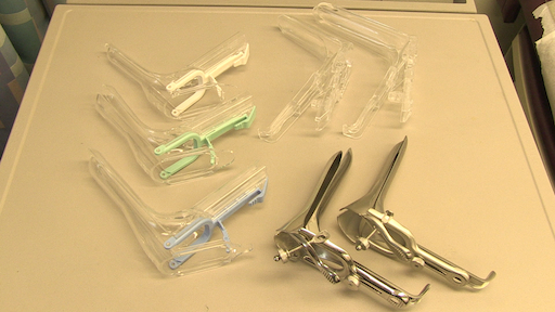
Hay dos tipos de espéculos: metal y plástico (figura 1). Esta demostración utiliza plástico, espéculos de plástico son más comúnmente utilizados en las clínicas para las pruebas de rutina. Cuando se utiliza un espéculo de metal, recomienda el uso de un espéculo de Graves si el paciente ha dado a luz por vía vaginal y un espéculo Pederson si el paciente no tiene. Espéculos de Pederson y tumbas son formas diferentes, y ambos vienen en muchos tamaños diferentes (medio se utiliza más a menudo). Antes de colocar un espéculo de metal, es útil realizar un examen cervical digital para evaluar el tamaño de espéculo adecuado. La profundidad y la dirección de la cerviz se calcula colocando un dedo en la vagina. Si el cuello uterino de la paciente puede localizar mientras el paciente está sentado, es probable que el paciente tiene una vagina poco profunda y por lo tanto debe ser más cómodo con un espéculo de metal corto.

Figura 1. Una fotografía de espéculos disponibles comercialmente en diferentes tamaños.
Espéculos de plástico están en forma de Pederson espéculos de metal y vienen en diferentes tamaños. Para determinar el tamaño adecuado para un espéculo plástico, el examinador coloca dos dedos en la vagina del paciente, Palma abajo e intenta separar los dedos: Si no hay ningún espacio entre los dedos, debe utilizarse un espéculo plástico pequeño; Si hay espacio entre los dedos, debe utilizarse un medio. El examen no debe realizarse con un espéculo grande (como lo es significativamente mayor) sin antes de determinar la longitud de la vagina.
El espéculo se usa para realizar la prueba de Papanicolaou como parte de exámenes de detección del cáncer de cuello uterino. Cáncer de cuello uterino fue la principal causa de muerte por cáncer para las mujeres en los Estados Unidos, pero en las últimas décadas el número de casos y muertes ha disminuido significativamente de1. Este cambio se le atribuye el descubrimiento hecho por Georgios Papanicolaou en 1928 que el cáncer de cuello uterino podría ser diagnosticado por frotis vaginales y cervicales. La prueba de Papanicolau, como se le llama ahora, detecta las células anormales en el cuello uterino, canceroso y precanceroso. Las guías actuales para los intervalos de detección recomendadas pueden encontrarse a través de la Task Force de servicios preventivos Estados Unidos (USPSTF) sitio Web2.
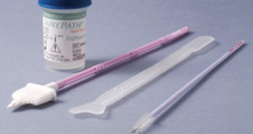
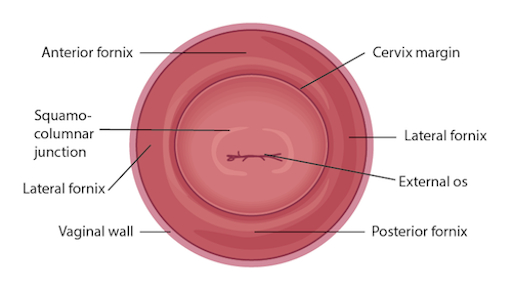
La prueba puede realizarse utilizando cualquiera de los dos 1) una diapositiva de cristal convencional y fijador con un cepillo espátula y endocervical (el tradicional "citología vaginal") o 2) más comúnmente utilizados de citología de base líquida con una escoba cervical o un cepillo espátula y endocervical (figura 2). No importa qué herramientas se utilizan, las muestras se recogen desde dentro el os externo y escamoso o zona de transición en el sistema operativo (figura 3). Este video muestra la espátula y cepillo endocervical con citología de base líquida, como la preparación de líquidos es una técnica más efectiva para la detección de lesiones cervicales, y el cepillo espátula y endocervicales mejorar la recogida de la muestra.

Figura 2. Herramientas de prueba de Papanicolaou. Se muestra en la secuencia son: un frasco de citología líquida cervical escoba, espátula y cepillo endocervical.

Figura 3. Diagrama de la cerviz conestructuras relevantes etiquetadas.
Procedure
El examen de espéculo comienza inmediatamente después del final del examen de los genitales externos; por lo tanto, el paciente ya ha proporcionado una historia y está en la posición de litotomía modificada. Asegúrese de que el paciente está sentado en el extremo de la mesa, como el espéculo no se puede insertar totalmente cualquier otra forma.
1. preparación
- Coloque los suministros para la prueba de Papanicolau.
- Etiqueta de la lata de citología líquida con la información del paciente.
- Desenrosque la tapa de la lata hasta que descansa en la parte superior y puede ser levantado.
- Coloque el espéculo en la mano no dominante y coloque el dedo índice por encima de las cuentas, el dedo medio por debajo de las cuentas y el pulgar en la parte posterior del espéculo (figura 4).
- Utilice su mano dominante para difundir el lubricante a base de agua (o agua caliente, de otra manera) en el exterior de las cuentas.
- Introducir el espéculo y que el paciente sabe qué esperar: "se trata de un espéculo. Estas son las cuentas, que estará poniendo en tu vagina para ver el cuello uterino y tomar algunas muestras. Este es el mango, el cual no se insertará".

Figura 4. Cómo llevar a cabo un espéculo plástico.
2. colocación
- Como siempre, introducir su toque al paciente antes de comenzar el examen.
- Deje que el paciente sabe que está a punto de colocar el dorso de la mano en el muslo del paciente y luego comenzará el examen.
- Coloque el dorso de la mano en el interior del muslo del paciente, sobre el cobertor, a continuación, comenzar a examinar: esto prepara al paciente y comienza con el contacto no invasivo, que puede ayudar a pone al paciente cómodo.
- Usando las teclas del índice y dedo medio de su mano dominante, separar los labios menores justo encima del perineo para obtener una visión clara del introitus vaginal.
- Guía del paciente a través de una técnica de relajación: "voy a enseñarte cómo hacerlo más cómodo para ti. Por favor, toma una respiración profunda en y al exhalar, llevan hacia abajo como si estás haciendo un movimiento de intestino. (Si el paciente no entiende esto, pida al paciente que empuje contra los dedos.)"
- Como el paciente lleva hacia abajo, se abre el introito vaginal. Deje que el paciente lo sepa, "Sentirán me insertar el espéculo".
- Introducir suavemente el espéculo sobre a mitad de camino en la vagina en un ángulo oblicuo (aproximadamente 45°), pesca las facturas debajo de donde se espera que el cuello uterino (basado en el examen digital) manteniendo la presión posterior.
- Utilice los dos primeros dedos de su mano dominante para borrar los labios por un lado, por lo que los labios no se tiró junto con el espéculo.
- Utilice su mano dominante para borrar los labios en el otro lado.
- Llevar su mano no dominante a la manija de la parte inferior del espéculo, rote el espéculo plano e Inserte el, hasta que el mango esté al ras contra la pelvis del paciente y es perpendicular al piso.
- Coloque un dedo de su mano dominante dentro de las cuentas del espéculo y utilice el dedo para aplicar fuerte presión posterior como su mano no dominante se tira hacia abajo el mango del espéculo al mismo tiempo. Aplique suficiente presión posterior para que espacio en la vagina puede verse sobre el espéculo.
- Coloque el pulgar de su mano no dominante sobre la palanca y presione suavemente.
- No continuar Oprima una vez la resistencia, o puede abrir las cuentas demasiado y provocar el malestar del paciente.
- Mantenga el espéculo y compruebe para ver si el cuello uterino ha sido localizado.
3. prueba de Papanicolaou
- Si hay suficiente descarga en la cara del cuello uterino para ocultar el sistema operativo e interferir en la recogida de muestras, utilice un hisopo de algodón grande cervical suavemente claro exceso mucoso.
- Utilice su mano dominante tome la espátula (o el acompañante mano a usted).
- Introduzca la espátula en la vagina, teniendo cuidado de no dejarlo tocar las paredes, hasta que el extremo largo se basa en el sistema operativo y la depresión y el extremo corto se presionan contra la unión escamoso-cilíndrica.
- Girar 360°, manteniendo una presión constante y el contacto con el exocervix.
- Retirar la espátula, teniendo cuidado de no tocar las paredes de la vagina.
- Coloque la espátula en el frasco abierto y enjuague bien agitando vigorosamente en el líquido (diferentes marcas de citología de base líquida recomienda se arremolinan para diversas longitudes del tiempo; estar familiarizado con las recomendaciones del fabricante antes de empezar).
- Deseche la espátula y colóquela nuevamente en la bandeja.
- Utilice su mano dominante para recoger el cepillo endocervical.
- Inserte el cepillo endocervical en la vagina, teniendo cuidado de no dejarlo tocar las paredes y suavemente lo inserte en el sistema operativo hasta que solo las cerdas de la parte inferior.
- Lentamente gire 180° en una sola dirección. No lo rote demasiado.
- Si se usa una escoba cervical, en lugar de un cepillo endocervical y espátula, inserte el cepillo hasta que el centro son en el sistema operativo y las cerdas más cortas descansan en la unión escamoso-cilíndrica, luego girar cinco veces de lleno en una dirección antes de retirar.
- Retire el pincel, teniendo cuidado de no tocar las paredes de la vagina.
- Coloque el cepillo endocervical en el frasco abierto y enjuagar vigorosamente se arremolinan y presionando repetidamente contra los lados de la lata para liberar el material.
- Desechar el cepillo endocervical.
- Coloque y apriete la tapa de la lata de citología.
4. retirar el espéculo
- Coloque su pulgar no dominante en la palanca y mantenga la presión mientras se suelta el mecanismo de bloqueo.
- Continúan presionado la palanca y retire el espéculo para aproximadamente una pulgada hacia fuera para permitir que el cuello uterino limpiar la punta de las cuentas.
- Completamente retire su dedo pulgar de la palanca y coloca sobre el mango del espéculo.
- Girar el espéculo 45° mientras que suavemente se quita el resto del camino hacia fuera, permitiendo que las paredes vaginales para cerrar las cuentas.
- Coloque la mano dominante por debajo del espéculo para atrapar cualquier descarga.
- Deseche el espéculo de plástico, si disponible.
El examen del espéculo se utiliza en una amplia variedad de procedimientos ginecológicos y puede proporcionar una gran cantidad de información de diagnóstico. El espéculo es un instrumento bivalvo, que se utiliza para separar las paredes de la vagina. Esto no sólo permite la inspección visual del cuello uterino, sino que también proporciona un acceso a esta región para recogida de muestras durante procedimientos de diagnóstico como el Papanicolau o prueba de Papanicolaou, que se realiza para detectar cambios precancerosos. Este video ilustra la técnica correcta de utilizar el espéculo para la inspección cervical y el método apropiado para la recogida de muestras para la prueba de Papanicolau.
Vamos a empezar con la revisión de los pasos involucrados en la preparación para el examen de espéculo y la prueba de Papanicolau, seguida por una discusión de lo que debe buscar un médico mientras la inspecciona el cuello uterino con el espéculo. Hay diferentes tipos de espéculos disponibles comercialmente. Algunos se componen de plástico desechable, mientras que los metales son reutilizables. En esta demostración, vamos a utilizar un espéculo plástico.
Antes de comenzar el examen, es imprescindible para familiarizarse con el instrumento a utilizar y entender cómo funciona. Después de se iniciará con el examen. Recuerde que este procedimiento generalmente viene después de la inspección externa de la pelvis, así que en este punto la historia del paciente se ha obtenido, y ya están en la litotomía modificada posición.
Asegúrese de que el paciente está sentado en el extremo de la mesa para permitir la inserción del espéculo de competir. También ponen a los suministros para la prueba de Pap, incluyendo un bote de citología; lubricante exprimido en una bandeja limpia con un trazador de líneas, y una espátula y endocervicales, cepillan o simplemente una escoba cervical para recolectar la muestra de células. Etiqueta de la lata de citología líquida con la información del paciente. Desenroscar la tapa de la lata hasta que se apoye en la parte superior que se puede levantar fácilmente.
Es fundamental para comprender cómo llevar a cabo un espéculo. En caso de que usted está usando un espéculo plástico, coloque en su mano no dominante y coloque el dedo índice por encima de las cuentas, el dedo medio por debajo de las cuentas y el pulgar en la parte posterior del espéculo, evitando la palanca abría el espéculo. Con la mano dominante separó el lubricante a base de agua en el exterior de las cuentas. Mostrar el espéculo al paciente sin apuntar directamente a ellos y hacerles saber qué esperar durante el examen, "Diálogo".
Comience por dejar que el paciente sabe que primero se coloque el dorso de la mano en su muslo. Esto se hace para preparar al paciente para el examen por establecer primero un contacto no invasivo. Ahora separe los labios menores con los pads dominantes del índice y el dedo medio para obtener una visión clara del introitus vaginal. A continuación, explicar la técnica de relajación para el paciente, "Diálogo". Debe abrir el introito vaginal, el paciente lleva hacia abajo. Que el paciente conozca que usted está a punto de introducir el espéculo, "Diálogo" y coloque hasta la mitad en el canal vaginal, mantener las cuentas en ángulo de unos 45 °. A continuación, llevar su mano no dominante a la manija inferior y girar el espéculo plano, mientras al mismo tiempo limpiar los labios en ambos lados. Entonces ángulo de la punta de las cuentas hacia el piso e inserte completamente, tal que la punta termina por debajo del cuello uterino. Deténgase cuando el espéculo esté lavado contra la pelvis del paciente.
A continuación coloca uno de los dedos dominantes dentro de las cuentas y aplicar fuerte presión posterior mientras tira hacia abajo la palanca con la otra mano hasta que quede perpendicular al piso. Asegúrese de que usted aplique suficiente presión para ver el espacio sobre el espéculo. Ahora, mientras mantiene la presión posterior con el dedo dentro del espéculo, puede presione suavemente la palanca para abrir las cuentas. Deje de una vez resistencia. Luego enganchar la cerradura, empujando la palanca hacia arriba uno o dos clics y retire el dedo de dentro el espéculo. Mantenga el espéculo y, usando una fuente de luz, comprobar si el cuello uterino es visible. Tenga en cuenta el tono, color y posición del cuello uterino y observar para descarga, lesiones, pólipos, ulceraciones y las masas.
Puede visualmente la parte inferior del intravaginal del cuello uterino. Esto incluye el exocérvix, que es normalmente de 2-3 cm de diámetro, color de rosa en color y tiene una superficie lisa; el orificio externo, que es la abertura del endocervix en la vagina; y las cuatro bóvedas, que son los huecos entre el margen del cuello uterino y la pared vaginal.
Después de la inspección visual del cuello uterino, proceder a la recogida de muestras para la prueba de Papanicolau. Con el espéculo en su lugar, introduzca la espátula en la vagina, teniendo cuidado de no tocar las paredes. Posición la espátula con su extremo más largo en el sistema operativo y el extremo corto se presiona contra el cruce. Ahora girar 360°, manteniendo la presión constante y el contacto con el exocervix. Retire con cuidado la espátula evitando las paredes vaginales. Coloque la espátula en el frasco abierto citología y enjuague bien agitando vigorosa en el líquido siguiendo las instrucciones del fabricante. A continuación, inserte el cepillo endocervical en la vagina, evitando el contacto con las paredes. Empuje el cepillo en el sistema operativo hasta que solo las cerdas de la parte inferior se exponen y lentamente giran 180° en una sola dirección. No sobre gire. Con cuidado retire el cepillo evitando los muros y colóquelo en el recipiente de la citología. Bien enjuagar el cepillo agitando vigorosa y presione repetidamente contra los lados del frasco para soltar el material. En lugar de utilizar la espátula y cepillo, se puede usar sólo el cepillo endocervical, que tiene las cerdas formando un patrón triangular de diferentes tamaños. Si se usa, entonces insertarlo más cerdas el resto en el sistema operativo y el resto más corto en la zona de transición y gírelo aproximadamente cinco a diez veces, dependiendo de las instrucciones del fabricante. El lanzamiento de la muestra es el mismo en cuanto a para el cepillo endocervical.
Una vez finalizada la recogida de muestras, suelte el mecanismo de cierre presionando hacia abajo la palanca de pulgar y manteniendo la palanca hacia abajo y cuentas abrir, quitar el espéculo alrededor de dos a tres pulgadas, para asegurarse de que el cuello uterino se borra de la punta de las cuentas. Retire su dedo pulgar de la palanca y coloca sobre el mango. Finalmente, gire el espéculo por 45° mientras suavemente para retirarlo completamente. Tienen la mano por debajo del espéculo para recoger cualquier posible descarga y deseche el espéculo, si disponible. Al final, vuelva a colocar la tapa de la lata. Ahora la muestra está lista para posterior análisis citológico.
"Examinador explicar diferentes tipos de espéculos las similitudes y diferencias"
Sólo ha visto ilustración de Zeus del examen de espéculo y la prueba de Papanicolau. Ahora debería entender cómo utilizar el espéculo vaginal y cómo recoger la muestra de células cervicales para evaluación diagnóstica. ¡Como siempre, gracias por ver!
Subscription Required. Please recommend JoVE to your librarian.
Applications and Summary
Este vídeo repasa las técnicas para realizar un examen de espéculo cómodo y recoger las muestras para la prueba de Papanicolau. Antes de inicia el examen, el examinador debe garantizar todos los suministros están preparados y que el paciente sabe qué esperar. Ser capaz de realizar un examen cómodo espéculo es una habilidad importante para cualquier practicante, ya que se utiliza en una amplia variedad de procedimientos ginecológicos y puede proporcionar una gran cantidad de información. Cuando se inserta el espéculo, es posible observar el cuello uterino y las paredes vaginales para una variedad de señales incluyendo tono, color, secreción, lesiones, pólipos, ulceraciones, y mucho más-todo de que puede ser clínicamente significativa y puede ayudar con el proceso de diagnóstico. Un espéculo insertado bien también permite un fácil acceso para el endocérvix, momento en el que se pueden tomar muestras para la prueba de Papanicolau (así como para otras proyecciones, como clamidia y gonorrea). Es necesario utilizar un espéculo a la cerviz para muchos otros procedimientos, como insertar o retirar un dispositivo intrauterino (DIU) y un procedimiento de escisión electroquirúrgica (LEEP).
Muchos pacientes pueden sentir ansiedad sobre el espéculo y experimentarlo como la parte más invasiva de examen ginecológico. El proveedor puede ofrecer el apoyo general al paciente y la empatía, junto con herramientas específicas para hacer el examen más cómodo para ellos mismos. Pidiendo al paciente que respire profundamente y luego llevar hacia abajo como si tener una deposición antes de inserción puede abrir el introito vaginal y ayuda mucho confort. El examinador puede ofrecer a un paciente especialmente la oportunidad de insertar el espéculo al colocarlo boca abajo con el mango apuntando hacia el techo como el proveedor les habla a través de la apertura3. A menudo es más fácil de obtener una visión clara del cuello uterino con una inserción invertida, pero no es una técnica que deben emplear los médicos, porque coloca la mano del examinador directamente contra el clítoris de la paciente.
Hay muchas cosas que el médico puede hacer para asegurar que el examen sea cómodo. El espéculo debe insertarse en un ángulo oblicuo para evitar poner demasiada presión directa sobre la uretra. Cuando el espéculo se inserta completamente, las facturas deben orientarse debajo de donde se encuentra el cuello uterino durante el examen digital. El médico entonces puede aplicar suficiente presión posterior con espéculo, así que hay espacio visible en la vagina sobre los proyectos de ley; Esto permite que las facturas se abra sin ejercer presión sobre las delicadas estructuras anteriores. Lo más importante es nunca insertar o quitar un espéculo, mientras que las cuentas están abiertas. Esto es muy doloroso y el riesgo de lesionar al paciente. El examinador no debe tocar la palanca hasta que el espéculo es totalmente colocado y listo para ser abierto. El bloqueo del espéculo debe libera completamente antes de retirar, y cualquier presión mantenida manualmente. Una vez que la cerviz está libre, la palanca debe ser totalmente liberada y el espéculo suavemente quita el resto del camino, permitiendo que las paredes vaginales cerrar las cuentas en la salida.
Subscription Required. Please recommend JoVE to your librarian.
References
- Cervical Cancer Statistics. U.S. Preventive Services Task Force. Centers for Disease Control and Prevention (2014).
- Cervical Cancer: Screening. Recommendation Summary. U.S. Preventive Services Task Force (2012).
- Wright, D., Fenwick, J., Stephenson, P., Monterosso, L. Speculum 'self-insertion': a pilot study. Journal of Clinical Nursing. 14(9): 1098-1111 (2005).


