Blood Withdrawal I

All animal procedures described here must be conducted in accordance with institutional animal ethics guidelines and approved by IACUC. All procedures must follow the principles of the 3Rs—Replacement, Reduction, and Refinement—and must be performed by trained personnel.
1. Tail vein blood collection
- Equipment
- Plastic restraining holder
- Vacuum blood collection tubes with rubber stoppers
- Double-pointed (butterfly) needle (22G)
- Eyeglasses magnifier
- Cotton balls soaked in 70% ethanol
- Warm water (20–30°C)
- Restraint
- The mouse is placed into a plastic restraining holder.
- The front and hind limbs are positioned comfortably to minimise stress and movement.
- The tail is extended and kept straight for sampling.
- Blood withdrawal
- The animal is anesthetized with an inhalation anesthetic, such as isoflurane, in a bell jar or gas anesthesia induction chamber, to effect.
- Once the animal is fully anesthetized, it is removed and placed in lateral recumbency.
- The eye is protruded by placing a finger on the top of the head and along the jawline, and pulling the skin back and down.
- Avoid applying pressure to the trachea, as that may collapse or occlude the airway causing death by asphyxia.
- Blood withdrawal
- The tail is washed with warm water (20–30°C) to promote vasodilation. DO NOT use hot water.
- The tail is then wiped with 70% ethanol to clean the surface and further enhance vein visibility.
- One of the lateral tail veins is selected for sampling. The lower portion of the tail is gently grasped and stabilised. An eyeglasses magnifier may be used to improve visualisation of the vein.
- A 22G butterfly needle is inserted into the lateral tail vein approximately halfway from the tip of the tail at an angle of about 10°, with the needle directed towards the base of the tail for repeated sampling.
- Blood is collected into a vacuum tube by inserting the rubber end of the needle into the collection tube.
- Multiple samples can be obtained from the tail in accordance with approved protocols. For blood microsampling, the lateral tail vein is punctured using a fine-gauge needle to obtain a small, controlled volume of blood.
2. Tail snip
- Equipment
- Small animal restrainer (disinfected)
- Sharp surgical scissors (or preferably a sterile scalpel blade)
- Heparinized capillary tubes
- Bulb syringe
- Potassium EDTA microtainer tubes (labelled)
- Isopropyl alcohol
- Topical anesthetic cream
- Sterile gauze or swabs (for haemostasis)
- Restraint
- The mouse is gently placed into a disinfected restrainer.
- The restrainer is secured to minimise movement during the procedure.
- The tail is positioned to allow easy access to the tip.
- Blood withdrawal
- The tail is cleaned using fresh isopropyl alcohol. A topical anesthetic cream is applied to the tail tip as required.
- Using sharp scissors (or preferably a scalpel blade), approximately 0.5–1 mm of the tail tip is removed.
- Blood is collected directly into heparinized capillary tubes (approximately 80 µL).
- The collected blood is transferred using a bulb syringe into labelled potassium EDTA microtainer tubes. The tubes are gently inverted (~20 times) to prevent coagulation.
- Hemostatis and recovery
- Bleeding is controlled by applying gentle pressure to the tail tip using sterile gauze until bleeding stops.
- If required, additional methods such as cautery or styptic agents may be used cautiously to achieve haemostasis.
- Animals must be monitored to ensure haemostasis before and after returning to the home cage.
3. Cardiac blood collection
- Equipment
- For a mouse, a 3 cc syringe with a 22-25 gauge x 1" needle is preferred. Smaller syringes do not have the same back pressure and can make blood withdrawal more difficult. Needles smaller than 25 gauge restrict the flow of blood, leading to increased clotting and damage to the blood cells. Needles shorter than 1" may not reach the level of the heart when approaching from the diaphragm.
- For a rat, a 10-12 cc syringe with an 18 gauge x 1.5" needle is preferred. Depending on the size of the rat, a smaller syringe may not hold the entire blood volume to be collected, and thus the syringe would have to be changed during the procedure. Needles smaller than 20 gauge restrict the flow of the blood, leading to increased clotting. Needles shorter than 1.5" may not reach the level of the heart when approaching from the diaphragm.
- A blood collection tube of sufficient size is used to hold the blood collected.
- Restraint
- Proper restraint is essential to the success of this method. The animal is held by the scruff with the body hanging vertically. It is important that the body be straight to prevent deflection of the heart or a twisting of the chest.
- An alternative position is dorsal recumbency when placing the needle between the ribs on the animal's left side. This is especially useful for very large rats or when multiple animals are to be bled.
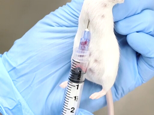
Figure 1. Cardiac blood withdrawal with mouse held vertically.
- Blood withdrawal
- The approach from the posterior aspect, puncturing the diaphragm is more easily accomplished when the mouse or rat is held vertically by the scruff.
- The needle is advanced in the notch just to the left of the animal's xiphoid.
- The needle should be parallel to the spine and placed just under the ribs.
- The heart is located approximately at the level of the elbow.
- Place the needle, bevel up, into the chest, and puncture the heart.
- Apply slight back pressure with the syringe. If the needle is in the heart, blood will flow into the syringe.
- Wait until blood has filled the syringe before adding additional back pressure on the syringe.
- The lateral approach from the animal's left side requires positioning the animal in dorsal recumbency.
- The point of entry is measured against the point of the elbow on the chest wall. The heart is located approximately at the level of the elbow.
- The needle is inserted perpendicular to the plane of the table at a point midway on the chest wall as measured dorsoventrally.
- Place the needle, bevel up, into the chest, and puncture the heart.
- Apply slight back pressure with the syringe. If the needle is in the heart, blood will flow into the syringe.
- Wait until blood has filled the syringe before adding additional backpressure on the syringe.
- The approach from the posterior aspect, puncturing the diaphragm is more easily accomplished when the mouse or rat is held vertically by the scruff.
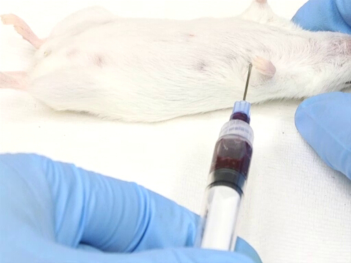
Figure 2. Cardiac blood withdrawal with mouse in dorsal recumbency position.
- Technical tips
- The normal heart is situated with the apex pointing to the left. In rare instances, the heart may be reversed, resulting in difficulty in puncturing the heart.
- Excessive back pressure on the syringe may collapse the heart, occluding the needle bevel and stopping blood flow into the syringe.
- Applying back pressure and releasing it repeatedly will initiate clotting in the syringe.
- Gently applying pressure to the liver can force additional blood volume into the circulatory system, making it available for withdrawal.
4. Retro-orbital bleed
Retro-orbital bleeding is an invasive technique and should generally be performed under appropriate anesthesia to minimize pain and distress, as significant adverse effects can occur even in skilled hands. In many protocols, particularly when the procedure is terminal, deep (terminal) anesthesia is required. For non-terminal or repeated sampling, lighter anesthesia may be used instead of terminal anesthesia; however, such procedures must be carefully justified and explicitly approved by the institutional ethics committee/IACUC, with consideration of animal welfare and cumulative impact.
- Equipment
- Prepare an anesthetic induction chamber, to administer an anesthetic gas such as isoflurane. When using a bell jar, it is imperative that the liquid anesthetic does not come into contact with the animal, to avoid absorption through the skin. A platform with small holes can be used.
- Microhematocrit tubes that hold 50-75 microliters are preferred. Mylar wrapped tubes are less likely to break between the fingers of the operator and should be considered as a safety measure.
- Several paper towel thicknesses, or other insulating materials, are placed on the work surface to maintain the animal's body heat during the procedure.
- Preparation and positioning of the animal
- The animal is anesthetized with an inhalation anesthetic, such as isoflurane, in a bell jar or gas anesthesia induction chamber, to effect.
- Once the animal is fully anesthetized, it is removed and placed in lateral recumbency.
- The eye is protruded by placing a finger on the top of the head and along the jawline, and pulling the skin back and down.
- Avoid applying pressure to the trachea, as that may collapse or occlude the airway causing death by asphyxia.
- Blood withdrawal
- The microhematocrit is placed in the medial canthus of the eye and directed caudally at a 30-45° angle from the plane of the nose.
- Apply pressure while gently rotating the hematocrit tube. This will cut through the conjunctival membranes and rupture the ocular plexus.
- The blood will flow into the hematocrit tube by capillary action.
- Avoid pushing so deep that you hit the bone at the back of the ocular cavity.
- Once blood begins to flow, maintain pressure to keep the eye protruded.
- To collect multiple tubes of blood, it is not necessary to place the next tube into the ocular plexus, as the blood will continue to flow and can be collected as it comes from the medial canthus.
- To stop bleeding, release the skin and allow the eye to return to the normal position. Close the eyelids and gently apply pressure to the underlying eye for ~30 seconds to facilitate hemostasis.
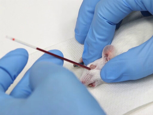
Figure 3. Retro-orbital blood withdrawal in mice.
Key Terms and Definitions
Prior to the bleeding procedure, the type of sample required must be determined. Experimental procedures could require whole blood, plasma, or serum. For whole blood, an anticoagulant must be added to the sample. Plasma, which contains fibrinogen and other clotting factors when separated from the red blood cells, can be extracted from an anticoagulated sample. Serum is obtained through blood collection without an anticoagulant. The serum will result from centrifugation of the sample once a clot has formed. As the sample has clotted, the serum will not contain fibrinogen or other clotting factors. Both plasma and serum are obtained through the use of a centrifuge run at 2200-2500 RPM for a minimum of 15 minutes.
For a sample that must yield whole blood or plasma, an appropriate anticoagulant must be used. Commonly used anticoagulants for laboratory animals are heparin, sodium citrate, and ethylenediamine tetraacetic acid (EDTA); selection of which is based on research needs. Sequester-a liquid form of EDTA, heparin, and sodium citrate-can be loaded directly into the syringe to coat the surfaces. This allows contact of the anticoagulant directly as the blood is drawn, aiding in the prevention of clotting. As rat blood clots faster than most mammalian blood, it is essential that the correct ratio of anticoagulant to blood be used for blood collection.
Needle selection is based on the size of the animal and the site of the venipuncture. In general, the larger the bore of the needle, the more rapidly the sample can be collected. Less damage to the blood cells is another benefit to larger needles. However, the main disadvantage to large-bore needles is the potential damage to the vessel. On mice and rats, the choices of size range from 20-29 gauge needles that are 0.5-1.5 inches in length. If a needle is too long, not only is it awkward to use, but having the extra space in the needle could result in clotting. The appropriate needle size is listed for each method in the procedures section.
The size of the required sample must also be predetermined. Due to the small size of the mouse or rat, the maximum amount of blood collection must be calculated for a survival bleed. An average mouse weighing 25 grams has a total blood volume of 1.8 ml; the average rat weighing 250 grams has a total blood volume of 16 ml. For a single blood sample on a mouse or rat without fluid replacement, the maximum blood volume that can be safely removed is 10% of the total blood volume, or 7.7-8 µl/g. Thus for an average mouse, 10% of its blood volume is 193-200 µl. For an average rat of 250 grams, this is equivalent to 1.9-2.0 ml. Studies have shown that removing more than 15% of the blood volume can cause hypovolemic shock. However, with fluid replacement, up to 15% of the total blood volume-or 12 µl/g-can be removed. For a 25 gram mouse, this is equivalent to 300 µl; for a 250 gram rat, it is equivalent to 3 ml. For fluid replacement, the fluids should be warmed and given subcutaneously.
If it is necessary to take multiple samples, the blood volume drawn is reduced. The maximum blood volume that may be drawn per week is no more than 7.5% of the total blood volume, or 6 µl/g. For a 25 gram mouse, this is equivalent to 145-150 µl per week. For a 250 gram rat, this is equivalent to 1.45-1.50 ml per week. If sampling will occur every 2 weeks, up to 10% of the total blood volume (8 µl/g) may be drawn. This is equivalent to 200 µl every 2 weeks for an average mouse, and up to 2.00 ml every 2 weeks for a 250 gram rat. One study, performed on rats with the average weight of 250 grams, revealed that when blood volumes of 15-20% were removed, it took more than 29 days for blood levels to normalize. For repeated blood collection, fluid replacement does not allow for a larger blood volume or more frequent blood collection, as it only replaces volume. The animal will need time to replenish blood cells.
The use of the retro-orbital plexus has been a common practice in the past. However, many concerns about the humaneness of this procedure have arisen. During the procedure, excessive movement of the hematocrit tube once placed in the medial canthus of the eye can cause damage to the surrounding tissues, resulting in swelling of the eyelids and/or conjunctival membranes. The swollen tissues can cause the eyeball to protrude far enough so that closure of the eyelid is impeded, potentially resulting in corneal drying and damage. Pain from swelling can trigger scratching and self-mutilation that results in enucleation of the eye. Improper placement of the hematocrit tube during a retro-orbital bleed can sever the optic nerve, resulting in blindness. If the hematocrit tube is advanced at an improper angle, the eye can be forced out of the orbit, allowing the eyelids to fall behind the eyeball. If this occurs, it is very difficult to correctly replace the eye into the socket. Other issues that can arise include fracturing of the fragile orbit bones, penetration of the eye globe that results in the loss of vitreous humour, or the formation of a hematoma behind the eye that can result in extreme pain due to the pressure on the eye and surrounding structures. Despite all of these concerns, if a skilled technician performs the procedure and the animal is fully anesthetized with a general anesthetic, such as isoflurane inhalant anesthesia, retro-orbital bleeding has been shown to be an effective method of blood collection in rodents.
The anatomical structure of the orbital area is different between the mouse and rat. The mouse has the retro-orbital sinus collection of vessels that create a sinus in the orbital area. In the orbit of the rat eye, there is a plexus of vessels that flow behind that eye; however, they do not form a sinus, as in the mouse. Consequently, it is easier to perform this procedure on mice. For repeated sampling collection via the retro-orbital plexus, a minimum of 10 days between bleeds is required to allow the tissues in the area to heal. Although general anesthesia is recommended, the procedure can be performed in mice without general anesthesia if a topical ophthalmic anesthetic, such as proparacaine or tetracaine, is applied prior to the procedure. As rats do not have the retro-orbital sinus, and because their membranes around the orbit are much stronger, it is mandatory to anesthetize them for this procedure.
The sample collected from a tail snip can contain both arterial and venous blood, along with tissue product contamination. The sample quality decreases if the tail is stroked or "milked" to obtain more blood. To increase blood flow, the tail can be heated with warm compresses, a heat lamp, or submersion in warm water. Pressure should be applied to the tail tip for hemostasis, and animals should be checked every 5-10 minutes to ensure hemostasis has been achieved. Hemostasis is often delayed with repeated sampling. A styptic powder may be used for hemostasis. For the initial amputation, anesthesia (general or local) is recommended. Subsequent bleeding should not require anesthesia, especially as the animals become habituated to the procedure. Anesthesia will cause a drop in blood pressure, making blood collection with this technique difficult.
Often, studies require a nonsurvival, large blood sample that is collected through exsanguination via an intracardiac bleed. Approximately half of the total blood volume can be collected from a mouse or rat by cardiac puncture. This is equivalent to 40 µl/g or approximately 1 ml for an average 25 gram mouse. A 250 gram rat would yield approximately 10 ml of blood. The animal must be anesthetized for exsanguination. Inhalant anesthesia can be used by a proficient technician; injectable anesthesia can also be used. However, there may be a decrease in blood pressure and circulation, which could decrease the amount of blood collected.
The intracardiac method can be performed either with the animal restrained manually once it is anesthetized (closed method), or the heart can be surgically exposed as per the protocol for caudal vena cava blood collection method (open method). For the closed method, the landmarks for needle placement are the groove formed by the rib cage at the xiphoid process, on the animal's left side.
Questions that this video will help you answer
Blood collection for mice and rats can be accomplished with a variety of techniques. Although many factors, such as sample size, frequency of sampling, and the size and age of the animal influence this, the most essential component is the skill level of the technician performing the sample collection. For the methods described here, the proper use of anesthetics is also crucial for quality samples and the wellbeing of the animals.
Source: Kay Stewart, RVT, RLATG, CMAR; Valerie A. Schroeder, RVT, RLATG. University of Notre Dame, IN
Blood collection is a common requirement for res…
Chapters in this video
0:00
Overview
0:56
General Considerations for Blood Withdrawal
4:00
Tail Vein Bleed
5:11
Tail Snip
5:56
Cardiac Blood Collection
8:23
Retro-Orbital Bleed
10:38
Applications
Copyright © 2026 MyJoVE Corporation. All rights reserved.