Dissociating cells from specific tissue types requires specific parameters for tissue aggitation to obtain a high volume of viable, culturable cells. The Miltenyi gentleMACS Dissociator optimizes this task with a simple, practical protocol. In this publication the use of this apparatus on nerual tissue is explained.
Method Article
Generation of Single-Cell Suspensions from Mouse Neural Tissue
In This Article
Summary
Abstract
Within the nervous system, hundreds of neuronal and glial cell types have been described. Each specific cell type in the brain or spinal cord has a repertoire of cell surface molecules, or molecular determinants, through which it can be identified and characterized. Currently, robust cell identification and separation technologies require single-cell preparations to be generated while simultaneously limiting cell death and destruction of characteristic surface protein. The gentleMACS Dissociator, when used in combination with trypsin or papain-based dissociation kits, can effectively and gently dissociate brain tissue while preserving antigen epitopes and limiting cell loss. Standardized preparation of single-cell suspensions is achieved using C Tubes and optimized, preset gentleMACS Programs. Once generated, single-cell suspensions can be treated with monoclonal conjugates like Anti-Prominin-1 MicroBeads, which identify neural progenitors, or purified further using Myelin Removal Beads.
Protocol
To work under sterile conditions, it is recommended to perform all steps in a laminar flow hood.
1. Materials
- Neural Tissue Dissociation Kit (P) (Components: Solution 1,2 , 3, and 4, Storage Buffer)
- Pre-Separation Filters
- (Optional) Myelin Removal Beads
- Beta-mercaptoethanol
- gentleMACS Dissociator
- C Tubes
- MACSmix Tube Rotator
- HBSS (w/o)
- HBSS (w)
- Prepare the following solutions prior to beginning the protocol:
- Hanks’ Buffered Salt Solution without divalent cations Ca2+ or Mg2+ (HBSS (w/o)
- HBSS, standard, i.e. with Ca2+ and Mg2+ (HBSS (w)
- Depending on the antigen epitope of interest in subsequent applications, use either the Papain-based Neural Tissue Dissociation Kit (P) or the Trypsin-based Neural Tissue Dissociation Kit (T).
- Prepare the following solutions from the Neural Tissue Dissociation Kit:
- Solution 2: add beta-mercaptoethanol to 0.067 mM
- Solution 4: dissolve powder in 0.7 ml Storage Buffer (provided in kit), gently mix (do not vortex)
- Enzyme Mix 1: Pipette 1.9 mL Solution 2 and 50 μl Solution 1 (both from kit) into a C Tube and incubate for 10-15 min at 37 °C. This is sufficient to dissociate 400 mg of brain tissue.
2. Dissociating the neural tissue
- Weigh brain tissue in a tube containing 1 ml HBSS (w/o)
- Transfer mouse brain into the C Tube containing 1950 μL of preheated Enzyme Mix 1. (NOTE: this protocol is written for ≤ 400 mg, but solution volumes can be scaled to accommodate up to 1600 mg of brain tissue per C Tube)
- Place the C Tube onto the gentleMACS Dissociator and run program “m_brain_01”.
- Incubate with rotation (4 rpm using a MACSmix Tube Rotator) for 15 min at 37 °C.
- Place the C Tube onto the gentleMACS Dissociator and run program “m_brain_02”.
- During the dissociation step prepare 30 μl of Enzyme Mix 2 per 400 mg of tissue by gently mixing 20 μl of Solution 3 and 10 μl of Solution 4.
- Add Enzyme Mix 2 to the C Tube via the septum-sealed cap and mix gently without vortexing.
- Incubate the C Tube with rotation for 10 min at 37 °C in an incubator.
- Place the C Tube onto the gentleMACS Dissociator and run program “m_brain_03”.
- Incubate the C Tube with rotation for 10 min at 37 °C in an incubator.
- Centrifuge briefly to collect the sample at the bottom of the tube.
3. Filtration
- Select a filter large enough to allow passage to the cells of interest. For instance, Purkinje cells are too large to pass through a 30 μm filter, but Prominin-1+ cells will.
- Use a suitable1000 μl pipette to remove the cells form the C Tube through the septum-sealed cap and apply it to the Pre-Separation Filter on a 15 ml collection tube. (NOTE: for larger sample sizes use a 50 ml collection tube).
- Wash the filter with 10 ml of HBSS (w).
- Centrifuge the filtered cells at 300 x g for 10 min at RT.
- Resuspend the supernatent in your medium or buffer of choice for subsequent applications.
- To remove myelin debris, use Myelin Removal Beads.
4. Representative Result: Please See Figures 1-3
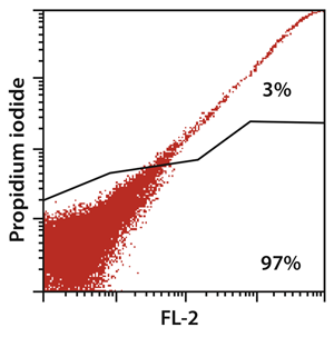
Figure. 1 The brain dissociation with the gentleMACS Dissociator resulted in 97% viable cells, as flow cytometric analysis shows.
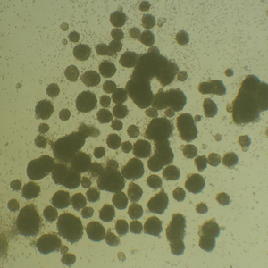
Figure. 2 Light microscope picture of neurosphere formation after magnetic cell sorting using Anti-Prominin-1 MicroBeads after 7 days of cultivation in MACS® NeuroMedium supplemented with MACS Supplement B27 PLUS. Cells were prepared from CD1 mouse brain (P3) using the Neural Tissue Dissociation Kit (P).

Figure. 3 Myelin debris in single-cell suspensions considerably impairs cell isolation, and removal of myelin debris by Myelin Removal Beads increases efficiency cell separations. For MACS Separations using Anti-Prominin-1 MicroBeads, P22 mouse brain was dissociated using the Neural Tissue Dissociation Kit (P). Cells from the single-cell suspension were either directly used for separation, or were submitted to myelin depletion using Myelin Removal Beads. Comparing the separation from samples without and with myelin removal, demonstrates that the purity is higher for samples with previous myelin removal.
Discussion
The gentleMACS Dissociator facilitates the standardized preparation of single-cell suspensions from neural tissues in a closed system. Neural Tissue Dissociation Kits are optimized to preserve antigen epitopes needed for further applications like immunostainings and immunomagnetic cell separation1-5. In this protocol we show the gentle enzymatic dissociation of mouse brain using the gentleMACS Dissociator and the Neural Tissue Dissociation Kit (P), yielding in 97% viable cells. 100 mg of neural tissue yields between 5x10⁶ and 1x10⁷ cells, depending on the age of the host tissue. The targeted Prominin-1+ cells were isolated using Anti-Prominin-1 MicroBeads and subsequently taken into culture. The protocol is shown to be even more effective with the additional use of Myelin Removal Beads when working with neural tissue derived from mice >P7.
Disclosures
The authors are employees of Miltenyi Biotec GmbH, Germany.
Materials
References
- Hardt, O., Scholz, C., Küsters, D., Yanagawa, Y., Pennartz, S., Cremer, H., Bosio, A. Gene expression analysis defines differences between region-specific GABAergic neurons. Mol Cell Neurosci. 39, 418-428 (2008).
- Lee, J. K., McCoy, M. K., Harms, A. S., Ruhn, K. A., Gold, S. J., Tansey, M. G. Regulator of G-protein signaling 10 promotes dopaminergic neuron survival via regulation of the microglial inflammatory response. J Neurosci. 28, 8517-8528 (2008).
- Környei, Z., Gócza, E., Rühl, R., Orsolits, B., Vörös, E., Szabó, B., Vágovits, B., Madarász, E. Astroglia-derived retinoic acid is a key factor in glia-induced neurogenesis. FASEB J. 21, 2496-2509 (2007).
- Meylan, F., Davidson, T. S., Kahle, E., Kinder, M., Acharya, K., Jankovic, D., Bundoc, V., Hodges, M., Shevach, E. M., Keane-Myers, A., Wang, E. C., Siegel, R. M. The TNF-family receptor DR3 is essential for diverse T cell-mediated inflammatory diseases. Immunity. 29, 79-89 (2008).
- Skog, J., Würdinger, T., van Rijn, S., Meijer, D. H., Gainche, L., Sena-Esteves, M., Curry, W. T. Jr, Carter, B. S., Krichevsky, A. M., Breakefield, X. O. Glioblastoma microvesicles transport RNA and proteins that promote tumour growth and provide diagnostic biomarkers. Nat Cell Biol. 10, 1470-1476 (2008).
Reprints and Permissions
