Lentiviral expression vectors are the most effective vehicles for stably expressing different effector molecules or reporter constructs in dividing and non-dividing mammalian cells and whole organisms. Here we provide a protocol on how to package lentivector expression constructs in pseudoviral particles and to transduce target cells using the pseudoviral particles.
Method Article
Packaging HIV- or FIV-based Lentivector Expression Constructs & Transduction of VSV-G Pseudotyped Viral Particles
In This Article
Summary
Abstract
As with standard plasmid vectors, it is possible to transfect lentivectors in plasmid form into cells with low-to-medium efficiency to obtain transient expression of effectors. Packaging lentiviral expression constructs into pseudoviral particles, however, enables up to 100% transduction, even with difficult-to-transfect cells, such as primary, stem, and differentiated cells. Moreover, the lentiviral delivery does not produce the specific cellular responses typically associated with chemical transfections, such as cell death resulting from toxicity of the transfection reagent 1, 2. When transduced into target cells, the lentiviral construct integrates into genomic DNA and provides stable expression of the small hairpin RNA (shRNA), cDNA, microRNA or reporter gene 3, 4. Target cells stably expressing the effector molecule can be isolated using a selectable marker contained in the expression vector construct such as puromycin or GFP. After pseudoviral particles infect target cells, they cannot replicate within target cells because the viral structural genes are absent and the long terminal repeats (LTRs) are designed to be self-inactivating upon transduction 5, 6.
There are three main components necessary for efficient lentiviral packaging 1, 5, 6, 7.
1. The lentiviral expression vector that contains some of the genetic elements required for packaging, stable integration of the viral expression construct into genomic DNA, and expression of the effector or reporter.
2. The lentiviral packaging plasmids that provide the proteins essential for transcription and packaging of an RNA copy of the expression construct into recombinant pseudoviral particles. This protocol uses the pPACK plasmids (SBI) that encode for gag, pol, and rev from the HIV or FIV genome and Vesicular Stomatitis Virus g protein (VSV-G) for the viral coat protein.
3. 293TN producer cells (derived from HEK293 cells) that express the SV40 large T antigen, which is required for high-titer lentiviral production and a neomycin resistance gene, useful for reselecting the cells for maintenance.
An overview of the viral production protocol can be seen in Figure 1. Viral production starts by co-transfecting 293TN producer cells with the lentiviral expression vector and the packaging plasmids. Viral particles are secreted into the media. After 48-72 hours the cell culture media is harvested. Cellular debris is removed from the cell culture media, and the viral particles are precipitated by centrifugation with PEG-it for concentration. Produced lentiviral particles are then titered and can be used to transduce target cells. Details of viral titering are not included in this protocol, but can be found at:
http://www.systembio.com/downloads/global_titer_kit_web_090710.pdf 8.
This protocol has been optimized using the specific products indicated. Other reagents may be substituted, but the same results cannot be guaranteed.
Protocol
1. Transfection of 293TN Cells with Packaging Plasmids and the Expression Construct
- Seed 7.0 - 8.0 x 10⁶ 293TN cells per 15 cm2 culture plate in 20 ml of culture medium containing DMEM medium supplemented with 4 mM L-glutamine, 4.5 g/l glucose, and fetal bovine serum (10%) without antibiotics. Grow for 18-24 hours at 37 °C with 5% CO2 so that the cell density reaches 60-80% confluency at the time of transfection. In some cases, it may be necessary to seed several plates of cells to obtain a high enough titer for transduction of target cells.
- For each plate of cells, add 1.6 ml serum-free DMEM to an autoclaved 2 ml Eppendorff tube. Add 45 μl pPACKH1 or pPACKF1 and 4.5 μg of your lentivector construct to the DMEM and mix by pipetting up and down.
- Add 55 μl of PureFection into the same tube. Vortex for 10 seconds. Incubate the DMEM-Plasmid-PureFection mixture at room temperature for 15 min.
- Add the DMEM-Plasmid-PureFection mixture to the tissue culture plate drop-wise and swirl gently to evenly disperse throughout the plate. Return the dish to the incubator and grow at 37 °C with 5% CO2.
- There is no need to change the media after transfection. If your lentivector construct expresses a gene encoding a fluorescent protein like GFP, check your cells under a microscope 12-24 hours after transfection. You should be able to see >90% GFP-positive cells, indicating that the transfection was successful.
- Collect the cell culture supernatant after 48 and 72 hours into a 50 ml sterile centrifuge tube. This cell culture supernatant now contains infectious pseudoviral particles. (Follow the recommended guidelines for working with BSL-2 safety class.) Centrifuge at 1,500 x g for 15 min at room temperature to pellet the cellular debris. Transfer the viral supernatant to a new tube, ensuring not to disturb the pellet.
- 293TN cells that have been used for viral packaging should be disposed of in a biohazard container and are not to be used for further rounds of viral packaging.
2. Concentration of the Viral Supernatant
- Add 1 volume of cold (4 °C) PEG-it Virus Precipitation Solution to every 4 volumes of lentiviral particle-containing supernatant. Precipitation of lentiviral particles from large volumes can be achieved with Corning 250 ml polypropylene centrifuge tubes. Mix the solution by inverting the tubes several times, but do not vortex the mixture.
- Refrigerate the solution at 4 °C for at least 12 hours (up to 4 days is acceptable). Do not shake or rotate the tubes during the refrigeration step.
- Centrifuge the supernatant/ PEG-it mixture at 1,500 x g for 30 minutes at 4 °C. After centrifugation, the lentiviral particles will appear as a beige or white pellet at the bottom of the tube. Pour out the supernatant.
- Remove all traces of fluid by aspiration, taking care not to disturb the precipitated lentiviral particles in the pellet. Traces of residual PEG-it will dilute the virus (thus reducing the titer) but will not affect the integrity of the target cells since PEG-it is inert and not toxic.
- Resuspend and combine the lentiviral particles in 1/100 of the original volume using cold (4 °C), sterile phosphate buffered saline or DMEM containing 25 mM HEPES buffer.
- Aliquot into sterile cryogenic vials and store at -70 °C until ready for use.
3. Viral Titering and Transduction of Target Cells
- We recommend titering the lentiviral particles before transducing the target cells-of-interest and calculating the appropriate multiplicity of infection (MOI) for the target cells for consistency between experiments. For viral titering, we recommend using HT1080 cells as an easy-to-infect positive control line. Please note that the titering protocol involves transducing HT1080 cells with a small aliquot of the lentiviral particles you have packaged. Once the titer is known, the target cells-of-interest can also be transduced with the produced lentiviral particles. A recommend titering protocol can be found at: http://www.systembio.com/downloads/manual_titer_kit_web.pdf 7.
- Plate 50,000 target cells-of-interest per well in a 24-well plate in cell culture media. Use the media that the cells are normally cultured in. Grow the cells overnight at their specified culture conditions. Cells should be between 50 to 70% confluent on the day of transduction.
- On the day of transduction, aspirate the media from the cells. Combine culture medium with TransDux to a 1 x final concentration (for example, 2.5 μl TransDux to 500 μl of culture medium). Then transfer the TransDux-cell culture medium mixture to each well.
- Pipette the appropriate volume of virus which corresponds to the optimal MOI for the target cells into each well. If the optimum MOI is unknown, we recommend trying a range of different MOIs (such as an MOI of 1, 2 and 5 for easy-to-infect tissue culture cells, and an MOI of 1, 10 and 20 for more difficult-to-infect primary cells). If the concentration of the lentiviral particles is not known (as determined through a virus titering protocol), different volumes of virus can be tried, such as 1, 2 and 5 μl of virus. Virus can remain in contact with the target cells for up to 72 hours.
- Further experiments with the transduced target cells-of-interest may be commenced 72 hours after transduction, once the viral construct has integrated into the host cell genome. Change the medium and passage the target cells-of-interest according to their specific needs.
4. Representative Results
Transfection of 293TN cells
The starting density of the 293TN cells is critical for successful viral packaging. 293TN cells must be between 60 -80% confluent the day of transfection (Figure 2). If the 293TN cells are less than 60% confluent, allow them to grow for a few more hours before attempting the transfection. If the 293TN cells are >80% confluent, they should be replated before transfection.
If using a lentivector expressing GFP, at least 90% of the 293TN cells should fluoresce green 24 hours after the transfection, indicating a successful transfection (Figure 3). If less than 90% of the cells are GFP-positive, do not proceed to the next step because this indicates that the transfection was not successful, and the viral titers will be low. Instead, plate new 293TN cells and start over, making sure that the 293TN cells are at the proper cell density before transfecting.
293TN cells may pull off the tissue culture plates 48-72 hours after transfection. This does not negatively affect the production of lentiviral particles.
Titering with HT1080 Cells
Viral titers can be calculated using SBI's Global UltraRapid Titering kit according to the manufacturer's instructions. This system utilizes qPCR to measure viral integration of the WPRE sequence from the lentiviral vector into HT1080 cells and compares this to a standard curve, based on a genomic reference sequence to accurately measure infectious units per ml (Figure 4).
Transduction of Target Cells
If using lentiviral particles expressing a constitutively active GFP marker, GFP positive cells should start to appear within 12-24 hours of transduction, if the transduction reaction is working. GFP expression should continue after the lentiviral construct stably integrates into the host cell genome. If transducing with different MOIs, the GFP fluorescence should approximately match the MOI, where low MOIs show less GFP fluorescence and higher MOIs show increased GFP fluorescence (Figure 5).
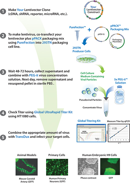
Figure 1. Overview of the viral production protocol. The lentivector and pPACK packaging plasmids are mixed and co-transfected into 293TN cells. After 48 -72 hours, the viral supernatant is collected, and viral particles are precipitated with PEG-it. After titering the lentiviral particles, they can be used to transduce the target cells-of-interest. Click here to view larger figure.

Figure 2. Phase contrast image of 293TN cells. Cell density should be between 60-80% confluent the day of transfection.

Figure 3. Fluorescent image of GFP-positive 293TN cells 24 hours after transfection with Purefection, pPACKH1, and a lentivector construct encoding GFP.

Figure 4. WPRE qPCR results and standard curve obtained using SBI's Global UltraRapid Titering kit to calculate MOI and corresponding titer of packaged viral particles.

Figure 5. Fluorescent images of HT1080 cells transduced with increasing amounts of lentivirus. Images are from 72 hours post-transduction.
Discussion
This viral packaging and transduction of target cells protocol has been optimized for use with SBI's viral packaging reagents. Substitution of other reagents may affect the outcome of the protocol. This protocol has only been optimized for lentiviral production and may not be suitable for producing other types of viruses or pseudoviruses.
Successful viral packaging is dependent upon several key steps. The 293TN cells express the SV40 large T antigen, which is absolutely necessary for secretion of the lentiviral particles into the cell culture supernatant. The density at which the 293TN cells are at the time of transfection is especially important in order for the PureFection reagent to sufficiently transfer the lentivector and packaging plasmids into the cells. Adding the PureFection mixture to the tissue culture plate in a drop-wise manner followed by thoroughly swirling the plate is important for dispersal of the packaging plasmids and lentivector construct throughout all of the cells. Failure to appropriately distribute the vectors may result in insufficient lentiviral particles being produced. The viral packaging reaction can be scaled up for production of high titer virus, based on the surface area of the tissue culture plates. One can choose to plate several tissue culture dishes of 293TN cells per construct that needs to be packaged. Concentration of the viral supernatants is especially helpful in creating high-titer virus. Viral supernatant from multiple plates of cells can be combined and concentrated for use in in vivo experiments, or for cells that are difficult to transduce.
Titering the produced lentiviral particles is important for calculating an accurate MOI to use for transduction of target cells. Knowing the MOI that was used in a given transduction enables troubleshooting in the event that the transduction is not successful. For example, using an MOI that is too low may result in very few target cells expressing the construct-of-interest. Using an MOI that is too high, however, may result in target cells that show signs of stress. The goal is to use an MOI that maximizes expression of the construct-of-interest while preserving the health of the cells. While we recommend a qPCR-based titering protocol that measures integrated copies of the lentiviral construct in HT1080 cells, other titering approaches can also be used, such as p24 ELISA or estimation of the percent of GFP-positive cells.
Successful transduction of target cells is also dependent upon several key factors. TransDux is a unique infection reagent that enables high transduction efficiencies, but that is not toxic to cells. TransDux can be used instead of polybrene, which is often toxic to the target cells. Inclusion of TransDux in the transduction of the target cells helps to neutralize charges on the viral particles and allows the virus into the cells. Since TransDux is not toxic to the cells, one can allow the lentiviral particles to remain in contact with the cells for up to 72 hours (the time at which the viral constructs have integrated into the host cell genome), thus increasing the probability of the viral particles entering the cell. For some difficult-to-transduce cells, transductions can be repeated on successive days to boost the transduction efficiency. For example, transduce the cells on day 1, allow the cells to rest on day 2, and then transduce the cells again on day 3. It is important to wait 72 hours after the last transduction before beginning selection, further experiments or characterization of the target cells.
Efficient viral packaging is also dependent upon the size of the lentiviral construct, between the 5' and 3'LTR regions. This section of the lentiviral construct should be approximately 9 kb or less, which corresponds to the size of the native HIV genome. Exceeding 9 kb may decrease the titer of the produced pseudoviral particles. New technology, such as piggyBac transposon vectors, allow for reliable, stable integration of transgenes up to 100 kb through a single transfection. This approach may be used for constructs that exceed the size limit for lentiviral vectors, in cases where it is possible to transfect target cells, and provides the additional advantage of not requiring a viral packaging step 9,10.
The pseudoviral particles produced using this protocol are infectious in that they can infect most mammalian cell types. The products used in this protocol, however are third-generation biosafe in that they produce non-replicative and self-inactivating pseudoviruses. Therefore, once transduced into target cells or animals, the target cells or animals are not infectious or contagious. Substitution of other lentivector plasmids that are not third-generation biosafe is possible although not recommended from a biosafety perspective. The viral production protocol and subsequent transduction of target cells should be carried out under Biosafety Level 2, according to the NIH guidelines 11, and researchers should always wear a lab coat and gloves when handling the viral particles. Used 293TN producer cells, tubes of viral particles, and disposable pipettes should be disposed of in an appropriate biohazard container. Reusable pipettes and glassware should be decontaminated with bleach and autoclaving to decrease exposure of personnel.
Disclosures
Production and Free Access to this video article is sponsored by System Biosciences.
Acknowledgements
We would like to acknowledge the staff and scientists at SBI for their support in the development and testing of these protocols.
Materials
| Name | Company | Catalog Number | Comments |
|---|---|---|---|
| DMEM High Glucose Medium | GIBCO, by Life Technologies | 11995-073 | |
| 293TN Cells | SBI | LV900A-1 | |
| pPACKH1 | SBI | LV500A-1 | For HIV-based lentivectors |
| pPACKF1 | SBI | LV100A-1 | For FIV-based lentivectors |
| Any Lentiviral vector | SBI | Various | cDNA, shRNA, microRNA, transcription reporters |
| Purefection | SBI | LV750A-1 LV750A-5 | |
| PEG-it | SBI | LV810A-1 LV825A-1 | |
| Global UltraRapid Titering kit | SBI | LV961A-1 | Not described in this protocol, but needed to titer the viral particles |
| TransDux | SBI | LV850A-1 |
References
- RNA Viruses. A Practical Approach. Cann, A. J. , Oxford Univ. Press. (2000).
- Gould, D. J., Favorov, P. Vectors for the treatment of autoimmune diseases. Gene Therapy. 10, 912-927 (2003).
- Naldini, L., Blomer, U., Gage, F. H., Trono, D., Verma, I. M. Efficient transfer, integration, and sustained long-term expression of the transgene in adult rat brains injected with a lentiviral vector. Proc. Natl. Acad. Sci. U.S.A. 93, 11382-11388 (1996).
- Naldini, L., Blomer, U., Gallay, P., Ory, D., Mulligan, R., Gage, F. H. In vivo gene delivery and stable transduction of nondividing cells by a lentiviral vector. Science. 272, 263-267 (1996).
- Dull, T., Zufferey, R. A third-generation lentivirus vector with a conditional packaging system. J. Virol. 72, 8463-8471 (1998).
- Federico, M. Lentivirus Gene Engineering Protocols. , Humana Press. New Jersey. (2003).
- Curran, M. A., Kaiser, S. M., Achacoso, P. L., Nolan, G. P. Efficient transduction of nondividing cells by optimized feline immunodeficiency virus vectors. Mol. Ther. 1, 31-38 (2000).
- Manual Titer Kit. , Available from: http://www.systembio.com/downloads/manual_titer_kit_web.pdf (2011).
- Kahlig, K. M., Saridey, S. K., Kaja, A., Daniels, M. A., George, A. L. Jr, Wilson, M. H. Multiplexed transposon-mediated stable gene transfer in human cells. Proc. Natl. Acad. Sci. U.S.A. 107 (4), 1343-1348 (2010).
- Kim, A., Pyykko, I. Size matters: versatile use of PiggyBac transposons as a genetic manipulation tool. Mol. Cell Biochem. , (2011).
- NIH Office of Biotechnology Activities. Guidance on Biosafety Considerations for Research with Lentiviral Vectors. , Available from: http://oba.od.nih.gov/oba/rac/Guidance/LentiVirus_Containment/pdf/Lenti_Containment_Guidance.pdf (2011).
Reprints and Permissions
