Simple and reproducible procedures are described for making three structurally distinct collagen assemblies from a common commercially available Type I collagen monomer. Native type, fibrous long spacing or segmental long spacing collagen can be constructed by varying the conditions to which the 300 nm long and 1.4 nm diameter monomer building block is exposed.
Method Article
In vitro Synthesis of Native, Fibrous Long Spacing and Segmental Long Spacing Collagen
In This Article
Summary
Abstract
Collagen fibrils are present in the extracellular matrix of animal tissue to provide structural scaffolding and mechanical strength. These native collagen fibrils have a characteristic banding periodicity of ~67 nm and are formed in vivo through the hierarchical assembly of Type I collagen monomers, which are 300 nm in length and 1.4 nm in diameter. In vitro, by varying the conditions to which the monomer building blocks are exposed, unique structures ranging in length scales up to 50 microns can be constructed, including not only native type fibrils, but also fibrous long spacing and segmental long spacing collagen. Herein, we present procedures for forming the three different collagen structures from a common commercially available collagen monomer. Using the protocols that we and others have published in the past to make these three types typically lead to mixtures of structures. In particular, unbanded fibrils were commonly found when making native collagen, and native fibrils were often present when making fibrous long spacing collagen. These new procedures have the advantage of producing the desired collagen fibril type almost exclusively. The formation of the desired structures is verified by imaging using an atomic force microscope.
Introduction
Collagen is a class of structural proteins that have a triple helical structure formed from three polypeptide chains. These triple helical monomers further assemble in a hierarchal manner to form progressively larger structures. There are at least 28 genetically different collagen types that have been identified 1. Combined, collagens make up 30% of the total protein found in animals, with Type I the predominant form accounting for up to 90% of all the collagen proteins 2. Type I collagen is found as fibrillous structures in skin, ligaments, tendons and bones. Atomic force microscope (AFM) and electron microscope (EM) images of Type I native collagen fibers typically show banding with a characteristic period of ~67 nm (often referred to as D-banding) 3-5.
Type I collagen monomer is a triple helical heterotrimer consisting of two identical α1(I) chains and one α2(I) chain, each more than a thousand amino acids long. It is long and thin with dimensions of 300 nm in length and 1.4 nm in diameter. Type I collagen monomer can be readily obtained by breaking down naturally formed higher order structures 6. We 7 and many others 8-10 have shown that these soluble monomer building blocks can then be used to reconstruct D-banded fibrils in vitro (Figure 1).
In addition to native collagen fibrils, two other higher order collagen structures have been found to form in vitro using the same monomer building block. The two structures are fibrous long spacing (FLS) and segmental long spacing (SLS) collagen (Figure 1). FLS collagen is a fibrillous construct like native collagen, but is characterized by a larger banding periodicity than native collagen 11. In vitro, FLS collagen forms when the collagen monomer is combined with α1-acid glycoprotein 12. FLS collagen has been observed in vivo as well, although rarely 13. SLS collagen is described as crystallites because the monomers are aligned in register like in a crystal lattice 14. SLS collagen forms when the collagen monomer is combined with adenosine triphosphate (ATP) 15. Naturally occurring SLS collagen has been observed in the culture medium of fibroblasts but not in extracted tissue samples, likely due to its small size and lack of distinguishable topographical features 16.
The goal of this manuscript is to present detailed procedures for making native collagen fibrils as well FLS and SLS collagen that even the most inexperienced researcher can reproduce. Using commercially available advanced starting materials, we have tried to make the protocols as simple as possible and still have them work reliably. We also detail how to use an AFM to characterize the different collagen structures that are made. This work will be beneficial to researchers who want to study one or more of these higher order collagen structures and/or use them as precursors in other applications, but currently lack of the expertise or simply do not wish to work with tissue samples.
Protocol
NOTE: The water used in all the protocols has a resistivity >18 MΩ.cm.
1. Native Collagen Fibrils
- Combine 6 μl of water, 20 μl of 200 mM Na2HPO4 adjusted to pH 7 with HCl and 10 μl of 400 mM KCl in a microfuge tube and mix.
- Add 4 μl of collagen monomer (~3 mg/ml in 0.01 N HCl) to the microfuge tube and mix. The solution should be clear and colorless.
- Place the microfuge tube containing the reaction mixture in a heat block that has been pre-warmed to 37 °C and leave for 3 to 4 hr. At this point, the solution should be slightly cloudy and contain mainly native collagen fibrils.
2. Fibrous Long Spacing Collagen
- Dialyze 1 ml of collagen monomer (~3 mg/ml in 0.01 N HCl) using 12-14 kDa MWCO membrane against 400 ml of water, changing the water four times over a period of 24 hr at room temperature. The dialyzed collagen monomer that is not used right away can be stored at 4 °C for future use.
- Combine 20 μl of water, 20 μl of 3 mg/ml α1-acid glycoprotein in water and 20 μl of dialyzed collagen monomer in a microfuge tube. The solution should be clear and colorless.
- Leave the microfuge tube containing the reaction mixture at room temperature for 30 min. At this point, the solution should be cloudy and contain predominantly FLS collagen fibrils.
3. Segmental Long Spacing Collagen
- Combine 67 μl of water, 60 μl of 100 mM glycine-HCl buffer at pH 3.3 and 40 μl of 10 mg/ml ATP in water in a microfuge tube and mix.
- Add 33 μl of collagen monomer (~3 mg/ml in 0.01 N HCl) to the microfuge tube and mix. The solution should be clear and colorless.
- Leave the microfuge tube containing the reaction mixture at room temperature for 2 hr. At this point, the solution will still appear clear, but should contain mostly SLS collagen.
4. AFM Characterization
- Cleave the surface of a piece of mica attached to an AFM substrate by covering the mica surface with a piece of sticky tape and then peeling it away. Check the tape afterwards to confirm that an entire layer of mica was cleaved in order to reduce irregularities on the surface and ensure that the old sample is removed if the mica is being reused.
- Apply 20 μl of the collagen fibril solution to the freshly cleaved mica substrate. Leave for 5 min.
- Gently rinse off solution of collagen fibril with water. It is advisable not to apply the water directly onto the center of the mica substrate but at the edge and to allow it to flow across the sample.
- Dry the surface under a gentle stream of nitrogen gas. It is advisable to direct the stream at the edge and not at the center of the mica substrate.
- Under an optical microscope at 200× magnification, the native and FLS collagen samples should show clumps of fibrils. SLS collagen will not be visible.
- The unique characteristics of the three different collagen structures are observable by AFM. Generally, we image native collagen fibrils with an AFM in intermittent contact mode using silicon AFM probes, whereas FLS and SLS collagen fibrils we typically image with an AFM in contact mode using silicon nitride AFM probes. We do this because of time and cost considerations. Silicon AFM probes are usually sharper than silicon nitride AFM probes, which make them particularly useful for imaging the narrower banding structure of native collagen fibrils. However, silicon AFM probes are much more prone to contamination by collagen samples than silicon nitride AFM probes and need to replaced frequently. Therefore, we reserve the use of silicon AFM probes mostly for imaging native collagen fibrils where resolution is a greater necessity and we operate in intermittent contact mode to prolong their lifetime of usefulness.
We recommend an initial 100 × 100 μm2 scan which should show at least a few collagen constructs. From there, zoom in to scan sizes of 10 × 10 μm2 and then 2 × 2 μm2 to observe the banding periodicity of native and FLS collagen, or the finer features of an SLS collagen. It is also a good idea to check at least two other regions on the collagen sample to verify that the initial scans are representative.
Results
Native collagen
The reaction mixture of ~0.3 mg/ml collagen monomer in 100 mM phosphate and 100 mM KCl at pH 7 left at 37 °C for 3-4 hr will yield a solution containing native type collagen fibrils with clear ~67 nm D-banding, without unbanded fibrils.
Under an optical microscope at 200× magnification, clumps of several fibrils can be normally seen on the mica substrate particularly when viewed by differential interference contrast (DIC) microscopy (Figure 2). In a typical sample, a random 100 × 100 μm2 scan by AFM will usually show at least a few fibrils that are 5-50 microns long (Figures 3a-c). Individual, separated collagen fibrils can be easily identified at this stage. With a 512 × 512 pixel2 image scan, zooming into a 10 × 10 μm2 scan size shows that all the fibrils are banded (Figures 3d-f). In order to accurately measure the banding periodicity, it is best to zoom into a 2 × 2 μm2 scan size (Figures 3g-i).
Banding periodicity can be determined by simply measuring and averaging the longitudinal distance between several peaks or valleys. Alternatively, if the AFM software allows, a one dimensional Fourier transform along a longitudinal cross-section can also be used. Figure 4 shows an AFM height image of a fibril and a corresponding longitudinal cross-section.
FLS collagen
The reaction mixture of 1 mg/ml μ1-acid glycoprotein and ~1 mg/ml collagen monomer in water left at room temperature for 30 min will yield a solution of FLS collagen fibrils.
Clumps of fibrils, similar to what is observed in the case of native collagen, can be easily seen on the mica substrate under an optical microscope at 200× magnification. In a typical sample, a random 100 × 100 μm2 scan by AFM will usually show at least a few fibrils. With a 512 × 512 pixel2 image scan size, the banding periodicity can be measured by zooming into a 10 × 10 μm2 scan (Figure 5a) or more accurately with a 2 × 2 μm2 scan (Figure 5b). Figure 5c shows a longitudinal cross-section of a FLS fibril.
SLS collagen
The reaction mixture of 2 mg/ml ATP and ~0.5 mg/ml of collagen monomer in 100 mM glycine-HCl buffer at pH 3.3 left at room temperature for 2 hr will yield a solution of SLS collagen.
Typically, SLS crystallites will not be visible under an optical microscope. An AFM is required to confirm the presence of SLS crystallites. In a typical sample, a random 100 × 100 μm2 scan by AFM will usually show many dots. Zooming into a 10 × 10 μm2 scan will usually show several SLS crystallites (Figure 6a). And a 2 × 2 μm2 scan (Figure 6b) shows the finer structure of an SLS crystallite.
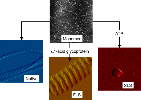
Figure 1. The three structurally different forms of collagen fibrils that can be formed in vitro from collagen monomer. AFM images depicting collagen monomer and the three higher order collagen structures all on the same size scale (2 × 2 μm2).
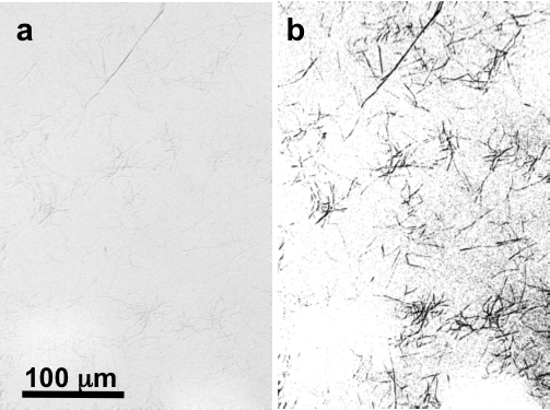
Figure 2. Digital image at ~200× magnification by optical DIC microscopy of native collagen fibrils on mica (a) untouched and (b) thresholded and sharpened to highlight the fibrils.
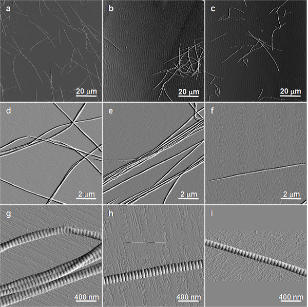
Figure 3. AFM images of native collagen fibrils on mica. Intermittent contact mode AFM amplitude images are shown.
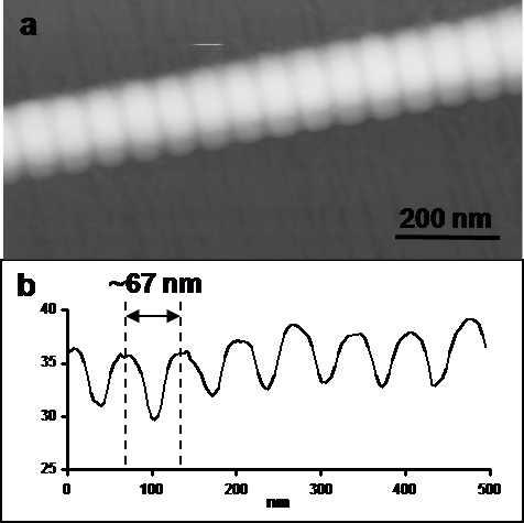
Figure 4. (a) 0.5 × 1 μm2 intermittent contact mode AFM height image (vertical scale = 100 nm) and (b) longitudinal cross-section of a native collagen fibril.
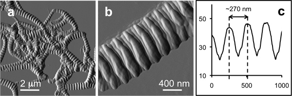
Figure 5. AFM images of FLS collagen fibrils on mica. (a) 10 × 10 μm2 contact mode AFM deflection image, (b) 2 × 2 μm2 contact mode AFM deflection image and (c) longitudinal cross-section of a FLS collagen fibril.
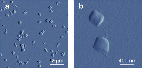
Figure 6. AFM images of SLS collagen on mica. Intermittent contact mode AFM amplitude images are shown.
Discussion
Purified collagen monomer is stable at low pH and temperature and the formation of native collagen fibrils essentially involves raising the pH and temperature of the collagen monomer solution. Procedures for the reconstitution of banded collagen fibrils from monomers have existed for more than 50 years. The procedure that our laboratory used in our first studies with collagen 15 years ago 7 was based on procedures summarized by Chapman and co-workers in 1986 10. The conditions for collagen fibril formation in that earlier work were 0.18 mg/ml of collagen monomer in Na2HPO4/KH2PO4 (I = 0.2, pH 7.4) buffer at 34 °C. The drawback of this and other procedures that we have tried is that there are always unbanded fibrils formed alongside the desired banded fibrils. Our new conditions are ~0.3 mg/ml of collagen monomer in 100 mM phosphate and 100 mM KCl at pH 7 left at 37 °C for 3-4 hr. The procedure described herein differs in every aspect from the earlier procedure for making native collagen fibrils. But most noticeably, we have doubled the ionic strength by raising the buffer concentration and adding 100 mM KCl. The increase in ionic strength results in more consistent formation of exclusively banded collagen fibrils.
In our experience, the only times that this procedure has not produced native type collagen was when the collagen monomer solution was past the manufacturer's recommended date of use. When this occurs, what is observed ranges from fewer fibrils that are all unbanded to many fibrils still observed but predominately unbanded. Note that expired collagen monomer can often still be successfully used to make native collagen, but when it fails, new collagen monomer should definitely be purchased.
FLS was first identified by Highberger et al. 17 in collagen preparations credited to Orekhovich et al. 18. Further studies led Highberger and Schmitt to conclude that it was α1-acid glycoprotein that promotes the formation of FLS 12. We were later able to replicate the procedure for making FLS collagen by combining commercially available collagen monomer and α1-acid glycoprotein at low pH and slowly allowing the pH to rise by dialyzing the mixture against water for 24 hr 11. We now present a modification of that procedure by dialyzing the collagen monomer against water first and simply combining it with α1-acid glycoprotein in water to form FLS collagen. The conditions for FLS collagen assembly described here are much less time consuming. The collagen monomer still needs to be pre-dialyzed against water, but it can be done in bulk and then stored at 4 °C stably until it is used. This new procedure yields predominately FLS banded collagen fibrils.
From our experience, α1-acid glycoprotein with a lower combined protein and water content, and by inference higher sugar content, is critical for successfully making FLS fibrils. We typically check with the manufacturer that the α1-acid glycoprotein lot we use has a combined % protein and water content of 82 or less. And as with the procedure for native collagen synthesis, collagen monomer that is too old can also result in the failure to produce FLS collagen. In addition, the purity of the water used for the dialysis is critical in this procedure. For example, dialysis of collagen monomer against even ~8 MΩ.cm rather than >18 MΩ.cm water results in a collagen solution that will not form FLS collagen.
Of the three collagen fibril types, the easiest to make is SLS collagen. The earliest preparation of SLS was described by Schmitt and co-workers 15. We published an adaption of that procedure 12 years ago for making SLS collagen using commercially available collagen 14. The procedure simply entailed combining 2 mg/ml ATP and 0.5 mg/ml of collagen monomer in 0.05% (v/v) acetic acid at pH 3.5, and leaving the mixture at room temperature overnight.
In our experience, the critical aspect of SLS assembly that must be controlled is the pH of the reaction solution. SLS assembled in more basic conditions (~pH 3.6-3.9) results in aggregated clumps of crystallites. More acidic conditions (~pH 2.9-3.2) results in thinner, more separated crystallites than ones assembled under the ideal conditions of pH 3.3-3.5. Reaction pHs outside this range (< 2.8 and > 4) do not yield any SLS collagen. In the past, we adjusted the pH of the reaction mixture by the addition of acetic acid. To simplify the procedure even further, in this manuscript we introduced a glycine-HCl buffer to control the pH.
The three different collagen structures formed from the protocols described above have characteristic features that can only be discerned by instruments capable of nanometer resolution. Native and FLS collagen are characterized by banding periodicities of ~67 nm and ~270 nm. The longest dimension of SLS collagen is just ~360 nm. We have described specifically how we use an AFM to characterize the three different collagen structures. However, any AFM operating in contact or intermittent contact mode with any AFM probe having <10 nm radius should also be able to image these collagen structures. In addition, electron microscopy can be and has been used to characterize these collagen structures 19 .
The major advantage of these procedures is that they produce predominantly the desired collagen construct. The only other form of collagen that is found in these reactions is the starting monomer, which is often visible in the background of the AFM images. In the case of native and FLS collagen, they can be easily separated from most of the unreacted monomer by repeated centrifugation and washing of the resulting native or FLS collagen pellet with water.
We have presented simple and reliable procedures for making native, FLS and SLS collagen using advanced, commercially available starting materials. The collagen monomer used in all these procedures is commercially available in a purified form. α1-Acid glycoprotein and ATP are also both available commercially.
Disclosures
No conflicts of interest declared.
Acknowledgements
We would like to thank all the many past undergraduate, graduate and post-doctoral students who have contributed their time and effort to the study of collagen fibrogenesis in our laboratory. The Natural Sciences and Engineering Research Council of Canada has continuously provided funding for our collagen studies.
Materials
| Name | Company | Catalog Number | Comments |
|---|---|---|---|
| Type I collagen monomer (~3 mg/ml in 0.01 N HCl) | Inamed Advanced Biomatrix | 5409 5005-B | Lot 1261443 (2.9 mg/ml, pH 2.1) Lot 1629346 (3.2 mg/ml, pH 2.1) Lot 6006 (3.2 mg/ml, pH 2.1) |
| ATP | Research Biochemicals International | A-141 | Lot FBJ-1194A |
| α1-acid glycoprotein from bovine plasma | Sigma-Aldrich | G3643 | Lot 051K7440 (89.8%) Lot 011K7410 (82%) Lot 065K7405 (71.2%) Lot 063K7495 (71.9%) Lot 117K7535 (75.9%) (protein + water content) |
| a1-acid glycoprotein from human plasma | Sigma-Aldrich | G9885 | Lot 128H7606 (70.9%) Lot 049K7565V (68%) (protein + water content) |
| mica | Ted Pella | 53 | |
| Pointprobe - Silicon SPM-Sensor | Nanoworld | NHC | Silicon AFM probe that we use for intermittent contact mode |
| Veeco Nanoprobe Tips | Veeco (now Bruker AFM probes) | NP-S | Silicon nitride AFM probe that we use for contact mode |
References
- Kadler, K. E., Baldock, C., Bella, J., Boot-Handford, R. P. Collagen at a glance. J. Cell Sci. 120, 1955-1958 (2007).
- Abraham, L. C., Zuena, E., Perez-Ramirez, B., Kaplan, D. L. Guide to Collagen Characterization for Bio Studies. J. Biomed. Mat. Res. 87B, 264-285 (2008).
- Baselt, D. R., Revel, J. -P., Baldeschwieler, J. D. Subfibrillar Structure of Type I Collagen Observed by Atomic Force Microscopy. Biophys. J. 65, 2644-2655 (1993).
- Petruska, J. A., Hodge, A. J. A Subunit Model for Tropocollagen Macromolecule. Proc. Natl. Acad. Sci. U.S.A. 51, 871-876 (1964).
- Smith, J. W. Molecular Pattern in Native Collagen. Nature. 219, 157-158 (1968).
- Gross, J., Highberger, J. H., Schmitt, F. O. Extract of collagen from connective tissue by neutral salt solutions. PNAS. 41, 1-7 (1955).
- Gale, M., Pollanen, M. S., Markiewicz, P., Goh, M. C. Sequential assembly of collagen revealed by atomic force microscopy. Biophys. J. 68, 2124-2128 (1995).
- Wood, G. C., Keech, M. K. The formation of fibrils from collagen solutions. Biochem. J. 75, 588-597 (1960).
- Williams, B. R., Gelman, R. A., Poppke, D. C., Piez, K. A. Collagen Fibril formation. J. Biol. Chem. 253, 6578-6585 (1978).
- Holmes, D. F., Capaldi, M. J., Chapman, J. A. Reconstitution of collagen fibrils in vitro; the assembly process depends on the initiating procedure. Int. J. Biol. Macromol. 8, 161-166 (1986).
- Paige, M. F., Rainey, J. K., Goh, M. C. Fibrous long spacing collagen ultrastructure elucidated by atomic force microscopy. Biophysical Journal. 74, 3211-3216 (1998).
- Highberger, J. H., Gross, J., Schmitt, F. O. The interaction of mucoprotein with soluble collagen; an electron microscope study. PNAS. 37, 286-291 (1951).
- Ghadially, F. N. Ultrastructural pathology of the cell and matrix. , 3rd Edition, Butterworths. London. (1988).
- Paige, M. F., Goh, M. C. Ultrastructure and assembly of segmental long spacing collagen studied by atomic force microscopy. Micron. 74, 355-361 (2001).
- Schmitt, F. O., Gross, J., Highberger, J. H. A new particle type in certain connective tissue extracts. PNAS. 39, 459-470 (1953).
- Bruns, R. R., Hulmes, D. J. S., Therrien, S. F., Gross, J. Procollagen segment-long-spacing crystallites: their role in collagen fibrillogenesis. PNAS. 76, 313-317 (1979).
- Highberger, J. H., Gross, J., Schmitt, F. O. Electron microscope observations of certain fibrous structures obtained from connective tissue extracts. JACS. 72, 3321-3322 (1950).
- Orekhovich, V. N., Tustanovsky, A. A., Orekhovich, K. D., Plotnikova, N. E. O prokollagene kohzi. Biokhimiya. 13, 55-60 (1948).
- Lin, A. C., Goh, M. C. Investigating the ultrastructure of fibrous long spacing collagen by parallel atomic force and transmission electron microscopy. Proteins. 49, 378-384 (2002).
Reprints and Permissions
