The continuously growing mouse incisor provides a model for studying renewal of dental tissues from dental epithelial stem cells (DESCs). A robust system for consistently and reliably obtaining these cells from the incisor and expanding them in vitro is reported here.
Understanding the cellular and molecular mechanisms that underlie tooth regeneration and renewal has become a topic of great interest1-4, and the mouse incisor provides a model for these processes. This remarkable organ grows continuously throughout the animal's life and generates all the necessary cell types from active pools of adult stem cells housed in the labial (toward the lip) and lingual (toward the tongue) cervical loop (CL) regions. Only the dental stem cells from the labial CL give rise to ameloblasts that generate enamel, the outer covering of teeth, on the labial surface. This asymmetric enamel formation allows abrasion at the incisor tip, and progenitors and stem cells in the proximal incisor ensure that the dental tissues are constantly replenished. The ability to isolate and grow these progenitor or stem cells in vitro allows their expansion and opens doors to numerous experiments not achievable in vivo, such as high throughput testing of potential stem cell regulatory factors. Here, we describe and demonstrate a reliable and consistent method to culture cells from the labial CL of the mouse incisor.
One of the unique characteristics of vertebrates is the evolution of teeth. The tooth has become an important model system for many areas of research, as the molecular pathways and morphological specializations relevant to this organ have been investigated from several perspectives, including by developmental and evolutionary biologists5. More recently, the field of regenerative medicine has begun to gain valuable insights using the tooth as a model. In particular, the discovery of dental epithelial stem cells has been an important advance6-13.
All rodents possess continuously growing incisors whose growth is fueled by stem cells, making these teeth an accessible model system to study adult stem cell regulation. Labeling experiments in the 1970s10,11, followed by genetic lineage tracing experiments8,9,12,14, have demonstrated that DESCs reside in the proximal region of the incisor. The stem cell progeny in the epithelial compartment on the labial side move out of the putative niche, known as the labial cervical loop (CL), and contribute to a population of cells called transit-amplifying (T-A) cells (Figure 1). Specifically, DESCs reside in the outer enamel epithelium (OEE) and stellate reticulum (SR)8,9,14, and the inner enamel epithelium (IEE) gives rise to the T-A cells that progress through a limited number of cell cycles and then move distally along the length of the incisor (Figure 1). The differentiating ameloblasts in the mouse incisor continue to move distally along the incisor at a remarkable rate of approximately 350 µm in a single day15,16. As they move, the cells differentiate into mature ameloblasts and stratum intermedium (SI) cells. After depositing the full thickness of enamel matrix, many of the ameloblasts undergo apoptosis, and the remaining cells shrink in size and regulate enamel maturation17. The lineage of the other cell types in the labial CL, such as the SR, is less clear, and data regarding the stem cells in the mesenchyme18 and in the lingual CL are only beginning to emerge.
Using the mouse incisor model, a number of groups have worked to elucidate the genetic pathways and cell biological processes involved in natural stem cell-based organ renewal. However, the labial CL contains a relatively small number of cells, estimated to be about 5,000 per mouse incisor, which makes working with primary cells challenging. Therefore, efforts have been made to culture and expand these cells in vitro6,16,19,20 in order to open new doors to experimental approaches that are not achievable in vivo. The most recent study has demonstrated that these cells can both self-renew and differentiate into amelogenin expressing cells when cultured13. Here, we describe and demonstrate a method for the reliable and consistent culture of cells from the mouse labial CL.
1. Dissect Lower Hemi-mandibles from Adult Mouse
2. Isolate the Incisor
3. Microdissect the Labial Cervical Loop
4. Primary Culture of DESCs
5. Passage DESCs
The mouse hemi-mandible comprises one continuously growing incisor and three rooted molars (Figure 1A). All teeth consist of dentin and enamel, the two mineralized components of the tooth crown (Figures 1A and 1A'). The incisor houses two stem cell niches called the labial CL and lingual CL, and enamel is formed exclusively on the labial side (Figure 1A'). DESCs are responsible for incisor enamel formation and are housed in the labial CL, specifically the OEE and SR (Figure 1A'')8,9. The labial CL also contains the IEE, T-A cells, and the SI (Figure 1A''). Our technique is focused on the dissection and isolation of DESCs of the labial CL (Figure 1A''). Krt14-Actin GFP marks all epithelial-derived cells of the hemi-mandible (Figure 1B), and the labial CL can be clearly seen through the mandible (Figure 1B', white arrow). It should be noted that all cell types can be identified under higher magnification (compare Figures 1A'' and B'').
The procedure for the isolation of DESCs from the labial CL is summarized in Figure 2. Briefly, the hemi-mandible is first removed from the animal, mandibular bone is removed to expose the labial CL, and then the labial CL is treated with collagenase to separate the epithelium from the surrounding mesenchyme. The CL is microdissected, treated with cell dissociation buffer, and plated in tissue culture plates. Separation of the labial CL from the underlying jaw bone (Figure 3) and addition of proper volume of media are essential for isolation and the growth of the colonies, respectively. Small, tight epithelial colonies formed using this technique are visible by 7 days (Figure 4A), and these grow into large colonies within 2-3 weeks (Figure 4B).
Figure 1. Illustrations of the adult mouse incisor. (A) The adult mouse hemi-mandible showing the mineralized components, enamel and dentin, and the two types of teeth, molars and incisors. The proximal incisor region where the DESCs are housed is highlighted in A'. (A') Sagittal view of the proximal incisor showing the 2 stem cell niches, the labial and lingual cervical loop (laCL and liCL), ameloblasts that generate enamel, and the odontoblasts that form dentin. The laCL, which exclusively generates ameloblast progenitors cells and ultimately form incisor enamel is highlighted in A''. (A'') The laCL showing the outer enamel epithelium (OEE), stellate reticulum (SR), inner enamel epithelium (IEE), transit-amplifying (T-A) region, and the stratum intermedium (SI). The SR is represented in blue and dark pink to reflect the subpopulations with differing densities. (B) Visualization of the cervical loop using a fluorescent reporter mouse, KRT14-EGFP/Actb. Bone is relatively autofluorescent, and removal of the bone exposes the cervical loop (B') and the rest of the epithelium, which can then be easily excised. (B'') All structures of the cervical loop including the OEE, IEE, T-A and SI can be easily visualized with the reporter gene. Scale bars B', B” = 2 mm, B” = 50 μm. Please click here to view a larger version of this figure.
Figure 2. Overview of the dissection and isolation of the CL of the mouse incisor. Schematic representation of the procedure. The CL is located at the proximal end of the mandibular incisor. The first step is to remove surrounding jaw bone to expose the incisor with CL intact. Next, the tooth organ is isolated and placed in 2% collagenase to separate epithelium from mesenchyme. After 4 hr incubation, the CL is manually excised, dissociated into single cells, and cultured atop standard tissue culture plates.
Figure 3. Bone removal of the mouse mandible to expose the underlying CL region. Visual checkpoints for the bone removal of the dissection process are shown. (A) Shows the hemi-mandible after Step 1.5, once the muscle, tendon and ligament are removed from the bone. (B) Indicates the area to begin removing bone, starting from just below the 3rd molar, moving proximally towards the region containing the CL. Next, all bone proximal to the CL is removed (C), as noted in step 2.4. The next incision to remove the rest of the bone as stated in Step 2.5 is shown in (D). Finally, the CL is removed from the remaining bone as stated in Step 2.6 (E), magnified view (inset).
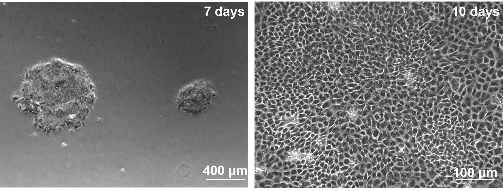
Figure 4. Representative results of successful colony formation in vitro. A. Phase contrast microscopy of DESCs, after plating in culture for 7 days. Tight colony formation indicates successful epithelial isolation with no mesenchymal contamination. Scale bar = 400 μm. B. After 10 days of incubation, colonies are larger in size and cells have a typical epithelial cobblestone morphology. Scale bar = 100 μm. Please click here to view a larger version of this figure.
Epithelial cells were first successfully cultured over 40 years ago21-24, and more recently, successful isolation of epithelial stem cells25-27 has advanced our knowledge of epithelial biology. We report a protocol for isolating the DESCs of the adult mouse incisor, a relatively understudied population of stem cells that has the potential to yield important insights into dental biology and enamel formation. This protocol was initially based on previous reports of epithelial stem cell isolation from the hair follicle28. Whereas many protocols use feeder layers and serum to maintain epithelial stem cells, our method is a feeder free, serum free system. However, our growth media does require the use of a cocktail of EGF, FGF2 and B27 supplement. EGF has long been used to maintain undifferentiated cells 29. A cocktail of EGF and FGF2 along with B27 was used previously to culture hair follicle stem cells27, and B27 is widely used to maintain neural stem cells in culture30,31. We have also previously measured the viability and growth rate characteristics of DESCs atop tissue culture plastic and on a variety of ECM substrates, and no differences were detected in the rates of proliferation19. Cells could be maintained for up to 10 serial passages19.
We use the lower incisors for this procedure because of the ease of removal as compared to the upper incisors. The most important steps within our protocol during isolation are the complete removal of the CL area from the mandibular bone before placing in collagenase and a clean microdissection of the CL. Because the proximal incisor is very delicate, it is important not to damage it during dissection, which could result in loss of the labial CL. Incomplete removal will lead to remnants of the labialCL and a lower yield of DESCs. In addition, it is important to avoid contamination with non-CL epithelial cells. Thus, after the epithelium has been separated from the mesenchyme, a clean V-shaped incision should be made to remove the labial CL from the neighboring epithelium. Finally, it is essential to plate these cells at a concentration of over 1 x 10⁴ cells per ml and leave half of the conditioned media for the first media change.
The major benefit of this protocol to dental research is that it allows efficient production and manipulation of DESCs in vitro. Although the mouse incisor is established as an in vivo model7-9,12,32-34, the development of an in vitro system advances the dental research field by opening the doors to experiments that are difficult to perform in vivo. Advantages of this system include the ability to readily manipulate the cells in various culture conditions, easy targeting of single or even multiple signaling pathways, and inhibition or activation of targeted molecules using siRNA or growth factors. Downstream applications of this in vitro system include using the above listed techniques as well as other cutting edge techniques to conduct functional and/or mechanistic studies, particularly at the single cell level.
Overall, information gleaned from DESC in vitro culture can help to unravel the intricacies of stem cell based tooth renewal.
The authors have nothing to disclose.
The authors thank Xiu-Ping Wang for initial help with DESC cultures. The authors are funded in part by fellowships and grants from the National Institutes of Health (K99-DE022059 to A.H.J., K12-GM081266 to M.G.C., K08-DE022377-02 to O.H., and R01-DE021420 to O.D.K.).
Forceps, size 5 | Fine Science Tools | 11251-30 | FST by Dumont, Dumoxel, |
Forceps Straight, fine, 3.5 | Roboz | RS5070 | |
Razorblades | Electron Microscopy Services | #71960 | |
Scalpel Handle No.3 | VWR | ||
Feather Blade No. 15 | Electron Microscopy Services | #72044-15 | Surgical Stainless Steel |
Collagenase Type-1 filtered | Worthington Biochemical Corporation | #4214 | |
Insulin syringe | BD | #329424 | |
Accumax | EMD Millipore | #SCR006 | |
DMEM/F12 | Gibco | 11320-033 | |
EGF | R&D | 2028-EG-200 | |
FGF2 | R&D | 233-FB-025 | |
B27 Supplement | Gibco | 10889-038 |