Molecular signaling through both estrogen and microRNAs are critical in breast cancer development and growth. Estrogen activates the estrogen receptors, which are transcription factors. Many transcription factors can regulate the expression of microRNAs, and estrogen-regulated microRNAs can be profiled using different large-scale techniques.
Method Article
Profiling of Estrogen-regulated MicroRNAs in Breast Cancer Cells
In This Article
Summary
Abstract
Estrogen plays vital roles in mammary gland development and breast cancer progression. It mediates its function by binding to and activating the estrogen receptors (ERs), ERα, and ERβ. ERα is frequently upregulated in breast cancer and drives the proliferation of breast cancer cells. The ERs function as transcription factors and regulate gene expression. Whereas ERα's regulation of protein-coding genes is well established, its regulation of noncoding microRNA (miRNA) is less explored. miRNAs play a major role in the post-transcriptional regulation of genes, inhibiting their translation or degrading their mRNA. miRNAs can function as oncogenes or tumor suppressors and are also promising biomarkers. Among the miRNA assays available, microarray and quantitative real-time polymerase chain reaction (qPCR) have been extensively used to detect and quantify miRNA levels. To identify miRNAs regulated by estrogen signaling in breast cancer, their expression in ERα-positive breast cancer cell lines were compared before and after estrogen-activation using both the µParaflo-microfluidic microarrays and Dual Labeled Probes-low density arrays. Results were validated using specific qPCR assays, applying both Cyanine dye-based and Dual Labeled Probes-based chemistry. Furthermore, a time-point assay was used to identify regulations over time. Advantages of the miRNA assay approach used in this study is that it enables a fast screening of mature miRNA regulations in numerous samples, even with limited sample amounts. The layout, including the specific conditions for cell culture and estrogen treatment, biological and technical replicates, and large-scale screening followed by in-depth confirmations using separate techniques, ensures a robust detection of miRNA regulations, and eliminates false positives and other artifacts. However, mutated or unknown miRNAs, or regulations at the primary and precursor transcript level, will not be detected. The method presented here represents a thorough investigation of estrogen-mediated miRNA regulation.
Introduction
Estrogen is a hormone that is important during mammary gland development. Estrogen also plays important roles in the development, maintenance, risk, and treatment of breast cancer1. Estrogen exerts its function by binding to ERs, which are transcription factors and regulate specific target genes. Of the two receptor variants, ERα is essential for estrogen-dependent proliferation of breast cancer cells. Most breast cancers are ERα-positive and depends on estrogen for growth. This has made estrogen signaling and ERα a target for treatment in hormone-receptor positive breast cancer. Understanding the underlying mechanism of ERα is important to improve treatment outcomes, overcome resistance to treatment, and understand how breast cancer develops.
miRNAs play critical roles in cell functions due to their major impact in post-transcriptional gene regulation. miRNAs are 19-24 nucleotides short, single-stranded, noncoding RNAs that are first transcribed by RNA polymerase II into primary miRNA transcripts (pri-miRNA). They are processed in the nucleus by Drosha into short-hairpin precursor-miRNAs (pre-miRNA) and then processed by Dicer and separated into single strands to form mature miRNAs in the cytoplasm. The single-stranded mature miRNAs are transferred to Argonaute proteins to form RISC complex. Then, the miRNAs can hybridize to the 3'-untranslated regions (3'-UTR) of a target mRNA, leading to post-transcriptional regulation by blocking translation or degrading the target mRNA2.
Due to the important role that both estrogen and miRNAs play in tumor progression, identifying miRNAs associated with normal or disrupted estrogen signaling is important in order to enhance our understanding of the development and improve treatment of breast cancer. While there is a good understanding of how ERα regulates protein-coding genes, the extension and details of noncoding RNA regulations remains to be thoroughly investigated. Initial studies aiming to elucidate ERα regulation of miRNA in breast cancers cell lines have yielded conflicting results, even when the same cell line has been analyzed3-6. This may be a result of varying treatments, biological variations, the use of different techniques, and the fact that the small size of miRNAs make them challenging to analyze. Here, a protocol that controls for variations and method artifacts is described.
To identify which miRNAs are regulated by estrogen, profiling of a well-defined breast cancer model of ERα activity is a first step. Several cell lines have been generated from human ERα-positive breast cancer tumors, which are dependent on estrogen similar to the majority of clinical breast cancers. The molecular properties of two of these cell lines, T47D and MCF7, including the expression of ERα and its downstream target, the progesterone hormone receptor (PR),a lack of expression of membrane receptor HER2, along with expression of estrogen-responsive and luminal-epithelial differentiation genes, make them suitable as models for the luminal subtype of breast tumors7-10. 17β-estradiol (E2) is the dominant form of estrogen, and the concentrations and time-points for optimal transcriptional activation of ERα have been characterized in multiple studies. In this protocol 10 nM E2 treatment for 24 hr is used and T47D and MCF7 as models for ERα activity in breast cancer cells1. In addition, time-dependent miRNA regulations can be specifically analyzed in a time interval of e.g. 1-72 hr.
Secondly, analyzing miRNA has separate challenges, in part due to their short sizes. miRNAs are not well retained in the total RNA preparation when regular Guanidinium thiocyanate-phenol/chloroform/isopropanol RNA precipitations are performed, or when standard column purifications are used. Special precautions need to be taken to maintain or enrich for the smaller fraction of RNAs. By increasing the volume of isopropanol, lowering the temperature before centrifugation (-80 °C), and omitting the washing in 70% ethanol, the retaining of miRNAs can be enhanced during precipitation. Or, specific columns and buffers for a high-quality and robust preparation of miRNA-containing total RNA can be used. Also for the analysis itself, their short sizes create challenges. For genome-wide miRNA screening, there are three common miRNA profiling techniques to choose from: microarrays, qPCR, and next-generation sequencing. Each technique can be performed using multiple different platforms, and different sample preparations are required, each with different risks of introducing artifacts. In miRNA microarray, a slide is spotted or synthesized with thousands of oligonucleotides. These oligonucleotides are used as probes, and each of the probes is designed to hybridize to a particular miRNA sequence. The sample preparation for microarrays, as performed in this study, first enriches for miRNAs and then introduces Cy5 and Cy3 labeling onto the miRNAs. Microarray gives the opportunity to observe the relative expression levels of a large number of genes simultaneously, is fast and suitable for screening of large number of samples, but can only analyze the sequences present on the microarray and will e.g. not detect changes in unknown or mutated miRNAs. Microarray analysis as performed in this protocol also requires relatively large amounts, about 5 µg, of total RNA per sample for analysis. Low-density qPCR miRNA profiling requires less material (700 ng/sample and replicate), and allows for the detection and quantification of miRNAs. Transcript levels can be determined, and the quantity can be an absolute amount or a relative amount. qPCR analysis first requires conversion of miRNA into cDNA, here by using a looped primer for each specific miRNA ensuring the analysis of only mature miRNA. This generates a longer template that can be amplified utilizing a miRNA-specific forward primer and a universal reverse primer complementary to the looped sequence, and can harbor the inclusion of a Dual Labeled Probe for specificity. qPCR can be used in a low-density format where hundreds of miRNAs can be detected in parallel using one or several 384-well plates with individual primer pairs in each well11. Next-generation sequencing, on the other hand, is the only of these techniques that allows for the discovery of novel, mutated or edited miRNAs, as all RNAs in a sample can be sequenced11. This technique, however, requires multiple steps to enrich small RNAs and produce a small RNA library using several steps of linker ligations and purifications with subsequent enhanced risks of modulating their relative expression levels between samples. It also requires significant bioinformatics analysis. Given the various techniques for miRNA profiling, the most appropriate technique depends on the applications. Microarrays are most suitable when material is relatively abundant and the interest is to define differential expression of already known miRNAs. Low-density qPCR arrays are most suitable when a limited amount of sample is available and a high sensitivity of low-expressed miRNAs is required. Sequencing is most suitable when the analysis of unknown miRNAs, mutated or different isoforms of a miRNA is required.
In the study of estrogen-regulated miRNAs in breast cancer, two model cells lines are used, T47D and MCF7, where large amounts of RNA are readily available. Each cell line was analyzed in replicated cell cultures using different passages, each in technical replicates of treatment. This allows for robust detection of reproducibly, ERα-regulated miRNAs. Relative miRNA expression levels were compared using both miRNA microarray and Dual Labeled Probes - low density arrays (DLP-LDA) and validated the results with specific qPCR using both Cyanine dye and DLP chemistries. miRNA regulations were then further analyzed in time-series to define their exact regulation over time, which can help differentiate random or circadian variations from estrogen-induced regulations, and indicate primary effects from secondary effects. Bioinformatical comparisons with chromatin-binding studies of ERα can further aid in the differentiation of primary versus secondary effects. The assay resulted in a reliable assessment of miRNA regulations where it could be established that after 24 hr E2 treatment protein-coding transcripts were readily regulated but mature miRNAs were not affected1. However, it is possible that miRNAs are regulated at later time-points, as demonstrated in the time-series analysis of selected miRNAs1. The details need to be further explored and the protocol presented here provides a robust way to study hormonal regulation of mature miRNAs in breast cancer cell lines.
Protocol
1. Preparation of Cell Culture Media
- For T47D cell line culture media:
- Mix 500 ml Dulbecco's modified Eagle's medium (DMEM) with 500 ml F12 [DMEM/F12 (1:1)] in an autoclaved 1 L bottle.
- Add 50 ml of fetal bovine serum (this makes 5% FBS), and then 10 ml of penicillin streptomycin (this makes 1% PEST). Mix contents completely.
- Store at 4 °C.
- For MCF7 cell line culture media:
- Mix 500 ml DMEM with 25 ml of the FBS (5% FBS), and then 5 ml of penicillin streptomycin (1% PEST).
- Store at 4 °C.
- For reduced-serum culture media:
- For T47D: Mix 500 ml phenol red-free DMEM with 500 ml F12 (1:1) in an autoclaved 1 L bottle, and add 50 ml of the dextran-coated charcoal-treated (DCC) FBS for 5% DCC-FBS. Make a separate bottle with 5 ml DCC-FBS for 0.5% DCC-FBS. Then, add 10 ml of PEST (1% PEST) to each bottle. Mix contents completely, store at 4 °C.
- For MCF7: Mix 500 ml phenol red-free DMEM with 5 ml of 100x L-glutamine (200 mM final concentration). Add 25 ml of DCC-FBS (5% DCC-FBS). Make a separate bottle with 2.5 ml DCC-FBS (0.5% DCC-FBS). Then, add 5 ml of PEST (1% PEST) to each bottle. Mix contents completely, store at 4 °C.
- For phosphate-buffered saline (PBS) solution:
- Fill a 1 L autoclaved glass bottle with ultrapure water or distilled water.
- Add one PBS tablet and shake occasionally until tablet has dissolved.
- Autoclave PBS solution and store at room temperature.
- Preparation of ligand stocks:
- Prepare a 100 mM stock solution of E2 by dissolving 2.72 mg of 17β-estradiol in 100 µl ethanol or DMSO in a sterile 1.5 ml microcentrifuge tube, and mix contents gently. Spin briefly and make serial dilutions (1:10) to yield 10 mM, 1 mM, and 0.1 mM stock concentrations using the solvent. Store all E2 stock solutions at -20 °C.
- Prepare a 100 mM stock solution of ICI by dissolving 6.07 mg of ICI 182,780 in 100 µl EtOH or DMSO in a sterile 1.5 ml microcentrifuge tube, and mix contents gently. Spin briefly and make serial dilutions (1:10) to yield 10 mM, 1 mM, 0.1 mM stock concentrations using the solvent. Store all ICI stock solutions at -20 °C.
- The vehicle is the solvent (ethanol or DMSO). Place 0.5-1 ml of 100% ethanol or DMSO in a sterile 1.5 ml microcentrifuge tube, and store at -20 °C.
2. Cell Culture and Treatment
Perform each treatment in double or triplicate plates for technical replicates. Repeat cell culture procedure and treatment using a different passage of the same cell line, which would serve as a biological replicate of this cell line. Repeat the procedure with a different cell line to replicate findings in more than one cell line. All cell culture techniques should be performed under sterile conditions in a laminar flow hood.
- Cell culture startup
- Warm the media to 37 °C in a sterile warm water bath.
- Thaw a frozen vial of T47D or MCF7 cells.
- Clean the outside of vial and media bottle with 70% ethanol, and then place both vial and bottle in sterile laminar flow hood.
- Label a sterile T-75 flask accordingly (in the hood).
- Add 12-15 ml of appropriate media into the flask using a sterile serological pipette (ensure that surface is completely covered with media).
- Transfer the cells from the vial to the flask, and mix gently.
- Place flask in a 37 °C incubator, supplied with 5% CO2.
- Preparation of cells for treatment
- Take cells out of the incubator and observe under a microscope to ensure that the cells are at least 80% confluent. This is to ensure that there are enough cells for experiment. Transfer flask to sterile hood.
- Remove media from flask using a sterile Pasteur pipette connected to a vacuum. Gently wash attached cells twice with PBS.
- Add 1 ml of warm trypsin-EDTA to the flask, and put flask in the incubator for 2 min. This is to make the cells detach from the flask.
- Add about 3-4 ml of appropriate warm media to the flask and triturate the detached cells using a 5 ml serological pipette. Triturate by taking up the cells in the serological pipette and releasing the cells with the tip of the pipette placed against the bottom of the flask to increase pressure. This is to break the cells apart into single cells.
- Count the cells, e.g. using a cell counter, according to manufacturer's protocol.
- Label 100 mm plates (as needed) and add about 10 ml media to the plates. Plate about 2.0 x 10⁶ cells/plate. Distribute cells by gentle swirling the plates.
- Incubate cells for 24-48 hr, until approximately 80% confluent.
- At 80% confluency, wash cells twice with PBS, and add the appropriate 5% DCC-FBS media. Then, incubate cells for 24 hr.
- After 24 hr, wash cells twice with PBS, and add the appropriate 0.5% DCC-FBS media. Then, incubate cells for additional 48 hr.
- Cell treatment
- After 48 hr, wash cells twice with PBS. Be sure to remove as much of the PBS as possible.
- In a 15 ml conical tube, add 10 ml of the appropriate 0.5% DCC-FBS media. Then, add 1 µl of the 0.1 mM ligand stock (E2 or ICI) for a final concentration of 10 nM. Also, add 1 µl of vehicle to 10 ml media in a separate tube for the control experiment.
- Mix contents in tube gently and add to cells in the plate. This should be one ligand per plate.
- Incubate cells for the chosen time point (0-72 hr).
3. RNA Extraction and Quality Control
- RNA extraction
- After treatment is finished for the desired period of time, wash cells twice with PBS, then add about 1-2 ml Guanidinium thiocyanate-phenol solution to the cells in the plate. CAUTION: Guanidinium thiocyanate-phenol solution is toxic by contact with skin or eyes, by inhalation, or if swallowed. Wear suitable protective clothing, gloves, and eye/face protection, and use fume hood.
- Ensure that the volume of cells is no more than 10% of the volume of Guanidinium thiocyanate-phenol solution, and that the plate is covered with the solution and allow to sit for 1 min. Then, scrape cells using a rubber scraper and transfer to a microcentrifuge tube. Cells can be stored in this solution at -80 °C for weeks.
- Extract and purify total RNA from each treatment using the guanidinium thiocyanate-phenol-chloroform method followed by spin column purification and DNase I DNA degradation method for animal cells.
- After extraction, cells are eluted in 60 µl RNase-free water. Aliquot 1-2 µl RNA for quantification analysis and store RNA at -80 °C.
- Measure the RNA concentrations using 1 µl of RNA and aspectrophotometer. Ensure that there is a blank taken before any measurement is done. Also, completely wipe the spectrophotometer reach time a measurement is taken. Concentrations are usually shown in ng/µl. Save results showing RNA concentrations.
- RNA quality control
- Take about 100 ng RNA of each sample and place in a microcentrifuge tube.
- Measure RNA integrity using a bioanalyzer. Usually 12 samples can be run at one time in a bioanalyzer. Run usually takes about 25 min.
- Compare results with the RNA ladder to ensure that the RNA is good for experiment. Save and print results.
4. Confirmation of Treatment: qPCR of Protein-coding Genes
- cDNA synthesis
- Take 500 ng or 1 µg of the total RNA (of each sample) and put in a 1.5 ml microcentrifuge tube. Bring the volume of each tube up to 10 µl using RNAse-free water.
- Add 2 µl of 50 µM random hexamer primers and heat tubes to 70 °C for 10 min.
- Transfer tubes to ice for 5min.
- For each sample, add 4 µl of 5x first strand buffer, 1 µl of 0.1 M DTT, 1 µl of 10 mM dNTPs, 0.5 µl of superscript III, and 1.5 µl RNase-free water.
- Place tubes in a 25 °C heat block or water bath for 10 min.
- Transfer tubes to a 46 °C heat block for 1 hr. Then, transfer tubes to a 70 °C heat block for 15 min to stop the reaction.
- Dilute the cDNA to a 5 ng/µl stock (calculated using total RNA input) using RNase-free water for dilutions and store at -20 °C.
- qPCR
- Obtain primers for genes of interest and reference genes. Primers can easily be designed using the primer designing tool on NCBI's Primer-BLAST program12. This usually generates a forward and reverse primer for each gene of interest. Primers should be 18-22 bp in length, have a melting temperature of 52-58 °C, have a GC content of 40-60%, and an amplicon length of 100-180 bp.
- For each qPCR reaction well, add 10 ng cDNA (2 µl from stock concentration from step 4.1.7), 1 pmol of each of the forward and reverse primers of the gene of interest (from step 4.2.1), and 5 µl of 2x Cyanine dye PCR master mix. The reaction for each well should have a 10 µl final reaction volume.
- Ensure that there are triplicate wells for each sample for each gene (for technical replicates). A 96-well reaction plate can be used.
- On the qRT-PCR system software, assign the reporter and target, enter reaction volume, select the comparative threshold cycle (ΔΔCt) method, and define sample wells.
- Make sure to perform melting curve analysis for all Cyanine dye runs to confirm the amplification of one specific fragment. Run plates using the default settings for the run.
- Save results after run, and export data (especially the CT values to MS Excel).
- qPCR data analysis using the ΔΔCT formula
The change in relative mRNA expression can be calculated as fold change relative to control on Excel for each sample using the following steps:- All exported results from the qPCR above should have the Ct values included. Calculate the ΔCT value using the formula below:
- ΔCT = CT (target gene) - CT (reference gene)
- Next, calculate the total standard deviation (SD) for each sample. First calculate the SD for the reference gene and then calculate the SD for the target gene (this can be done on excel using the STD DEV function on the CT values). Then calculate the total SD for each sample using this formula:
- Total SD = (SD2 (target) + SD2(reference))1/2
- Calculate the ΔΔCT of each sample within a gene (target) by:
- ΔΔCT = ΔΔCT (treated/test sample) - ΔΔCT(control/calibrator sample)
- Finally, calculate the fold-change (FC) values of each sample using the formula:
- FC = 2-ΔΔCT
- Student's t-test can be used for statistical analysis by using two-tailed distribution and two-sample unequal variance parameters: (P<0.001 (***), P<0.01 (**), and P<0.05 (*)). This can be achieved on Excel. Confirm that the treatment resulted in regulation of known targets before proceeding with miRNA profiling.
- All exported results from the qPCR above should have the Ct values included. Calculate the ΔCT value using the formula below:
5. miRNA Profiling Analysis
- miRNA microarray
- Take 5 µg of isolated RNA from cells treated with either vehicle or ligand, and place in a microcentrifuge tube. Adjust volume in tube to 20 µl. Each comparison should be performed in duplicates or triplicates.
- Perform the miRNA microarray using the RNA samples above. miRNA microarray expression profiles should be determined using human miRNA microarray dual-color sample array by µParaflo Microfluidic Biochip Technology (Sanger miRBase Release 14.0)13.
- Results should show differentially expressed miRNAs. Consider significant miRNA expressions when p<0.05.p-value <0.10 can also be considered for further confirmatory analysis using qPCR. Save this miRNA list for further validation with qPCR.
- miRNA profiling: DLP-LDA
- Each sample should be analysed in duplicates or triplicates. Take 700 ng of total RNA from cells treated with either vehicle or ligand, and place in a microcentrifuge tube. Adjust volume in tube to 3 µl.
- Perform the cDNA synthesis using the Dual Labeled Probes miRNA assay method and the 10x RT primers. Total volume for each reaction should be 7.5 µl.
- Place reaction tube in the PCR system Thermocycler, and set to recommended parameters as indicated in Dual Labeled Probes miRNA assay method. Start the run. This will generate cDNA. Note that the cDNA can be stored at -20 °C for at least 1 week.
- While RT reaction is running, take out the DLP-LDA plates and allow to sit at room temperature. Each array plate contains 384 wells that contain unique miRNA primers and control primers.
- Take 6 µl of the cDNA (from 5.2.3 ) and place in a 1.5 ml microcentrifuge tube. Add 450 µl of the Dual Labeled Probes 2x Universal PCR Master mix and add 444 µl RNase-free water.
- Invert tube about six times to mix contents, and centrifuge briefly.
- When the DLP-LDA plate has reached room temperature, load 100 µl into each of the eight 'Fill port' of the plate. Then, centrifuge the array plate.
- Open the qRT-PCR system and check that the block is that for the 384-well plate. Set up run using the SDS software. Select relative quantification (Ct), select the well number and array type, and enter sample description to define wells.
- Run array using the default thermal cycling conditions. When run is finished, save results and export to MS Excel and save for further analysis using the ΔΔCT formula (step 4.3).
6. Confirmation of miRNA Regulations using Separate qPCR Analysis
- Cyanine dye qPCR analysis
- Design primers for desired miRNA analysis. Sequence of the mature miRNA, which equals the forward primer, can be obtained from mirbase.org14. Convert all 'U' to 'T'.
- Prepare cDNA: Take 1 µg of total RNA and place in a 1.5 ml centrifuge tube. Follow the instructions for polyA tailing and first-strand cDNA synthesis of miRNA from a miRNA first-strand cDNA Synthesis and qRT-PCR protocol. Dilute the resulting cDNA (1:10) and store at -20 °C.
- Obtain one 96-well reaction plate.
- For each miRNA qPCR reaction well, add 16 ng of polyA cDNA (from step 6.1.2), 2 pmol of each of the specific forward primer (from step 6.1.1) and the universal primer (from the kit from step 6.1.2), and 5 µl of 2x qPCR master mix. Final reaction volume is 10 µl/well.
- Ensure that there are triplicate wells for each sample for each miRNA (for technical replicates). Also, make sure that a reference gene (usually U6 snRNA) is included.
- On the qRT-PCR system software, assign the reporter and target, enter reaction volume, select the comparative threshold cycle (ΔΔCT) method, and define sample wells.
- Run plates using the default settings for the run. Make sure to perform melting curve analysis for all Cyanine dye runs. Each melting curve should show only one specific peak to confirm the amplification of one specific fragment. If more than one peak or no clear peak is observed, the primers are not specific and the data cannot be used.
- Save results after run, and export data (especially the CT values to MS Excel).
- Dual Labeled Probes qPCR analysis
- Take 10 ng of each total RNA, each in a 5 µl volume and place in 0.2 ml microcentrifuge tube.
- Follow instructions from the protocol on performing the reverse transcription (RT) using the Dual Labeled Probes MicroRNA Reverse Transcriptase Kit15. Each total reaction volume should be 15 µl. Note that for the Dual Labeled Probes RT reactions, there are specific primers for each miRNA to be studied.
- Place reaction tube in the PCR system thermocycler, and set parameters according to the protocol indicated in step 6.2.2 above. Start the run. It should take about 70-80 min.
- After the RT reaction, dilute the sample cDNA in a 1:5 ratio, and store all cDNA in the dark at -20 °C.
- Obtain one 96-well reaction plate.
- For each miRNA qPCR reaction well, add the following: 10 µl of Dual Labeled Probes 2x Universal PCR Master Mix (same reagent as step 5.2.5), 7.67 µl of RNase-free water, 1 µl of 20× real-time assay primer (specific for each miRNA), and 1.33 µl of the cDNA from step 6.2.4 above. This should be a 20 µl final reaction volume. Mix contents gently, and centrifuge briefly.
- Ensure that there are triplicate wells for each sample for each miRNA (for technical replicates). Also, make sure that a reference gene (usually U6 snRNA) is included.
- On the qRT-PCR system software, assign the reporter (FAM) and quencher (NFQ-MGB) for each miRNA (target) of interest, enter reaction volume, select the comparative threshold cycle (ΔΔCt) method, and define sample wells.
- Follow the instructions for setting up the parameters for the qPCR run from the Dual Labeled Probes microRNA assay protocol. Then, start the run. Save results after run, and export data (especially the CT values to MS Excel).
Results
An overview of the approach is presented in Figure 1 and a comparison of the various sample preparations and nucleic acid modifications required for each technique is visualized in Figure 2.
The condition and environment of the cells before treatment with a hormone is important in determining the actual effects of the hormone16. Some medium and serum contain growth factors that may affect results, so cells are serum starved to ensure that all hormonal effects have been reduced to a minimum. Hence, when treating with E2, there is more certainty that the effects observed are E2-associated. An important factor is the confluency of cells, which influences cell-cell contact and behaviors such as cell-cell signaling and proliferation. Cells were allowed to be 80% confluent (Figure 3A) and well attached to allow growth and avoid loss of cells during subsequent washing and media change.
The conditions during RNA extraction and storage are important for the quality of RNA and subsequent analysis. It is also known that the number of cells, the extraction method, and the GC content of the miRNA can affect the yield and quality of both mRNAs and miRNAs17. Thus, it is necessary to perform RNA extraction during RNase-free conditions and to check the quality of the RNA before proceeding with experiments. After extraction, quantification of the RNA provides information on the concentration of the total RNA (containing both mRNAs and miRNAs), which enables observation of yield. It also provides a graphic representation of results as seen in Figure 3B, and this allows for the observation of the purity of the sample (usually indicated by one peak, top panel). Poor yield of RNA would produce no specific absorption at 260 nm (Figure 3B, bottom panel). To determine whether the RNA is of high quality, RNA integrity was measured. A RNA integrity number (RIN) of between 9-10 ensures that the RNA is not degraded and is of optimal quality. Also, the graphical representation of the RNA should be observed, and the 18S and 28S rRNA should have peaks as shown in Figure 4 (middle panel). Comparison with the ladder (Figure 4, top panel) identifies the size of the peaks. A degraded RNA would have less clear peaks and a reduced relative amount of the 18S and 28S rRNA (Figure 4, bottom panel). The fraction of small RNAs can further be ensured using the Agilent Small RNA Kit. It is recommended to validate estrogen response after treatment, under the specified experimental conditions, before proceeding to miRNA screening. A qPCR can be performed on a known ER target gene such as pS2 or SPINK4 18, using cDNA template. Such genes are usually upregulated within 1-24 hr after E2 treatment. Once the quality of the RNA and the estrogen response has been assessed, further experiment can be conducted.
Microarray is still a widely used technique for large-scale screening for miRNA expression in cells. For example, the miRNA microarray once used contained 894 mature miRNAs and 50 controls unique probes in quadruplets1. Hybridizations were performed in duplicates with dye swap procedure. The miRNA microarray results are usually graphically represented by heat maps. Figure 5A shows a heat map, representing data from a miRNA microarray comparison between the vehicle and E2 treated samples. The red indicates that the miRNA is upregulated in the E2-treated sample (higher expression), while the green indicated downregulation of the miRNA (lower expression). The miRNA selection usually depends on their significance in expression, and usually a p-value less than 0.05 is considered significant. The miRNAs that are indicated to be differentially expressed should be further validated with qPCR, to distinguish them from false positives.
The DLP-LDA array, on the other hand, uses a qPCR-based method to screen for regulated miRNAs in a 384-well format. Usually two 384-well plates (cards) are provided, pools A and B. Pool A usually contains better characterized and more highly expressed miRNAs than the pool B. The relative level of each miRNA are determined by qPCR, where one miRNA is analyzed per well (Figure 5B). A higher Ct-value indicates lesser miRNA expression. Much like the miRNA microarray, results attained from the DLP-LDA need to be further validated to ensure accuracy.
Validations can be performed using qPCR. Here, two qPCR detection methods are described to prevent method bias and to allow for thorough confirmation and investigation of miRNA regulations. The Cyanine dye-based detection chemistry works different than DLP that the DLP-LDA profiling is based on, and is suitable for confirmation purposes. It can be less specific as it detects all amplified double-stranded DNA, but the homogeneity of the sample can be assessed by performing a melting-curve analysis. Figure 6A (left panel) shows the melting-curve analysis of a miRNA, and a clearly defined uniform peak can be seen. Multiple peaks would be observed if the primer is nonspecific or significant amounts of primer-dimer is formed (Figure 6A, right panel). DLP miRNA assays on the other hand, is more specific as only amplification of the target miRNA is detected. Amplification plots of each target miRNA can be observed, as exemplified in Figure 6B. The option to control for the occurrence of multiple amplification products using the melting curve analysis is, however, not available with this chemistry and Cyanine dye qPCR is less expensive. Cyanine dye and DLP qPCR assays can sometimes generated slightly different results. For both qPCR assays, comparison to a reference gene is required to determine the relative expression of genes between two samples. The reference gene, also referred to as housekeeping gene, should be a gene whose expression is not changed and which is expressed at similar levels as the gene of interest. An example of a suitable reference gene in these breast cancer cell lines is ARHGDIA, a Rho GDP-Dissociation Inhibitor that functions GDP/GTP exchange reactions of the Rho proteins. As observed in Figure 6C (top), the mRNA level of ARHGDIA is not changed between the vehicle and the ligand-treated samples. The 18S rRNA can also be used, albeit its high expression requires a separate 1:500 dilution of the cDNA stock and is therefore less appropriate. For miRNA analysis, there is still a debate on a well-defined reference gene. So far, the U6 snRNA is a reference gene that is widely accepted for miRNA analysis. U6 snRNA is the ~110 nucleotide long, noncoding small nuclear RNA that function in nuclear pre-mRNA splicing19. As observed in Figure 6C (bottom), U6 expression levels are about the same across all samples shown, thus allowing for calculations of the variable levels of miRNA. A graphic representation of a miRNA qPCR result for a comparison between the vehicle and E2-treated cells that have been normalized to the reference U6 can be observed in Figure 6D. This illustrates a miRNA that were detected as nonregulated after 24 hr E2 treatment, but which harbors ER chromatin-binding sites close to its genomic location, and time-series analysis identified significant regulation 72 hr after treatment.

Figure 1. An overview of the approach employed for detection of hormonal regulation of miRNA in breast cancer cells. Process begins with cell culture and treatment, followed by large-scale miRNA profiling to identify possibly differentially expressed miRNAs, and ends with qRT-PCR validations of array results.
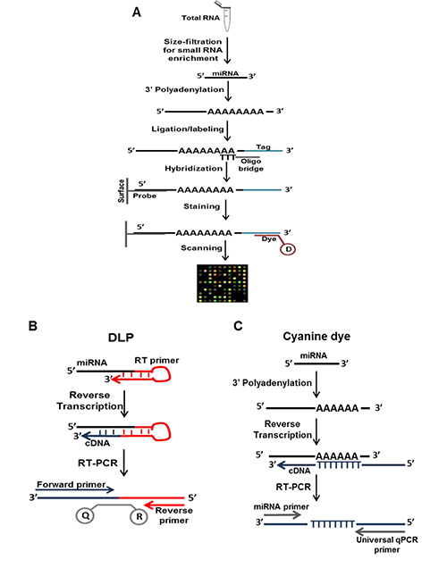
Figure 2. Comparison of the various sample preparations and nucleic acid manipulations for each miRNA detection technique. (A) miRNA microarray using the µParaflo technology method includes enrichment, poly-A tailing and ligation labeling. (B) DLP technology sample preparation and detection method includes a looped cDNA synthesis primer which extends after the denaturation step and provides a longer fragment harboring the reverse primer and probe sequence. (C) Cyanine dye technology sample preparation and detection method includes poly A-tailing, and cDNA synthesis with a oligo-dT primers including a universal 5' sequence.
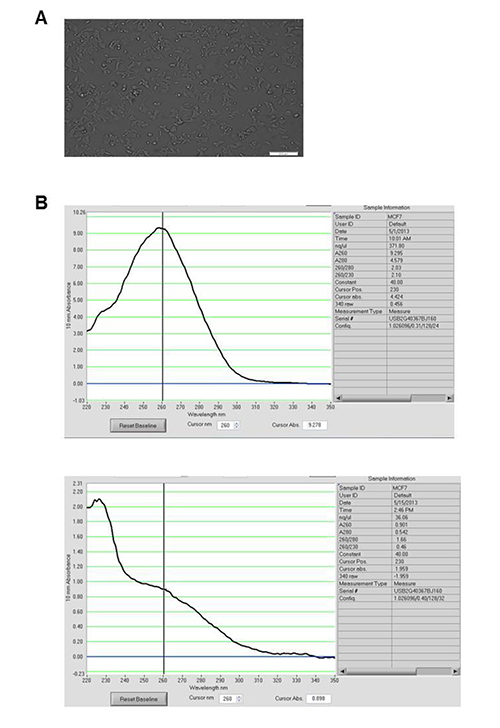
Figure 3. Cell culture and RNA extraction. (A) Cultured cells to illustrate 80% confluency needed before treatment with ligand. (B) Graphical representation of RNA concentration and purity after extraction. Each peak represents a sample and a successful extraction process yields pure RNA (top panel). A poorly extracted RNA shows no clear absorption peak at 260 nm (bottom panel).

Figure 4. RNA quality control. Results from RNA integrity analysis showing graphical representation for the ladder (top), a good quality RNA (middle) and a degraded RNA sample (bottom).
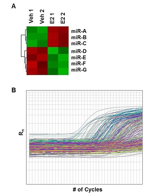
Figure 5. An illustration of miRNA profiling results. (A) A heat map representation of miRNA microarray results for differentially expressed miRNAs. miR-A, B, C are upregulated in the E2 treated-sample as indicated in red, while miR-D, E, F, G are downregulated in the E2-treated sample as indicated in green. (B) Amplification plots for each miRNA from the DLP-LDA results. miRNA detected are amplified as indicated by increase in the Rn value.
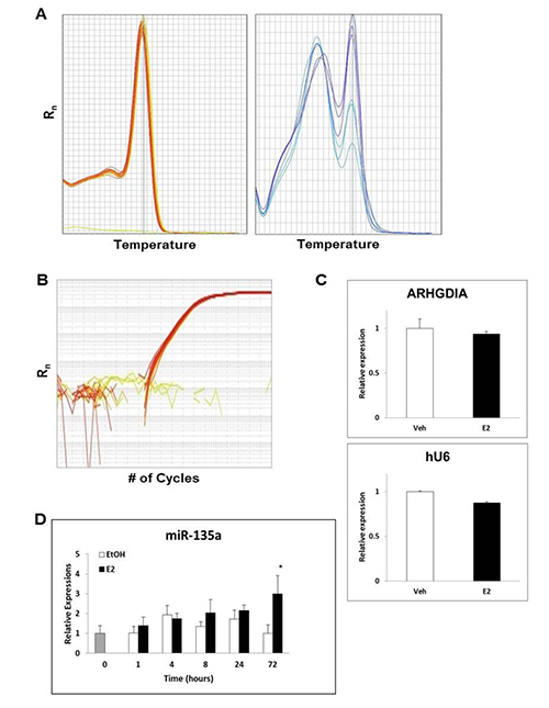
Figure 6. qPCR analysis of miRNA regulations. (A) Melting-curve analysis for a miRNA analyzed using Cyanine dye detection, showing homogeneity of all samples tested (left) and amplification of multiple fragments (right). (B) Amplification plots for each sample for a miRNA. This can be either from Cyanine dye or DLP detection methods. (C) Bar graph representation of suitable reference genes for mRNA analysis ARHGDIA (top)and for miRNA analysis snRNA U6 (bottom), showing no significant differential expression between samples. (D) miRNA qPCR results to illustrate comparison of relative expression levels of miR-135a over a time course. Figure 6D is reprinted from Katchy et al.1 with permission from Elsevier. Values are usually the average of separate experiments (biological duplicates) ±SD. Student's t-test is used to demonstrate significance: * p<0.05.
Discussion
Determining the mechanism for hormonal regulation of miRNA in breast cancer cells can provide a platform for understanding this disease and can provide potential treatment for breast cancer. Culturing these cells to provide the optimal condition for hormone action is very important. Here, a protocol that ensures that the hormone of interest (estrogen) was excluded before treatment and that an optimal dose of E2 was used for an appropriate time during treatment has been described. Other hormonal and growth factor effects were kept at a minimum by gradually lowering the serum levels and using DCC-FBS, which is FBS that have been stripped of most of its hormone, cytokines, and growth factors. Phenol red-free media was further used to minimize other nonhormonal effects, given that phenol red has been reported to have estrogenic activity and affect certain functions in cell lines20,21. The time of exposure of the cells to the hormone is of great importance. For estrogen-associated regulation of genes, it has been previously shown that 24-hrexposure produce significant response of direct target genes in breast cancer cells1,18. Since miRNAs are transcribed by RNA polymerase II, like mRNAs, it is conceivable that this is a suitable time point to detect direct regulation also of miRNAs. It is yet to be determined if and how these miRNAs affect the expression of these known ER targets in breast cancer cells.
miRNA expression profiling can be challenging because of the relative small size of the miRNA, the fact that the mature miRNA sequence can be found also in the pri-miRNA and the pre-miRNA, and because of heterogeneity in their GC content22. Several profiling techniques and chemistries have been employed to overcome this difficulty. Among these are different approaches of microarray and qPCR analysis (Figure 2). Although microarray is widely accepted for whole genome analysis, it can provide false negatives and there exist differences between the available platforms, ranging from the chemistry to the printing technology23. Also the normalization process presents a challenge when analyzing the relatively few miRNA genes, which are counted in thousands compared to the tens of thousands of mRNA transcripts. qPCR, on the other hand, is more sensitive, and is better applied when fewer genes are to be considered. However, when performed in large 384-well plates, with only one technical replicate per plate, multiple plates need to be analyzed for each sample which makes the analysis costly. Also, in our hands, many more false negative results were obtained using the DLP-LDA analysis compared to the microarray. Given that the mature miRNA sequence is present in the pri-miRNA and the pre-miRNA sequences, it is of interest to differentiate between these transcripts using PCR techniques24. DLP probes are available also for pre-miRNAs, and specific primers can also be designed to exclude different mature or precursor variants using Cyanine dye qPCR technology.
qPCR is a golden standard for validation of differential expression from profiling results. The effectiveness of the qPCR, however, depends on several parameters including RNA extraction, RNA integrity (quality), cDNA synthesis, primer design, detection method (chemistry), and the reference gene for data normalization1,22,25. The options for primer design for miRNA analysis is very limited, since the short miRNA sequence length does not give room for much alternative primer design, and only one primer can be harbored in this short sequence. Hence, the need for alternative manipulations as illustrated in Figure 2. Technical replicates are important to validate robustness of the amplification method and the process employed. Biological replicates are required for a representation of the general biological variation, including variable response to ligand treatment, and to show that effects seen in a cell are reproducible. Based on our experience, variations in miRNA expression between cell cultures can occur. Usually these differences are less than 1.3-fold, and the average of values and corresponding SD identifies the natural and technological variation. This variation could result from various factors including, cell density and the fact that cell functions could change from passage to passage. To identify statistically significant expressions resulting from ligand treatment, a widely accepted significance is when the p-value is less than 0.05. Here, a student's t-test on the biological and technical replicates using the two-tailed distribution and two-sample unequal variance parameters have been used. Other t-test parameters exist, and the choice depends on the experimental setup26.
A detailed protocol in evaluating hormonal regulations of mature miRNA in breast cancer cells has been provided. Important technologies and chemistry in profiling and validating these miRNA expressions have been clearly explained. The technology chosen for studies of miRNA in breast cancer cells ultimately depends on what exactly is being investigated.
Disclosures
The authors have nothing to disclose.
Acknowledgements
We are grateful to Dr. Xiaolian Gao, University of Houston, for providing advice to the LC Sciences miRNA microarray platform, to Dr. Karin Edvardsson, Karolinska Institute, Sweden, and Dr. Eylem Aydogdu, University of Houston, for sharing their miRNA expertise. Research reported in this publication was supported by the National Cancer Institute of the National Institutes of Health under Award Number R01CA172437. The content is solely the responsibility of the authors and does not necessarily represent the official views of the National Institutes of Health. This work was also supported by grants from the Texas Emerging Technology Fund, under Agreement no. 300-9-1958.
Materials
| Name | Company | Catalog Number | Comments |
|---|---|---|---|
| DMEM | Life Technologies | 11885092 | |
| F12 | Life Technologies | 11765054 | |
| FBS | Sigma-Aldrich | F0926-500ML | |
| Penicillin Streptomycin | Life Technologies | 15140122 | |
| Phenol red-free DMEM | Life Technologies | 11054020 | |
| Ultrapure Water | EMD Millipore | Specific equipment | |
| DCC-FBS | Sigma-Aldrich | F6765-500ML | |
| 100x L-glutamine | Life Technologies | 25030081 | |
| PBS tablet | Medicago, Accurate Chemicals | Accurate (BF092051-100), Medicago (09-9400-100) | |
| 17β-estradiol | Sigma-Aldrich | E2758 | |
| ICI 182,780 | Sigma-Aldrich | I4409 | |
| Ethanol | Sigma-Aldrich | E7023-600ML | |
| T47D or MCF7 cells lines | American Type Culture Collection (ATCC) | T47D (ATCC HTB-133) and MCF7 (ATCC HTB-22) | |
| T-75 flask | VWR International LLC | BD353136 (vented cap) | |
| Trypsin-EDTA | Life Technologies | 25300062 | |
| Serological pipet | VWR International LLC | 53300. For 5mL (53300-421) | |
| Countess Cell Counting chamber | Life Technologies | C10228 | |
| Countess Automated Cell Counter | Life Technologies | C10227 | |
| 100 mm plates | VWR International LLC | 25382-166 | |
| Guanidinium thiocyanate-Phenol solution (TRizol) | Life Technologies | 15596026 | |
| Rubber cell scraper | VWR International LLC | 82050-470 | |
| Guanidinium thiocyanate-Phenol-Chloroform method (miRNeasy Mini Kit/Trizol) | Qiagen | 217004 | |
| Rnase-Free DNase I kit (DNA degradation) | Qiagen | 79254 | |
| RNase-free water (Nuclease-free water) | Thermoscientific | SH30538.02 | |
| Nanodrop 1000 Spectrophotometer | Thermoscientific | ND-1000 | |
| Agilent RNA 6000 Nano Assay kit | Agilent | 5067-1512 | |
| Agilent 2100 Bioanalyzer | Agilent | G2938C and for software (G2946) | |
| All primers (except TaqMan primers) | Integrated DNA Technologies | Specific per order for each gene/miRNA | |
| First strand buffer, DTT, dNTPs, superscript III (Superscript III cDNA synthesis kit) | Life Technologies | 18080085 | |
| MicroAmp Fast 96-well reaction plate | Life Technologies | 4346906 | |
| Fast Optical Adhesive Covers | Life Technologies | 4311971 | |
| 2x SYBR green PCR master mix (Cyanine dye) | Life Technologies | 4385612 | |
| 7500 Fast Real-Time PCR System and software | Life Technologies | 4351106 | |
| μParaflo Microfluidic Biochip Technology | LCSciences | MRA-1001 and AS-2001(for result analysis) | |
| DLP-LDA (TLDA) Megaplex Pools Assay (Rt primers/Human pool set) | Life Technologies | 4400928 | |
| Dual Labeled Probes (TaqMan) miRNA RT kit | Life Technologies | 4366596 | |
| GeneAmp PCR System 9700 thermocycler | Life Technologies | 4310899 | |
| Dual Labeled Probes (TaqMan) 2x Universal PCR Master Mix-No AmpErase UNG | Life Technologies | 4324018 | |
| 7900HT Fast Real-Time PCR system | Life Technologies | 4329001 | |
| SDS and RQ manager softwares | Life Technologies | 4444202 | |
| NCode miRNA First-Strand cDNA Synthesis and qRT-PCR Kit | Life Technologies | MIRC-50 | |
| 10x DPL (TaqMan) RT primers | Life Technologies | Specific for each miRNA | |
| 20x DPL (TaqMan) real-time assay primer | Life Technologies | Specific for each miRNA |
References
- Katchy, A., Edvardsson, K., Aydogdu, E., Williams, C. Estradiol-activated estrogen receptor alpha does not regulate mature microRNAs in T47D breast cancer cells. J. Steroid Biochem. Mol Biol. 128 (3-5), 145-153 (2012).
- Bartel, D. P. MicroRNAs: genomics, biogenesis, mechanism, and function. Cell. 116 (2), 281-297 (2004).
- Wickramasinghe, N. S., Manavalan, T. T., Dougherty, S. M., Riggs, K. A., Li, Y., Klinge, C. M. Estradiol downregulates miR-21 expression and increases miR-21 target gene expression in MCF-7 breast cancer cells. Nucleic Acids Res. 37 (8), 2584-2595 (2009).
- Bhat-Nakshatri, P., Wang, G., Collins, N. R., Thomson, M. J., Geistlinger, T. R., Carroll, J. S., Brown, M., Hammond, S., Srour, E. F., Liu, Y., Nakshatri, H. Estradiol-regulated microRNAs control estradiol response in breast cancer cells. Nucleic Acids Res. 37 (14), 4850-4861 (2009).
- Klinge, C. M. miRNAs and estrogen action. Trends Endocrinol. Metab. 23 (5), 223-233 (2012).
- Gupta, A., Caffrey, E., Callagy, G., Gupta, S. Oestrogen-dependent regulation of miRNA biogenesis: many ways to skin the cat. Biochem. Soc. Trans. 40 (4), 752-758 (2012).
- Kao, J., et al. Molecular profiling of breast cancer cell lines defines relevant tumor models and provides a resource for cancer gene discovery. PLoS One. 4 (7), e6146(2009).
- Lacroix, M., Leclercq, G. Relevance of breast cancer cell lines as models for breast tumours: an update. Breast Cancer Res. Treat. 83 (3), 249-289 (2004).
- Ross, D. T., Perou, C. M. A comparison of gene expression signatures from breast tumors and breast tissue derived cell lines. Dis. Markers. 17 (2), 99-109 (2001).
- Neve, R. M., et al. A collection of breast cancer cell lines for the study of functionally distinct cancer subtypes. Cancer Cell. 10 (6), 515-527 (2006).
- Baker, M. MicroRNA profiling: separating signal from noise. Nat. Methods. 7 (9), 687-692 (2010).
- Ye, J., Coulouris, G., Zaretskaya, I., Cutcutache, I., Rozen, S., Madden, T. L. Primer-BLAST: a tool to design target-specific primers for polymerase chain reaction. BMC Bioinformatics. 13, 134(2012).
- Zhou, X., et al. MicroRNA profiling using microParaflo microfluidic array technology. Methods Mol. Biol. 822, 153-182 (2012).
- Griffiths-Jones, S., Grocock, R. J., van Dongen, S., Bateman, A., Enright, A. J. miRBase: microRNA sequences, targets and gene nomenclature. Nucleic Acids Res. 34, 140-144 (2006).
- Chen, C., et al. Real-time quantification of microRNAs by stem-loop RT-PCR. Nucleic Acids Res. 33 (20), e179(2005).
- Ruedl, C., Cappelletti, V., Coradini, D., Granata, G., Di Fronzo, G. Influence of culture conditions on the estrogenic cell growth stimulation of human breast cancer cells. J. Steroid Biochem. Mol. Biol. 37 (2), 195-200 (1990).
- Kim, Y. K., Yeo, J., Kim, B., Ha, M., Kim, V. N. Short structured RNAs with low GC content are selectively lost during extraction from a small number of cells. Mol. Cell. 46 (6), 893-895 (2012).
- Williams, C., Edvardsson, K., Lewandowski, S. A., Strom, A., Gustafsson, J. A. A genome-wide study of the repressive effects of estrogen receptor beta on estrogen receptor alpha signaling in breast cancer cells. Oncogene. 27 (7), 1019-1032 (2008).
- Yu, Y. T., Maroney, P. A., Darzynkiwicz, E., Nilsen, T. W. U6 snRNA function in nuclear pre-mRNA splicing: a phosphorothioate interference analysis of the U6 phosphate backbone. RNA. 1 (1), 46-54 (1995).
- Wesierska-Gadek, J., Schreiner, T., Maurer, M., Waringer, A., Ranftler, C. Phenol red in the culture medium strongly affects the susceptibility of human MCF-7 cells to roscovitine. Cell Mol. Biol. Lett. 12 (2), 280-293 (2007).
- Welshons, W. V., Wolf, M. F., Murphy, C. S., Jordan, V. C. Estrogenic activity of phenol red. Mol. Cell. Endocrinol. 57 (3), 169-178 (1988).
- Benes, V., Castoldi, M. Expression profiling of microRNA using real-time quantitative PCR, how to use it and what is available. Methods. 50 (4), 244-249 (2010).
- Git, A., et al. Systematic comparison of microarray profiling, real-time PCR, and next-generation sequencing technologies for measuring differential microRNA expression. RNA. 16 (5), 991-1006 (2010).
- Schmittgen, T. D., Jiang, J., Liu, Q., Yang, L. A high-throughput method to monitor the expression of microRNA precursors. Nucleic Acids Res. 32 (4), e43(2004).
- Setiawan, A. N., Lokman, P. M. The use of reference gene selection programs to study the silvering transformation in a freshwater eel Anguilla australis: a cautionary tale. BMC Mol. Biol. 11, 75(2010).
- Yuan, J. S., Reed, A., Chen, F., Stewart, C. N. Statistical analysis of real-time PCR data. BMC Bioinform. 7, 85(2006).
Reprints and Permissions
