The mouse model of renal ischaemia reperfusion injury described here comprises of a right nephrectomy that provides control tissue and clamping of the left renal pedicle to induce ischaemia that results in acute kidney injury. This model uses a midline laparotomy approach with all steps performed via one incision.
Renal ischaemia reperfusion injury (IRI) is a common cause of acute kidney injury (AKI) in patients and occlusion of renal blood flow is unavoidable during renal transplantation. Experimental models that accurately and reproducibly recapitulate renal IRI are crucial in dissecting the pathophysiology of AKI and the development of novel therapeutic agents. Presented here is a mouse model of renal IRI that results in reproducible AKI. This is achieved by a midline laparotomy approach for the surgery with one incision allowing both a right nephrectomy that provides control tissue and clamping of the left renal pedicle to induce ischaemia of the left kidney. By careful monitoring of the clamp position and body temperature during the period of ischaemia this model achieves reproducible functional and structural injury. Mice sacrificed 24 hr following surgery demonstrate loss of renal function with elevation of the serum or plasma creatinine level as well as structural kidney damage with acute tubular necrosis evident. Renal function improves and the acute tissue injury resolves during the course of 7 days following renal IRI such that this model may be used to study renal regeneration. This model of renal IRI has been utilized to study the molecular and cellular pathophysiology of AKI as well as analysis of the subsequent renal regeneration.
Ischaemia reperfusion injury (IRI) is a common mode of injury for multiple organs including the kidney, heart and brain. Renal IRI may lead to acute kidney injury (AKI) in patients and no specific treatment is available. AKI as a result of IRI has a complicated pathogenesis involving both the innate and adaptive immune response1. An experimental model of renal IRI offers the potential to dissect the key cells and mediators involved in the pathogenesis of AKI as well as the subsequent renal regeneration that ensues over subsequent days. Furthermore the effects of novel therapeutic agents upon disease processes can be assessed.
The overall goal of the experimental model of renal IRI described here is to induce both acute functional and structural kidney injury. Some investigators have utilized a model that involves the induction of bilateral IRI2. Although the bilateral renal IRI model is of use, the unilateral renal IRI model has the advantage of a right nephrectomy being undertaken at the time of surgery. The right nephrectomy tissue serves as valuable control tissue in studies involving a pretreatment step that either induces or suppresses the expression of a gene or protein. For example, we have used this model to assess the preconditioning effects of heme arginate (HA) injection 24 hours before renal IRI3. The successful induction of the cytoprotective enzyme heme-oxygenase-1 (HO-1) by HA before IRI was confirmed in the right nephrectomy control tissue4. HA reduced renal IRI in aged mice in part via an HO-1 dependent mechanism. Similarly, we have used the model in macrophage depletion studies to examine the role of macrophages in renal IRI5. Immunohistochemical analysis of the right nephrectomy tissue can be used to confirm the efficacy of the ablation methodology. The right nephrectomy tissue can therefore be used to both confirm and quantify the level of induction or inhibition of the molecule of interest in each individual experimental animal. This model will be of interest to researchers who are using drugs or other compounds to modulate the expression of genes or proteins etc. prior to the induction of renal IRI.
Other studies have used flank incisions to access the kidneys. The model described here uses a single midline abdominal surgery to perform both the right nephrectomy and induce ischaemia reperfusion of the left kidney. This surgical approach provides excellent visualization of the surgical field including the renal pedicles and color changes that follow renal pedicle clamping. Our published experience with the model4-6 indicates that mice quickly recover from the surgery with a near 100% survival rate.
Lastly, kinetic analysis of the model over a period of 7 days indicates that this model exhibits restoration of both renal function and tubular integrity, with significant tubular cell proliferation.
NOTE: Animal experiments were performed in accordance with the guidelines and regulations imposed by the Animals (Scientific Procedures) Act 1986. Procedures were conducted using sterile (autoclaved) surgical instruments and consumables. Whilst the murine model of renal IRI presented here was performed on an 8-week-old male Balb/c mouse it can be reproducibly performed on a variety of murine strains of either gender typically aged between 7 – 15 weeks, with the optimum age being 8 weeks. The data presented in the representative results section was obtained from both Balb/c and FVB mice. The application of warmed saline is used to keep the intestines and surgical area moist but it should be carefully monitored and kept to a minimum as the excessive application of fluids can lead to an artifactual lowering of the serum or plasma creatinine levels, which is an important experimental readout.
1. Animal Preparation and Laparotomy
2. Ureter Division and Right Nephrectomy
3. Left Kidney Ischaemia and Reperfusion
4. Post-operative Recovery and Care
5. Assessing Functional and Structural Kidney Injury and Regeneration
Tubular injury and recovery may be assessed by H&E or PAS staining of tissue sections following renal IRI. Tubules located within the OSOM are classified as healthy, injured, necrotic or recovering according to cell morphology, integrity and nuclei number (Figure 1). The functional and structural injury in this model is dependent upon the duration of ischaemia. A progressive increase in the severity of renal dysfunction, assessed by plasma creatinine and BUN, is evident as the duration of ischaemia is increased by 2min increments (Figure 2). The extent of structural renal injury, inferred from the ATN score, follows a similar trend with more severe ATN accompanying more prolonged ischaemia (Figure 3). Following an initial titration of varying durations of ischaemia, this model of renal IRI should achieve a near 100% success rate, with all mice developing both functional and structural injury following surgery. This model of renal IRI also enables a fine level of control over the degree of injury with limited variation observed between animals (Figures 2 and 3). The use of different durations of ischaemia enables therapeutic interventions to be examined for their ability to modulate different levels of injury severity. For example on the basis of the length of ischaemia the resulting injury can be classed as mild (20 min), moderate (22 min) or high (24 min). These values are for male Balb/c mice aged 8 weeks and are included as guidance only. Investigators should establish their own experimental conditions as these will differ according to mouse strain, age, sex and the biochemical assay used to assess renal function.
This model has been successfully used to study regeneration following renal IRI with both renal function and structure recovering over the ensuing days. A gradual decline in plasma creatinine and BUN levels are observed, with plasma creatinine returning to basal levels by day 7 (Figure 4). An assessment of H&E stained tissue sections indicates that an increased number of tubules are classified as healthy or recovering at day 7 compared to day 4 indicating that tissue regeneration is taking place (Figure 5). The administration of 5-bromo-2'-deoxyuridine (BrdU) prior to sacrifice facilitates the immunostaining of kidney tissue for BrdU and subsequent quantification of tubular cell proliferation. Dramatic nuclear BrdU expression is observed 4 days following renal IRI indicating that the tubules are undergoing marked cell proliferation in order to restore tubule integrity and function (Figure 6). The combination of renal function assessment, scoring for structural improvement and BrdU immunostaining enables this model to be used to study regeneration following renal IRI. This allows the long-term effects of therapeutic interventions to be investigated.
Lastly, the utility of this model is dependent upon the induction of global ischaemia to the entire kidney and this may be jeopardized by the presence of blood vessels supplying the kidney in addition to the main left renal artery. If these additional vessels are not clamped then part of the kidney will not be subjected to ischaemic injury (Figure 7).
Figure 1. Scoring of structural tubular injury and regeneration – Representative morphology of healthy, recovering, injured and necrotic tubules. Tissue sections from a kidney removed 4 days following renal IRI are stained with PAS for assessment. Tubules located within the OSOM are classified as healthy, injured, necrotic or recovering according to cell morphology, integrity and nuclei number with representative examples highlighted. Healthy tubules are intact with a normal cellular morphology. Necrotic tubules exhibit a compromised monolayer with evident cell loss and loss of cell morphology. Injured tubules exhibit a thinned cellular monolayer and contain fewer nuclei. In contrast, recovering tubules exhibit a more normal cellular morphology and a similar number of nuclei to healthy tubules. Magnification: 400x. Please click here to view a larger version of this figure.
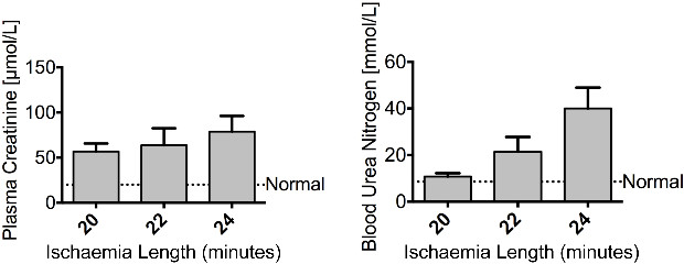
Figure 2. Injury – Renal ischaemia impairs renal function. Male Balb/c mice aged 8 weeks underwent a right nephrectomy and the left renal pedicle was clamped for 20, 22 or 24 min (n = 4 per group). Mice were sacrificed at 24 hr following renal IRI. Plasma creatinine and blood urea nitrogen show an increasing trend of severity as the length of ischaemia increased. The dashed line represents the levels of plasma creatinine and blood urea nitrogen found in normal control mice. Data presented as mean ± SEM and analyzed by one-way ANOVA. Please click here to view a larger version of this figure.
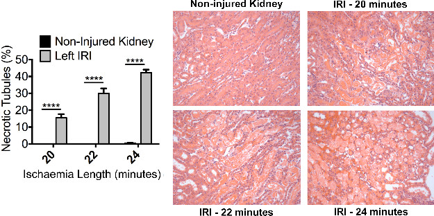
Figure 3. Injury – Renal ischaemia induces significant acute tubular necrosis. Male Balb/c mice aged 8 weeks underwent a right nephrectomy and the left renal pedicle was clamped for 20, 22 or 24 min (n = 4 per group). Mice were sacrificed at 24 hr following renal IRI. Representative photomicrographs (Magnification: 200x) of the OSOM from H&E stained kidney sections of control and injured kidneys illustrate ATN. Formal scoring of ATN (expressed as the proportion of necrotic tubules) confirms the increased level of injury with increasing duration of ischaemia. Data presented as mean ± SEM and analyzed by one-way ANOVA (**** = P ≤ 0.0001). Please click here to view a larger version of this figure.
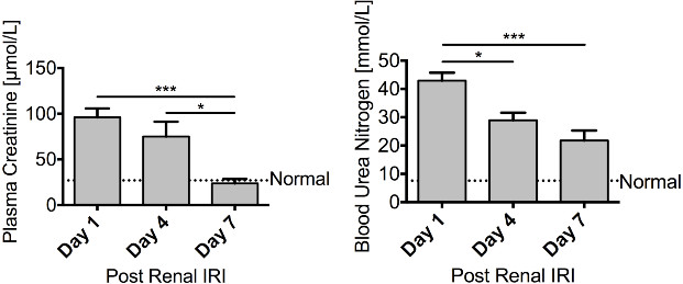
Figure 4. Regeneration – Renal function recovers following renal ischaemia. Male FVB mice aged 8 – 10 weeks underwent a right nephrectomy prior to 25 min of ischaemia to the left kidney. Mice were sacrificed 1 day (n = 10), 4 days (n = 10) or 7 days (n = 6) following renal IRI. Both plasma creatinine and blood urea nitrogen steadily decline over the course of 7 days, with plasma creatinine returning to basal levels, illustrating a recovery in renal function. Data presented as mean ± SEM and analyzed by one-way ANOVA (*** = P ≤ 0.001, * = P ≤ 0.05). Please click here to view a larger version of this figure.
Figure 5. Regeneration – Renal tissue shows signs of recovery following renal ischaemia. Male FVB mice aged 8 – 10 weeks underwent a right nephrectomy prior to 25 min of ischaemia to the left kidney. Mice were sacrificed 4 days (n = 10) or 7 days (n = 6) following renal IRI. Representative photomicrographs (Magnification: 200x) of the OSOM following the induction of ischaemia are shown. Formal scoring of the number of healthy, recovering, injured or necrotic tubules within the OSOM was assessed in PAS stained paraffin sections. At day 4 following renal IRI a large proportion of tubules within the OSOM still appear injured. However by 7 days there is a considerable increase in the proportion of tubules that are classified as healthy or recovering indicative of renal regeneration. Data presented as mean ± SEM. Please click here to view a larger version of this figure.
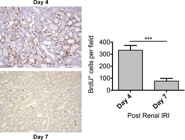
Figure 6. Regeneration – Dramatic tubular proliferation is evident 4 days following renal ischaemia. Male FVB mice aged 8 – 10 weeks underwent a right nephrectomy prior to 25 min of ischaemia to the left kidney. BrdU (50 mg/kg) was administered by intraperitoneal injection 24 hours prior to sacrifice. Mice were sacrificed 4 days (n = 10) or 7 days (n = 6) following renal IRI. Representative photomicrographs (Magnification: 200x) of the OSOM from mice sacrificed at day 4 and day 7 following the induction of ischaemia are shown. Tubular cell proliferation was assessed by immunohistochemical staining for BrdU on paraffin embedded kidney sections. Tubular cell proliferation was quantified by counting the number of BrdU positive nuclei in the OSOM with increased cell proliferation evident at day 4. Data presented as mean ± SEM and analyzed by students t-test (*** = P ≤ 0.001). Please click here to view a larger version of this figure.
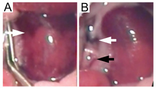
Figure 7. Left ischaemic kidney with non-occluded additional renal blood vessels. Failure to occlude additional blood vessels supplying the kidney leads to inconsistent ischaemia. (A) The absence of global kidney ischaemia is indicated by an uneven color change (white arrow) when the micro serrafine clip is in position. (B) Following removal of the micro serrafine clip, the main renal artery and vein are visible (white arrow) together with the additional blood vessel supplying the middle and lower pole of the kidney (black arrow). Please click here to view a larger version of this figure.
Renal IRI is an important cause of AKI with no specific treatment available. The experimental study of renal IRI has been highly informative with previous work demonstrating the role of macrophages, dendritic cells, lymphocytes, regulatory T-cells as well as other cells and mediators in the induction of both the acute injury and healing phase5,8-16. In addition, experimental renal IRI has been used to assess the effect of various therapeutic agents4,17-19.
The model of renal IRI detailed here uses a midline laparotomy approach to perform a right nephrectomy and induce ischaemia in the left kidney using clamps. As illustrated by the representative results, modifying the duration of ischaemia can control the severity of injury. Therefore this model can be adjusted to induce mild, moderate or a high level of kidney injury as required by the experimental question posed. However, a limitation of this model is that it is not suitable for the induction of very severe kidney injury by prolonged renal ischaemia. The resultant severe acute kidney dysfunction may lead to significant mortality.
There are several aspects of this model that are crucial in order to achieve predictable and reproducible renal injury. One major source of variability with this model can originate from the body temperature during the ischaemic period20. It is therefore essential that body temperature is maintained at a constant level and monitored throughout the ischaemic period. In this protocol a control unit with temperature probes and homoeothermic blanket was used to auto-regulate body temperature at 37 °C. The influence of body temperature on the susceptibility of the kidney to ischaemic injury is well described. Previous work has shown that body temperature affects the severity of renal IRI20 and variation in the body temperature of the mice is a potential confounding factor that may affect the interpretation of the results. Another important consideration is that mice may exhibit anatomical variation with additional blood vessels that supply the left kidney. It is critical that such additional blood vessels are identified and occluded when performing the right nephrectomy and inducing global ischaemia in the left kidney. Failure to do so will result in inconsistent ischaemia throughout the kidney. Similarly, the color change of the left kidney following clamping of the left renal pedicle should be carefully scrutinized as the absence of a global color changes suggests the presence of an additional renal artery that had not initially been identified.
In contrast to models that utilize bilateral kidney ischaemia, this model will be of interest to researchers who are using the prophylactic administration of pharmaceutical drugs or other agents to modulate the expression of genes, proteins etc. prior to renal injury. The right nephrectomy tissue can be used to both confirm and quantify the level of induction or inhibition of the molecule of interest in each individual experimental animal. This model will also be of interest to researchers studying renal regeneration as the acute injury phase is followed by prominent tubular proliferation.
Some experimental models of renal inflammation are limited as significant disease can only be induced in certain strains of mice. In contrast, the model of renal IRI described here is versatile and can be applied to both male5 and female4 mice, different strains of mice as well as aged mice4.
The authors have nothing to disclose.
The present study was supported by grants from Kidney Research UK (ST4/2011), the Cunningham Trust (CT11/14) and the Mrs EA Hogg’s Charitable Trust.
Name of Material/ Equipment | Company | Catalog Number | Comments/Description |
Tissue scissors | Fine Science Tools | 14072 – 10 | |
Micro-Adson forceps (Rat toothed) | Fine Science Tools | 11019 – 12 | |
S&T JFA-5bTC Forceps – SuperGrip Angled | Fine Science Tools | 00649-11 | |
Colibri retractor | Fine Science Tools | 17000 – 04 | |
Micro clip applicator | Fine Science Tools | 18057-14 | |
Micro serrafines | Fine Science Tools | 18055-04 | |
Olsen-Hegar needle holder | Fine Science Tools | 12002 – 12 | |
Hemoclip Plus Ligating Clips Small | Weck | 533837 | |
Autoclip Wound Clip System, 9mm | Harvard Apparatus | PY2 52-3748 | |
Silk Black Braided Suture, Size 6-0 | Harvard Apparatus | 723288 | |
Standard Heat Matt | |||
Homeothermic Blanket & Control Unit | Harvard Apparatus | ||
Lacri-Lube | Allergan | ||
Vetasept Chlorhexidine | AnimalCare | ||
Vetalar : Ketamine hydrochloride | 100mg/ml solution | ||
Domitor : medetomidine hydrochloride | 1mg/ml | ||
Vetergesic : Buprenorphine hydrochloride | 0.3mg/ml | ||
Antisedan : Atipamezole hydrochoride | 5mg/ml |