Primary cell culture using intact tissue organoids provides a model system that mimics the multi-cellular in vivo microenvironment. We developed a serum-free primary breast epithelium tissue culture model that perpetuates mixed cell culture lineages and exhibits differentiated morphology, without enzymatic tissue disruption. Breast organoids remain viable for >6 months.
Breast ductal carcinoma in situ (DCIS), by definition, is proliferation of neoplastic epithelial cells within the confines of the breast duct, without breaching the collagenous basement membrane. While DCIS is a non-obligate precursor to invasive breast cancers, the molecular mechanisms and cell populations that permit progression to invasive cancer are not fully known. To determine if progenitor cells capable of invasion existed within the DCIS cell population, we developed a methodology for collecting and culturing sterile human breast tissue at the time of surgery, without enzymatic disruption of tissue.
Sterile breast tissue containing ductal segments is harvested from surgically excised breast tissue following routine pathological examination. Tissue containing DCIS is placed in nutrient rich, antibiotic-containing, serum free medium, and transported to the tissue culture laboratory. The breast tissue is further dissected to isolate the calcified areas. Multiple breast tissue pieces (organoids) are placed in a minimal volume of serum free medium in a flask with a removable lid and cultured in a humidified CO2 incubator. Epithelial and fibroblast cell populations emerge from the organoid after 10 – 14 days. Mammospheres spontaneously form on and around the epithelial cell monolayer. Specific cell populations can be harvested directly from the flask without disrupting neighboring cells. Our non-enzymatic tissue culture system reliably reveals cytogenetically abnormal, invasive progenitor cells from fresh human DCIS lesions.
Proliferation of epithelial cells within the confines of breast ducts and alveoli (ductal carcinoma in situ) is recognized as an obligate precursor to invasive ductal and lobular breast carcinoma. Nevertheless, the molecular mechanisms and cell population dynamics that permit progression to invasive cancer are poorly understood. Elucidating the survival mechanisms used by pre-invasive breast carcinoma cells, or any pre-invasive tumor, may reveal therapeutic strategies for killing, or even preventing, pre-invasive neoplasms1. However, simple low-cost methods for functionally studying human pre-invasive lesions have been lacking. Although in vitro monolayer culture of transformed cell lines is an established laboratory method, the phenotype and genotype of these immortalized cell lines fails to recapitulate the molecular status of primary human tumor cells2. Furthermore, even the non-tumorigenic MCF-10A cell line, which recapitulates 3-D mammary gland architecture, fails to adequately represent the functional phenotype and molecular characteristics of an individual patient’s pre-invasive breast lesion3,4.
To determine if stem-like neoplastic cells capable of invasion existed within the ductal carcinoma in situ (DCIS) cell population, we developed a methodology for collecting and culturing sterile human breast tissue at the time of surgery (Figure 1)5. Our ex vivo breast organoid culture system does not rely on enzymatic tissue disruption, basement membrane extract matrix, or fibroblast depletion, for isolating and propagating mammosphere-forming cells from fresh human breast ductal carcinoma tissue6-8. Our new system is based on the principle of cell streaming/migration5. The discernible breast ducts, and surrounding stroma are submerged in a minimum volume of serum-free nutrient medium (just enough to cover the duct fragments) to maximize gas exchange, with the cut surface of the duct exposed to the culture medium, but in no specific orientation in the flask (Figure 1E-F). This culture system allows cells to migrate out of the duct and into/onto the autologous stroma and culture flask. The nutrient medium, supplemented only with Epidermal Growth Factor (EGF), insulin, and antibiotics, supports growth of mixed cell populations emanating from the organoid. The tissue culture flask has a removable, re-sealable lid that allows the organoids and/or cells to be harvested without disrupting the entire flask or neighboring organoids, while maintaining a sterile humidified environment.
Human breast tissue was collected from patients enrolled in a research study, with written informed consent, following Department of Defense, George Mason University, and Inova Health System Institutional Review Board’s approved protocols.
1. Prepare Nutrient Rich Medium with Growth Factors and Antibiotics
2. Tissue Acquisition and Grossing
3. Tissue Culture
4. Maintenance of Established Organoid/Epithelial Cell Colonies
Workflow for procuring and culturing sterile breast ductal carcinoma in situ tissue
Breast tissue sterility is maintained from the operating room to the cell culture lab via minor changes in typical hospital pathology workflow (Figure 1). Tissue is transported in a sterile container with a plastic film cover which allows radiologic assessment while maintaining tissue sterility. Gross tissue processing of a breast lumpectomy or mastectomy sample is performed with sterile gloves, blades, and tissue marking dyes. Gross morphologic appearance of breast ductal carcinoma in situ resembles pale, slightly raised areas with a gritty/firm texture, surrounded by reddish/yellow rubbery tissue. Areas of ductal hyperplasia and DCIS may feel “gritty” and firm due to calcifications. These areas of breast tissue often appear tan or a slightly different color than the surrounding breast tissue. However, ADH can only be distinguished after tissue collection and pathologic review of the stained tissue sections. Breast tissue remains viable by immersing the tissue and/or ductal organoids in serum-free nutrient medium supplemented with human recombinant EGF (10 ng/ml), insulin (10 μg/ml), streptomycin sulfate (100 μg/ml) and gentamicin sulfate (20 μg/ml)5. Cell culture flasks with removable/re-sealable lids permit periodic harvesting of cells/organoids (Figure 1E-F). This model successfully propagated human breast pre-invasive lesions from more than 20 patients diagnosed with atypical ductal hyperplasia (n = 2) and ductal carcinoma in situ (n = 18).
Spontaneous mammosphere formation in vitro and in vivo
Mammospheres and 3-D structures arose spontaneously from multiple, independent human DCIS duct tissue fragments from different patients diagnosed with atypical ductal hyperplasia or ductal carcinoma in situ (Figure 3 & 4)5,9. Enzymatic disruption of the breast tissue was not performed prior to tissue culture, which resulted in a mixed-cell type culture. Neither serum, basement membrane extract, nor gel-like matrices were required for spontaneous mammosphere formation (Figure 4). The mammospheres generated mammary xenograft tumors in a NOD/SCID mouse model with the same growth pattern as that of invasive cancer (Figure 5)5. These results demonstrate that progenitor cells with invasive potential pre-exist within the human breast DCIS duct but are apparently held in check by the ductal niche and can be coaxed to emerge in organoid culture. These cells constitute a new category of breast stem-like cells that exist prior to the overt manifestation of the invasive phenotype5,9,10.
Confirmation of epithelial derived mammospheres and xenografts
The mammospheres and xenografts derived from the mammospheres were confirmed by immunofluorescence as having epithelial origins. Epithelial cell adhesion molecule (EpCAM) is a membrane glycoprotein expressed on epithelial cells11. Immunofluorescence with a mouse monoclonal antibody reactive to human EpCAM (green) and a nuclear stain (DAPI, blue) showed EpCAM positive cells in the mammospheres in culture (Figure 6A) and the center of a NOD/SCID xenograft (Figure 6B).
Verification of intact basement membrane boundaries
The mammosphere forming, neoplastic epithelial cells in this culture system were derived from pre-invasive breast lesions that were devoid of frank invasion or microinvasion, as verified by independent pathologic analysis under standard of care histopathologic diagnosis. Multiple organoid structures from the same patient generated mammosphere forming colonies that proved to be tumorigenic. In addition, histopathologic examination of the tissue used for organoid culture revealed confluent intraductal lesions with intact basement membrane boundaries (Figure 7B)5. Thus it can be concluded that the spontaneous mammospheres formed in this culture system are derived only from pre-invasive neoplastic areas and are not a product of rare areas of microinvasion5.
Figure 1. Workflow for maintaining tissue sterility during radiological imaging and gross tissue dissection. (A) In the operating suite, breast tissue (lumpectomy sample shown) is placed in a sterile tray and covered with sterile plastic wrap. The tissue can be imaged directly in the plastic tray. (B) Tissue grossing to identify areas of DCIS. Single-use only tissue orientation dyes are painted onto the tissue surface using sterile cotton tipped swabs. Household distilled white vinegar is poured directly onto the tissue and blotted with sterile cotton gauze. (C & D) Breast tissue is transported to the tissue culture lab in nutrient rich medium supplemented with antibiotics. Tissue dissection, to isolate the areas of DCIS, is performed using sterile gloves and blades/scalpels/scissors. The DCIS tissue is cut into multiple organoids for culture. (E & F) In vitro culture of breast organoids. Human DCIS tissue is placed directly in tissue culture flasks with removable lids, without prior enzymatic digestion of the tissue. A minimal amount of serum-free culture medium supplemented with epidermal growth factor and insulin supports cellular growth while maintaining an air-liquid interface around the organoid. Please click here to view a larger version of this figure.
Figure 2. Illustration of gross tissue processing for breast DCIS tissue. The lumpectomy or mastectomy tissue is cut into thin sections by slicing the tissue vertically without cutting all the way through the specimen. This dissection method is often referred to as “bread loaf technique” since the cut tissue resembles a loaf of bread. The area(s) suspected of containing DCIS are cut out and sliced into 2 – 3 mm slices for diagnostic pathology and organoid culture.
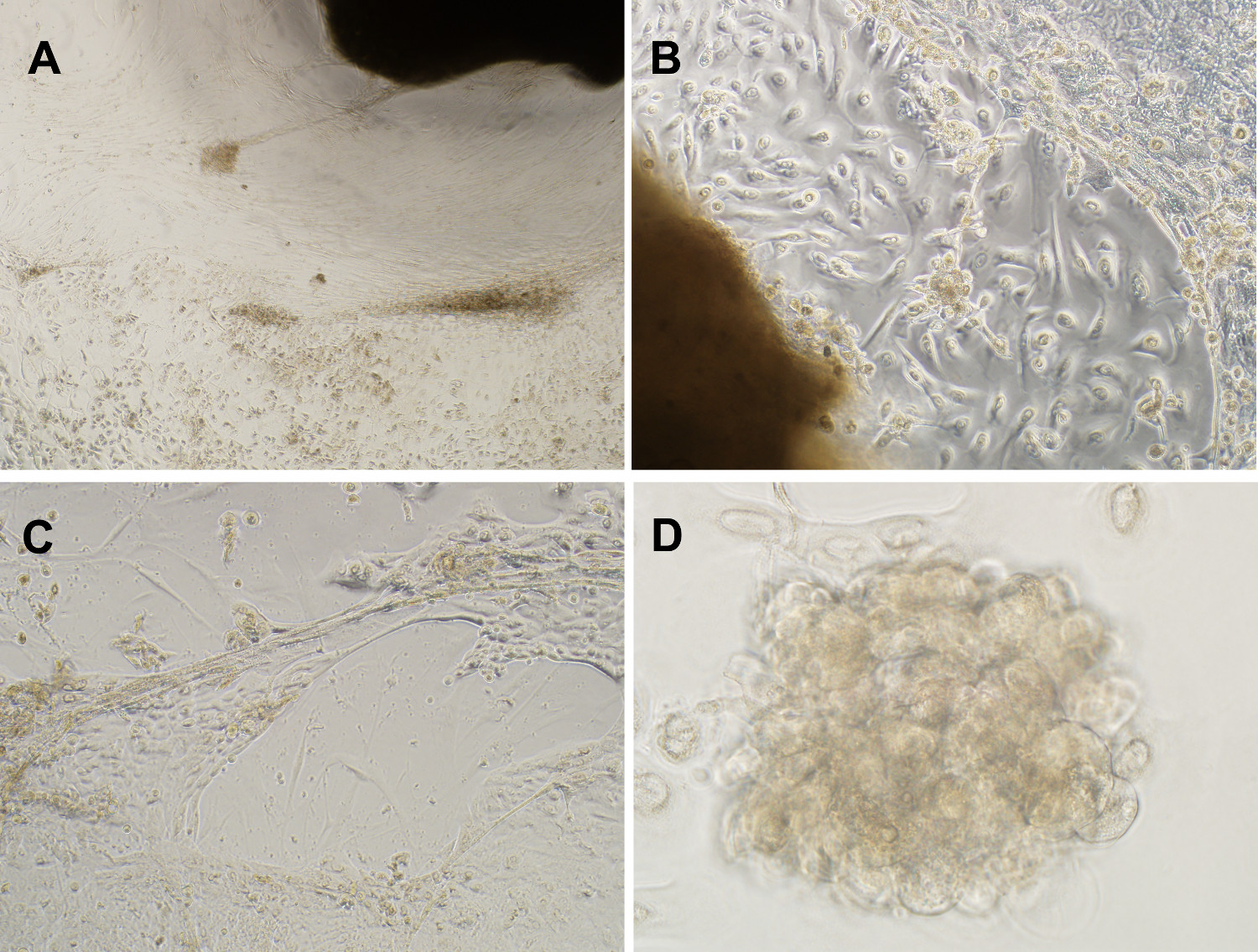
Figure 3. A mixed cell type culture maintains representative in vivo cell populations. (A) Phase contrast image of mixed cell culture generated from breast DCIS lesions over 11 weeks (4X magnification). (B & C) In vitro organoid cultivation successfully propagated DCIS derived epithelial cells with anchorage independent growth, defined as upward growing and expanding mammospheres, and lobulated, duct-like 3-D formations, in serum free medium supplemented with EGF, insulin, streptomycin and gentamicin (10X magnification). (D) Example mammosphere formed after 11 weeks in culture (40X magnification). Please click here to view a larger version of this figure.
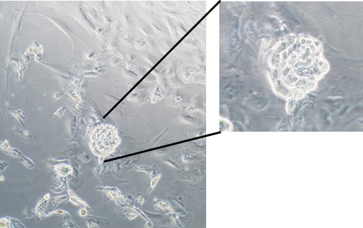
Figure 4. Spontaneous formation of mammospheres in serum free organoid culture. An example mammosphere formation following 33 days of culture (10X magnification, 20X inset). Please click here to view a larger version of this figure.
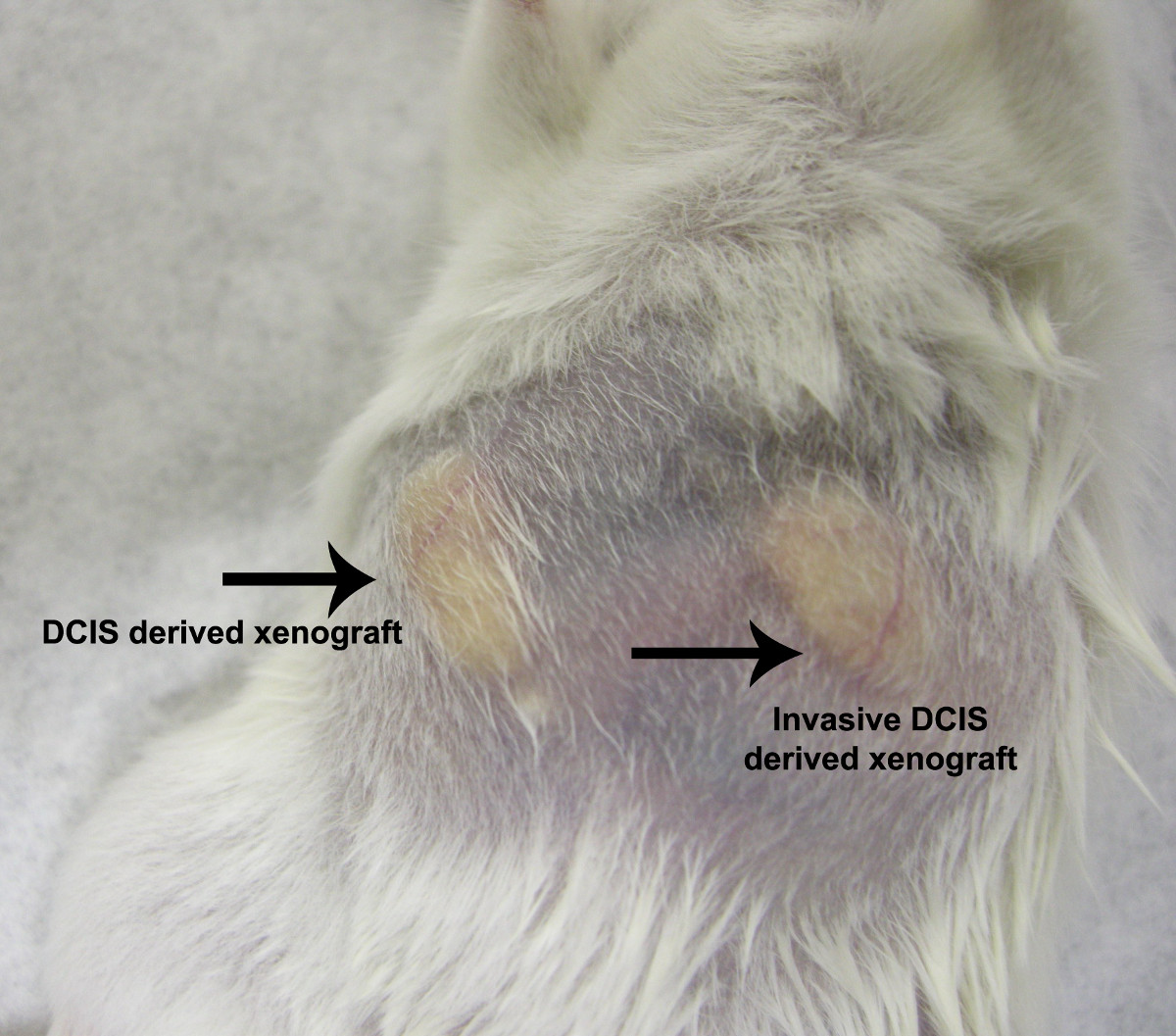
Figure 5. NOD/SCID mouse xenograft model. Xenografts were generated by injecting epithelial cells derived from a patient diagnosed with DCIS (mouse right mammary fat pad) or from a patient diagnosed with invasive DCIS (mouse left mammary fat pad). Xenografts derived from both pure DCIS and invasive DCIS revealed a similar growth pattern and rate. Please click here to view a larger version of this figure.
Figure 6. Mammospheres are confirmed to be of epithelial origin. Immunofluorescence with anti-EpCAM conjugated to FITC was used to confirm the epithelial origin of mammospheres and mouse xenografts generated from breast ducts containing DCIS. (A) EpCAM-FITC positive cells (pseudo-colored green, 488 nm) were only seen in mammospheres of the mixed cell cultures emanating from breast organoids (DAPI (pseudo-colored blue, 408 nm) nuclear stain). (B) In formalin fixed paraffin embedded mouse xenograft tissue sections, EpCAM positive cells were only detected in the center of the xenograft tumor section. (20X magnification) Please click here to view a larger version of this figure.
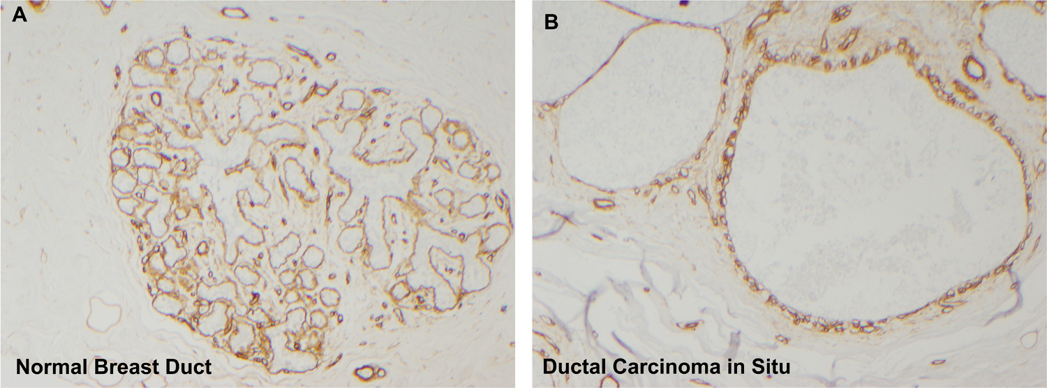
Figure 7. Collagen IV immunohistochemistry reveals intact basement membranes surrounding ducts. Normal breast ducts (A) are surrounded by intact basement membranes enriched in collagen IV (diaminobenzidine = brown staining). Following organoid culture, breast tissue also contains intact basement membranes confirming that the mammospheres are derived from areas of DCIS and not from invasive cancer (collagen IV immunohistochemistry, panel A 4X magnification, panel B 10X). Please click here to view a larger version of this figure.
The culture system described herein constitutes a new model for generating living pre-invasive neoplastic breast cells for basic and translational research studies. In the past, pre-malignant breast cancer progression has typically been studied using three different methods. The first method is histopathologic and genetic analysis of microdissected frozen or fixed human specimens12-14. The second method utilizes mouse models that contain hyperplastic alveolar nodules (HAN lesions) that are thought to be similar to human pre-invasive breast lesions15. The third model uses established breast carcinoma cell lines such as MCF7 sublines (MCF10A) that have a highly differentiated DCIS like morphology3,4. Although these three models have provided molecular clues to breast cancer progression, none of these methods are capable of assessing malignant potential or the molecular phenotype in an individual patient’s lesion(s). Histopathologic analysis does not provide information about the functional phenotype of the cells in the pre-invasive lesions. The mouse model of breast cancer progression may not accurately reflect the histomorphology and diversity of human atypical ductal hyperplasia, lobular carcinoma in situ, and ductal carcinoma in situ16-18. Furthermore, the stromal microenvironment and the deposition of extracellular matrix surrounding mouse precursor lesions are markedly different from the human counterpart16. Spontaneous murine precursor lesions can exhibit a very low level of progression to invasion and metastasis. The third method, cultured cell lines, can provide functional phenotypic information only if transplanted into immune suppressed hosts3,4. In addition the genetic abnormalities of a long passaged cell line may not represent spontaneous breast cancer progression in humans2. Finally, it is well established that each patient’s neoplasm has a unique combination of genetic and epigenetic abnormalities that drive the rate of growth, differentiated state, and progression to invasion and metastasis13,14,17. Human pre-invasive lesions are multifocal and heterogeneous in cell composition and histomorphology. Furthermore, the biologic malignant potential is unknown for an individual patient’s pre-invasive lesion.
Our primary tissue culture method overcomes the deficiencies of previous models of human pre-invasive breast cancer and provides the following advantages: 1) The organoid culture system supports growth of neoplastic cell populations within the native tissue microenvironment that spontaneously grow and generate mammospheres that will produce invasive tumors in mouse xenograft models. The cells represent the genotype and phenotype of an individual patient and thereby provide information for personalized therapy, or individual prognosis. 2) The organoid culture system maintains the native cellular subpopulations and provides a means to cultivate non-malignant epithelial cells, stromal cells and immune cells originally present in the primary breast tissue, and carried into culture within the organoid. The low volume of media in the culture system supports oxygen exchange encouraging spontaneous mammosphere formation and differentiated duct and alveoli like structures without the need for an artificial three dimensional scaffolding. 3) The primary cell culture is free from modifications and selection generated by enzymatic dissociation or exogenous genetic modification. Furthermore, the nutrient medium is low-cost and simple to prepare. 4) Molecular and genetic analysis can be conducted on specific cell populations and/or organoids from the culture at different points in time without disrupting the entire culture. 5) The organoid culture system permits the growth, differentiated morphology and cell-cell interactions of the native cell populations which can be studied before and after introduction of therapeutic agents into the culture media.
Although primary tissue culture has certain advantages, it is not without limitations. The organoid culture supports growth of mixed cell cultures, without overgrowth of any one cell type. However, the specific ratio of cell types cannot be controlled and thus may not recapitulate the exact cellular ratios found in vivo. Successful organoid cultivation requires sterile tissue collection and processing, both of which are not routine procedures in many community hospital pathology laboratories. Good communication among the clinical researchers, surgical staff, and pathology staff are essential for maintaining sample sterility within the continuum of patient care.
A further limitation of organoid culture is the effect of substratum firmness on cellular phenotype and gene expression16,19. Differentiation of stem cells in culture can be induced by addition of serum-containing medium or may be due to extended time in culture. A stem-like phenotype was maintained after several months in this non-enzymatic, serum-free organoid culture system5. However, at some point in time, the cells may differentiate which can be seen morphologically – the cells become smaller, denser, and fail to form mammospheres. To avoid potential issues with changes in cell phenotype over time, molecular experiments, such as transfections or knock down assays, should be performed with young cultures rather than with old cultures (more than 6 months).
The keys to successful organoid culture are using an appropriate volume of medium in the culture flask, and allowing the organoids time to adhere to the tissue culture flask. Excess medium in the flask limits oxygen diffusion, inhibiting mammosphere formation. The first 3 – 7 days of tissue culture are critical for the organoids to attach to the tissue culture flask. In general, if an organoid has not attached by day 14, it likely does not contain any viable DCIS ductal segments and will not become adherent. Organoids that are not adherent by day 14 should be removed from culture. The lack of viable breast DCIS ducts can be verified following culture by fixing the organoid in 10% formalin and processing the tissue into paraffin blocks for tissue staining and microscopic evaluation.
Our non-enzymatic, serum-free culture system findings support the hypothesis that genetically abnormal neoplastic precursor cells with invasive potential exist within pre-invasive human breast lesions5,9,20. This finding is in keeping with the previous work of Sgroi et al., who conducted genetic analysis of human breast pre-invasive lesions and Damonte et al. who studied the mammary intraepithelial neoplasia outgrowth (MINO) murine model of breast cancer progression12,14,21. Taken together with the conclusions of others, our culture model of individual patient pre-invasive lesions supports the concept that the aggressive phenotype of a patient’s invasive breast cancer may be largely pre-determined at the pre-invasive stage.
The authors have nothing to disclose.
This work was supported partially by (1) the Department of Defense Breast Cancer Research Program (US Army Medical Research Acquisition Activity) award #W81XVVH-10-1-0781 to LAL and VE, and (2) the Susan G. Komen Foundation grant IR122224446 to LAL and VE. Pathology support and tissue grossing was kindly provided by Inova Fairfax Pathology Department, Dr. Hassan Nayer, Dr. Geetha A. Menezes, and Dr. Charles Bechert. Patient consent and sample procurement was expertly guided by Inova Fairfax Hospital clinical research coordinators Holly Gallimore, Heather Huryk, and Emil Kamar.
Name of Reagent/ Equipment | Company | Catalog Number | Comments/Description |
Ethanol | Fisher | A405-P | prepare a 70% solution in Type 1 reagent grade water |
18 MΩ-cm water, sterile filtered | sterile filtered, Type 1 reagent grade water | ||
10cc plastic disposable syringe, sterile | BD | 305482 | |
0.2µm polyethersulfone (PES) syringe filter, sterile | Thermo Scientific | 194-2520 | |
15ml polypropylene conical tubes, sterile | Fisher | 14-959-49B | |
50ml polypropylene conical tubes, sterile | Fisher | 05-539-6 | |
1.5ml low retention microcentrifuge tubes,sterile | Fisher | 02-681-331 | |
nutrient medium, DMEM-F12/HEPES | Invitrogen | 11330-032 | with L-glutamine |
Insulin, human recombinant | Roche | 11376497001 | 10mg/ml stock |
Epidermal Growth Factor (EGF), human recombinant | Millipore | GF144 | 100µg/ml stock |
Streptomycin sulfate | Sigma-Aldrich | S1567 | 10mg/ml stock |
Gentamicin sulfate | Sigma-Aldrich | G19114 | 10mg/ml stock |
Filtration flask and filter top, sterile | Millipore | SCGPU02RE | 0.22µm PES membrane |
25ml sterile, disposable pipettes | Fisher | 4489 | paper-plastic wrapped |
10ml sterile, disposable pipettes | Fisher | 4488 | paper-plastic wrapped |
Tissue marking dyes (black, blue, red, green, yellow and orange) | CDI | MD2000 | after opening use only with single-use, sterile cotton tipped applicators, or use once and discard |
Cotton tipped applicators, sterile | Fisher | 23-400-115 | single use only |
Gauze pads, 10x10cm, sterile | Fisher | 2187 | |
Plastic transfer pipettes, sterile, disposable | Samco | 202-20S | |
Vinegar, white distilled | household use | 5% acetic acid; after opening use only with sterile pipettes | |
#10 scalpels, sterile, disposable | Thermo Scientific | 31-200-32 | |
petri dish, sterile | Fisher | FB0875713A | |
TPP 115cm2 flask, with removable lid | MidSci | 90652 | screw cap with filter |
CO2 incubator | Fisher | 13-998-074 | 5% CO2, 37 oC, humidified chamber |
inverted light microscope | Olypmus | IX51 | |
8M urea | Fisher | BP169-500 | optional, for mass spectrophotometric analysis of cultured cells |
2X SDS tris-glycine buffer | Life Technologies | LC2676 | optional, for proteomic analysis of cultured cells |
Cytocentrifuge | Thermo Scientific | A78300003 | optional, for preparing cell smears |