1,859 Views
•
08:18 min
•
May 12, 2023
DOI:
The significance of the protocol lies in its convenience in the short time needed for its completion. This enables a high throughput, a high concentration, and a high degree of the isolation of the stalked and sessile trichomes. The main advantage of this technique is its straightforwardness.
During this technique, one should consider the trichome density of the plant source and inspect the isolation degree at each step via microscope while performing the protocol for the first time. Demonstrating this technique will be Nurit Shalev, an expert on cannabis from Volcani Center. Begin by crushing a dry ice block into small, fine flakes using a hammer and a hard, flat object in a five liter plastic container.
Use a large strainer, and sieve the fine dry ice flakes from the non-crushed dry ice into another five liter plastic container. Pour approximately 200 cubic centimeters of the dry ice flakes into an upright one liter glass beaker. Add up to 10 grams of frozen cannabis sativa inflorescences onto the first layer of crushed dry ice, and cover it with an additional layer of 200 cubic centimeters of finely crushed dry ice.
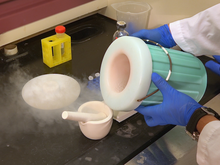
Then cover the opening of the one liter glass beaker with two to three layers of a one millimeter screen door mosquito net, and secure it with rubber bands. Next, pour liquid nitrogen into a large round-bottom stainless steel container. Insert a 350 micrometer mesh in a flour sifter or sieve strainer to cover the flour sifter mesh from below.
Place this flour sifter above the large round-bottom stainless steel container filled with liquid nitrogen. To minimize the loss of the sifted mass, ensure that the container’s width exceeds that of the flour sifter. Separate the trichomes from the plant material by shaking the one liter glass with its opening pointing downward toward the flour sifter.
Set aside the beaker every two to three minutes, allowing the sifting of the crushed dry ice and plant material accumulated on the flour sieve. Sift the flour sieve horizontally to facilitate the passage of the plant material into the liquid nitrogen in the round-bottom stainless steel container placed below. Next, add liquid nitrogen to the stainless steel container and the crushed dry ice to the one liter glass beaker when their levels run low.
When the plant material on the flour sieve is depleted, refill the used plant material in the glass beaker with an additional 10 grams of fresh plant material, and repeat the sifting process until the collection of a sufficient amount of enriched trichomes in the container. Verify the present of a white powder-like substance at the bottom of the stainless steel container submerged in liquid nitrogen before a microscope observation to confirm the collection of sufficient trichomes. Add a small amount of liquid nitrogen to a clean, small round-bottom stainless steel container.
Then fold a 40 by 40 centimeter microsieve having a 150 micrometer mesh size twice to obtain a 20 by 20 centimeter fold, and open it in a cone-like shape. Secure the mesh cone to the edge of the round-bottom stainless steel container with one or two clothespins so that the opened part of the cone is upright, and it’s pointed part is partly submerged in liquid nitrogen. Once done, gently pour the liquid nitrogen containing the white powder-like substance obtained earlier into the microsieve cone.
Using a wide brush, gather and transfer any remaining plant material from the first container into the 150 micrometer mesh cone. Next, carefully detach the clothespin from the lid of the container, and open the microsieve cone to locate the trichomes in the middle of the opened microsieve. Hold all four corners of the microsieve together, and submerge the middle part containing the trichomes in liquid nitrogen.
Gently dip and shake the microsieve in the liquid nitrogen for one minute. Using a sterile needle, make tiny holes in the cap of a 50 milliliter test tube to avoid pressure building. Scoop the non-sifted plant debris, dry ice, and larger trichomes wrapped in the center of the microsieve into a 50 milliliter test tube, and store it at minus 80 degrees Celsius for future use.
Transfer the sifted trichomes submerged in the liquid nitrogen sequentially through microsieves, having decreasing pour sizes from 105 to 80 to 65, and then to 50 micrometer. After each transfer, collect the different sized trichomes remaining on the microsieve separately, and label them. Make tiny holes in the cap of the tube using a sterile needle.
Transfer the desired trichomes submerged in liquid nitrogen and wrapped in the 50 micrometer microsieve to a clean plate using a pre-chilled spoon. Then quickly transfer the powder-like trichomes into a labeled pre-chilled 1.5 milliliter tube using a pre-chilled spatula, and immediately store the tubes at minus 80 degrees Celsius for further studies. In the initial stage of the isolation process, pieces of leaf tissue were predominant in the isolated portion, in contrast to the final isolation product, which was composed entirely of non-contaminated isolated trichomes.
The quality of the isolated granular capitate stalked and sessile trichomes was observed in the final isolation step. The purity of the trichomes isolated using this method was comparable with a conventional protocol. The delicate stalked trichome head structure was retained, and the whole heads of the isolated stalked trichome were visible, in contrast to the traditional method, where the complete trichome head structure was missing, and only disc cells were present.
The RNA extraction and estimation of RNA integrity of the isolated trichomes indicated high RNA integrity number or RIN values from 9.4 to 10, and gel chromatography indicated high RNA integrity. Carefully perform the T bag-like infusion motion of the 150 to 50 micro microsieved isolation. This method can provide insights into the transcriptomic and probably proteomic makeup of trichomes from cannabis and trichomes of other plant species.
Following this procedure, metabolic and transcriptomic characterizations can be performed. These additional methods can help to understand the expression levels of the genes in the isolated trichomes and their metabolic profiles. This technique has enabled the transcriptomic characterization of glandular trichomes from different cannabis varieties.
A protocol is presented for the convenient and high-throughput isolation and enrichment of glandular capitate stalked and sessile trichomes from Cannabis sativa. The protocol is based on a dry, non-buffer extraction of trichomes using only liquid nitrogen, dry ice, and nylon sieves and is suitable for RNA extraction and transcriptomic analysis.
08:56
Transient Expression in Nicotiana Benthamiana Leaves for Triterpene Production at a Preparative Scale
Related Videos
17122 Views
04:46
Isolation of Lymphocytes from Mouse Genital Tract Mucosa
Related Videos
14282 Views
10:54
Extraction of Plant-based Capsules for Microencapsulation Applications
Related Videos
11927 Views
05:46
Ultrasonic-Assisted Extraction of Cannabidiolic Acid from Cannabis Biomass
Related Videos
5956 Views
09:52
Isolation of Mouse Salivary Gland Stem Cells
Related Videos
23625 Views
15:53
Isolation and Biophysical Study of Fruit Cuticles
Related Videos
16595 Views
07:08
Isolation of Viable Multicellular Glands from Tissue of the Carnivorous Plant, Nepenthes
Related Videos
4746 Views
N/A
Comprehensive Approach for Microbial Isolation from Hidradenitis Suppurativa Tunnels
Related Videos
162 Views
10:53
Establishment of a Clonal Culture of Unicellular Conjugating Algae
Related Videos
13143 Views
06:53
Isolation, Characterization, and Total DNA Extraction to Identify Endophytic Fungi in Mycoheterotrophic Plants
Related Videos
2495 Views
Read Article
Cite this Article
Cohen, S., Itkin, M., Faigenboim, A., Davidovich-Rikanati, R., Bar, E., Hasson, D., Shalev, N., Koltai, H., Sagee, O., Lewinsohn, E., Spitzer-Rimon, B., Schaffer, A. A. Non-Aqueous Isolation and Enrichment of Glandular Capitate Stalked and Sessile Trichomes from Cannabis sativa. J. Vis. Exp. (195), e64798, doi:10.3791/64798 (2023).
Copy