Establishing primary endometrial stromal cell culture systems from hysterectomy specimens is a valuable biological technique and a crucial step prior to pursuing a vast array of research aims. Here, we describe two methods used to establish stromal cultures from surgically resected endometrial tissues of human patients.
Many efforts have been devoted to establish in vitro cell culture systems. These systems are designed to model a vast number of in vivo processes. Cell culture systems arising from human endometrial samples are no exception. Applications range from normal cyclic physiological processes to endometrial pathologies such as gynecological cancers, infectious diseases, and reproductive deficiencies. Here, we provide two methods for establishing primary endometrial stromal cells from surgically resected endometrial hysterectomy specimens. The first method is referred to as “the scraping method” and incorporates mechanical scraping using surgical or razor blades whereas the second method is termed “the trypsin method.” This latter method uses the enzymatic activity of trypsin to promote the separation of cells and primary cell outgrowth. We illustrate step-by-step methodology through digital images and microscopy. We also provide examples for validating endometrial stromal cell lines via quantitative real time polymerase chain reactions (qPCR) and immunofluorescence (IF).
The human uterus corpus is comprised of three layers, the perimetrium (or serosa), the myometrium, and the endometrium. Distinguishing each of these layers is an important step to establish endometrial cell lines. The perimetrium is the outer most layer of the uterus and composed of thin, serous cells. The myometrium is the thick, middle layer of the uterus and comprised of smooth muscle cells. The endometrium is identified as the inner layer of the uterus and includes epithelial and stromal cell populations.
The endometrium is further subdivided into the basalis layer whose stem cell population is hypothesized to repopulate the functionalis layer approximately every 28 days 1. The functionalis layer of the human endometrium undergoes significant biochemical and morphological changes in response to circulating hormones. These hormones are derived from the pituitary gland and the ovaries.
The coordinated production and release of hormones results in a reproductive cycle. The reproductive cycle is designed to prepare the endometrium for potential embryo implantation events. In humans, the reproductive cycle is known as “the menstrual cycle” and divided into three phases – proliferative, secretory, and menstrual. The proliferative phase involves the proliferation of the functionalis endometrial layer whereas the secretory phase is marked by functionalis maturation. Specifically, extracellular alterations, secretions, and cellular differentiation signal a potential implantation. If implantation does not occur before the end of the secretory phase, the functionalis endometrial layer is shed during the menstrual phase. The importance of menstruation and the events that trigger the shedding of the functionalis layer are still being debated. In humans, it has been posed that menstruation is the result of a specific mid-secretory phase differentiation event known as “spontaneous decidualization” 2. In this manuscript, we provide detailed methodology for both endometrial stromal cell isolation methods, and use a combination of immunofluorescence and digital images to demonstrate efficacy of these approaches. In addition, we apply a commonly used in vitro model of spontaneous decidualization to confirm endometrial stromal cell isolation.
Hysterectomy specimens used in this manuscript were collected in concordance with a University IRB-approved ethics protocol numbered IRB-HSR #14424.
1. Sample Acquisition from Clinical Source
2. Preparation of Primary Cell Lines using the Scraping Method
3. Preparation of Primary Cell Lines using the Trypsin Method
4. Saving Extra Tissue for Analysis (Snap Freezing and Formalin Fixation)
5. Immunofluorescence (IF)
6. RNA Extraction
7. Reverse Transcription
8. Real Time PCR
9. In vitro Decidualization Protocol (Derived from Ref 4 and 5)
As emphasized in the Protocol section, be sure to conduct all methods under government, institutional, and ethical guidelines when handling and preparing human tissue.
Included in this manuscript is an illustration of the general workflow of “the scraping method” (Figure 1A) and “the trypsin method” (Figure 1B) used to establish primary endometrial cultures. These methods are described in detail in the Protocol section (see parts 1. – 3.). Both methods prove successful in the growth of primary endometrial cultures. The advantage to “the scraping method” is the shortened preparation time; however, the time it takes to observe viable cells is usually 2 – 4 times longer compared to “the trypsin method.”
Provided are digital images of the initial human tissue received from tissue procurement. The uterine layers can be distinguished by the coarseness of the layer, i.e. the myometrium is thick and muscular and the endometrium is thin and more yielding. We have highlighted the thick, smooth muscle layer (myometrium) in white and outlined the endometrial layer of interest in black from two different hysterectomy samples (Figures 2A and 2B). In Figure 2C, tissues are oriented with the myometrium facing up while the endometrium is positioned down. The thin, perimetrial layer was surgically resected and not highlighted in these images. It is also important to note that the size of the endometrial layer in these digital 2-dimensional images is misrepresented as the actual 3-dimensional layer is very thin compared to the myometrium.
Cutting the appropriate tissue size is important for both “the scraping method” and “the trypsin method.” In Figure 2D, appropriate sizes are designated for each method above the respective sample. Using one 0.5×0.5×0.5 cm piece for “the trypsin method” [left] is sufficient for establishing a culture. The representative starting tissue for “the scraping method” [right] includes all uterine layers. The final product for “the scraping method” is represented in Figure 2E, and it is recommended that at least 1x1x1 cm of endometrial tissue be used to establish viable cultures. The requirement for this much tissue sample is a disadvantage of “the scraping method.”
Remaining tissue is often used in numerous ways including protein and RNA analyses. To ensure preservation of tissue quality, it is important to use a “snap freezing” method as in the Protocol section (see 4.). This method is also illustrated in Figure 2F and Figure 2G. Saving extra tissue by formalin fixation is also a common practice of pathologists and researchers. Preparing tissue for formalin fixation is described in the Protocol section (see 4.) and illustrated in Figure 2H.
Light microscopy is essential for monitoring primary endometrial cultures. Individual endometrial stromal cells should exhibit fibroblast morphology (Figure 3A). “The scraping method” typically generates clusters of early emerging cells, primarily along scalpel-induced scratches (Figure 3B, Day 2). “The trypsin method” generates both cluster and dispersed cell growth patterns (Figure 3C, Day 4). We have included several representative images from the initial plating (day 0) to the first passage of each method (Figure 3B and 3C).
Many tissues are defined by their expression of tissue-specific markers such as smooth muscle actin (SMA) for smooth muscle, CD34 for immune stem cells, etc. While gene and protein expression experiments have been conducted for the endometrium 6-11, an endometrial stromal cell specific marker has yet to be discovered. Instead, researchers commonly assess endometrial stroma purity by measuring markers from potential cell contaminants. For example, cytokeratin markers are used to show epithelial populations. However, it is less of a concern, because establishing and maintaining endometrial epithelia are difficult and usually selected against (Ref 12 and Figure 4A). If samples were to be obtained during pregnancy, positive expression of HLA-A, -B, -C demonstrates germ, trophoblast cells 13. On the other hand, like other mesenchymal fibroblasts, endometrial stromal cells express vimentin (CDH1). We demonstrate with immunofluorescence, the expression of vimentin and the absence of cytokeratin and E cadherin (CDH1) in our established endometrial stromal cells (Figure 4B).
Finally, we provide functional evidence of primary endometrial culture via an in vitro spontaneous decidualization assay. This method was adapted from Ref 4 and 5, and involves treatment of cultures with a combination of Medroxyprogesterone acetate (MPA) and 8- bromoadenosine 3’,5’-cyclic monophosphate (cAMP) (described in 9. In vitro Decidualization Protocol and modeled in Figure 5A). In the presence of MPA and cAMP, endometrial stromal cells exhibit significantly altered gene expression profiles (reviewed in Ref 14). The frequently published spontaneous decidualization markers are Prolactin (PRL) and Insulin-like Growth Factor Binding Protein 1 (IGFBP1) (reviewed in Ref 14). The response of these two genes to MPA and cAMP are strong indicators of both spontaneous decidualization and successful establishment of an endometrial stromal culture. We demonstrate this in Figure 5B.
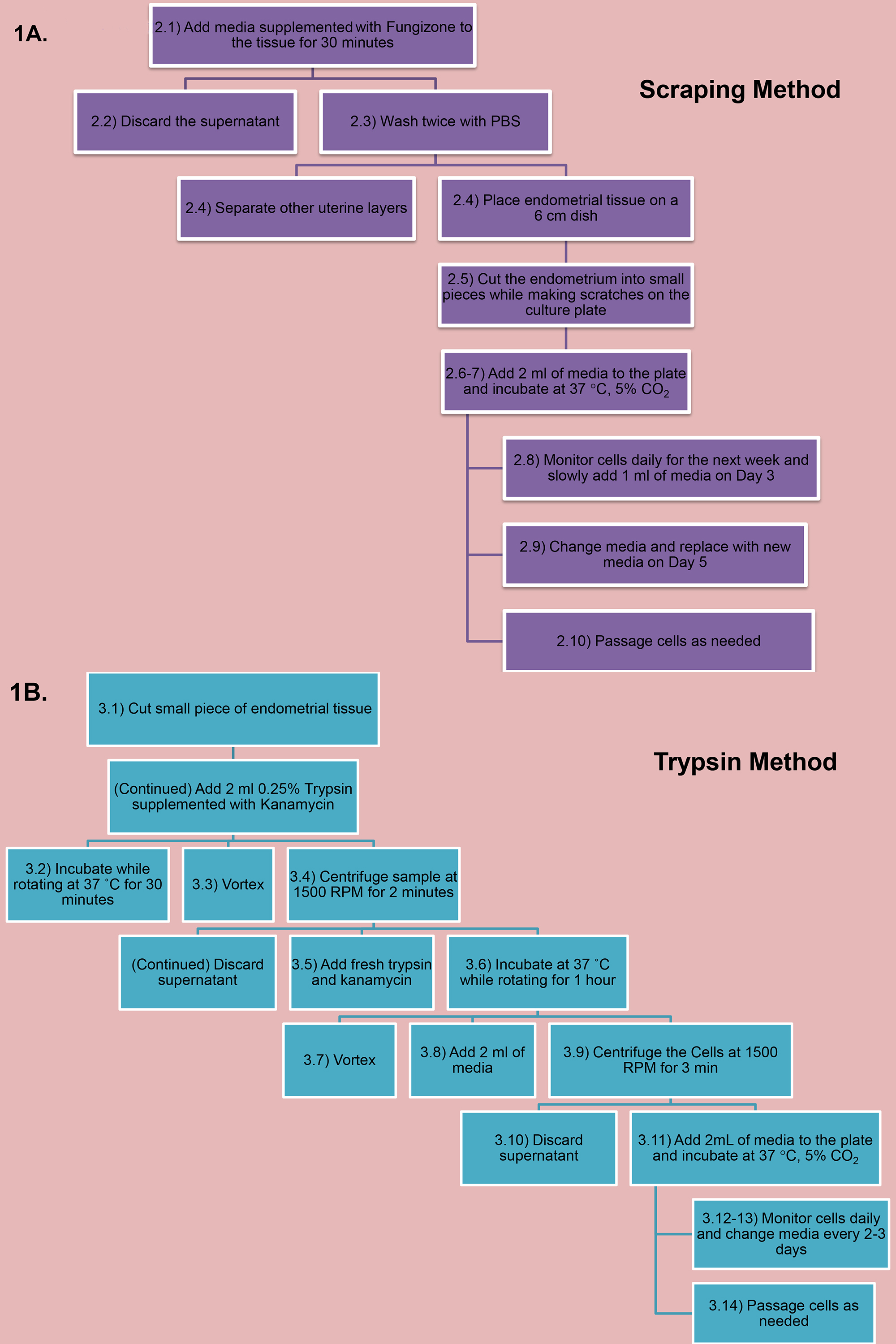
Figure 1. Schematic of the two primary endometrial isolation methods. (A) Steps 2.1-2.10 of the scraping method displayed in an organization chart. (B) Steps 3.1-3.14 of the trypsin method displayed in an organization chart. Click here to view larger image.
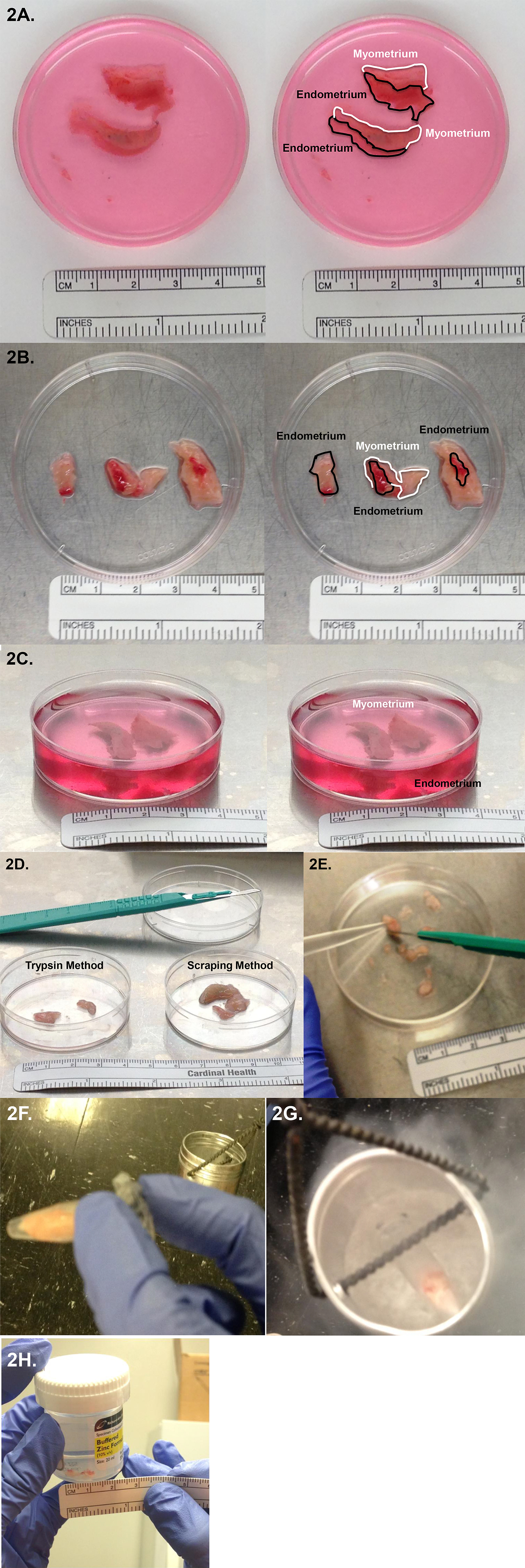
Figure 2. Processing uterine tissue. (A-C) Clinical uterine samples from two female patients. A & C are derived from Patient 1 and B is derived from Patient 2. Surgically resected uterine tissue (left) and highlighted uterine layers (right). (A & B) The myometrium is circled in white and the endometrium in black. (C) The myometrium is facing the camera. (D) Initial representative endometrial sample sizes for both isolation methods are indicated. The trypsin method requires pure endometrium before addition of the trypsin whereas the scraping method can uses the entire uterine specimens. (E) Example of the processed endometrial sample of the scraping method. (F & G) Image of remaining uterine tissue being prepared for Snap Freezing. Shown is a lab-made metal container utilized for this method. (H) Formalin fixation of endometrial sample. Click here to view larger image.
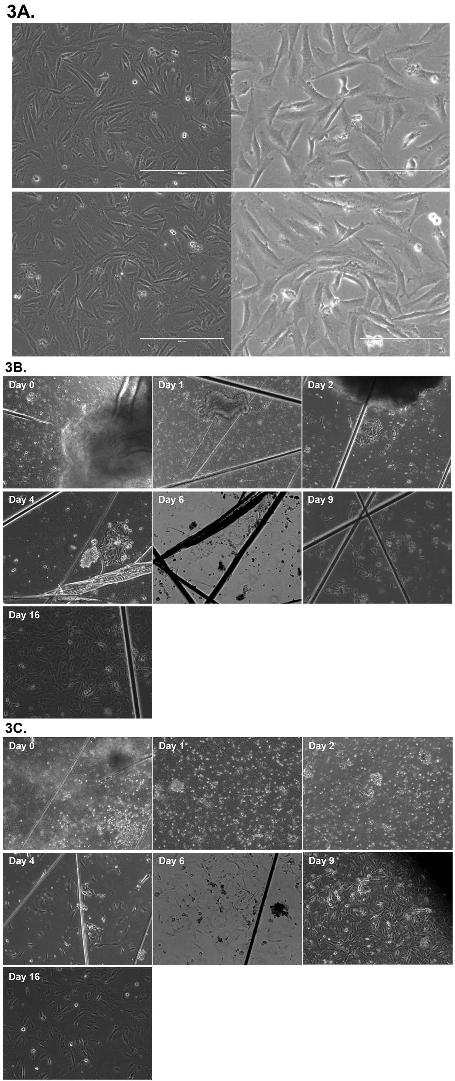
Figure 3. Light microscopy images of primary endometrial cultures. (A) Representative images of endometrial stromal cell cultures taken at various powers and confluency. (B) Representative images of the scraping method (Days 0 – 16) taken of the same culture dish, powered at 10x. Note: Visual lines indicate scratches from surgical blade. (C) Representative images of the trypsin method (Days 0 – 16) taken of the same culture dish, powered at 10x. Click here to view larger image.
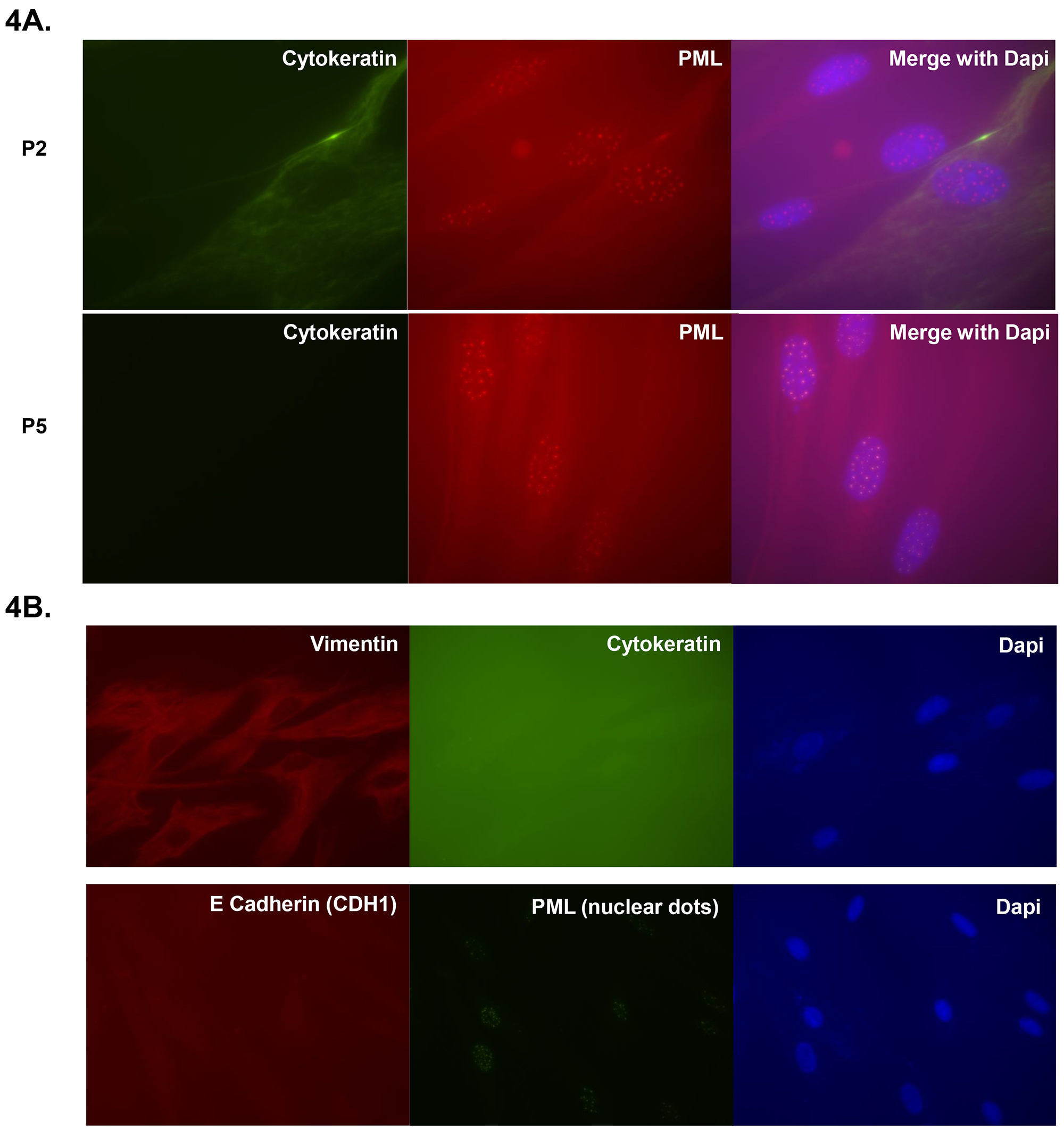
Figure 4. Immunofluorescence images of endometrial stromal cells. (A) Immunofluorescence of primary endometrial cultures isolated via the scraping method. Images demonstrate loss of cytokeratin between passage 2 (P2) and passage 5 (P5). Promyelocytic leukemia protein (PML) nuclear protein and DAPI staining were used as controls. (B) Images demonstrate positive staining for the mesenchymal marker Vimentin. Images also show negative staining for E cadherin (CDH1) and Cytokeratin. Staining for DAPI and Promyelocytic leukemia protein (PML) were provided as controls. Click here to view larger image.
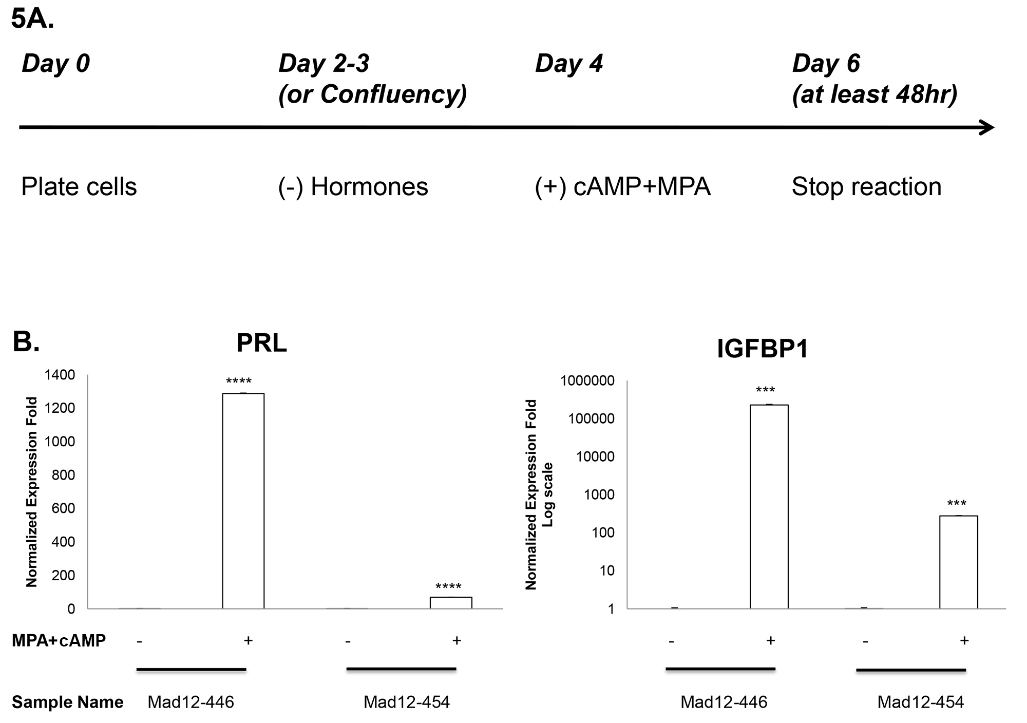
Figure 5. Spontaneous decidualization of two primary endometrial stromal cells. (A) Outline of experimental procedure. (B) Upregulation of Prolactin (PRL) and Insulin-like Growth Factor Binding Protein 1 (IGFBP1) gene expression in response to MPA and cAMP. Results were measured via RT-qPCR, and expression was normalized to GAPDH. Significance was calculated using students t-test (P < 0.001 *** and P < 0.0001 ****). Both cell lines were isolated using the scraping method. Click here to view larger image.
Primer | Sequence | Annealing Temperature (°C) | Cycles | Assay |
Prolactin (PRL) Forward | CATATTGCGATCCTGGAATGAGC | 60 | 40 | Sybrgreen |
Prolactin (PRL) Reverse | TCCTCAATCTCTACAGCTTTGGA | 60 | 40 | Sybrgreen |
Insulin-like growth factor binding protein 1 (IGFBP1) Forward | TCCTTTGGGACGCCATCAGTAC | 60 | 40 | Sybrgreen |
Insulin-like growth factor binding protein 1 (IGFBP1) Reverse | GATGTCTCCTGTGCCTTGGCTA | 60 | 40 | Sybrgreen |
GAPDH | Unspecified (see reagent list) | 60 | 40 | Taqman |
Table 1. PCR conditions for decidualization markers.
Other groups have described and adapted methodology for the preparation of endometrial stromal cultures, most of which utilize collagenase 4,12,13,15-18. In this manuscript, we have provided methodology and evidence for two simplified primary endometrial stromal culture methods, both of which are utilized by our lab for economical reasons and the convenient availability of trypsin and/or a razor blade.
When comparing our two methods, both successfully generate viable primary cultures. Our preferred method is the trypsin method as there is typically a higher yield with a smaller sample size requirement. However, if preparation time is limited, the scraping method requires 30 minutes whereas plating cells using the trypsin method takes more than an hour and a half.
Also noteworthy is that we demonstrate these methods with hysterectomy samples as opposed to endometrial biopsies. Endometrial biopsies are taken without significant intrusion and tend to produce smaller specimens. Still, the methods described in this manuscript (especially the trypsin method) should yield cultures from endometrial biopsies.
There are numerous applications for primary endometrial stromal cells. Comparing endometrial stromal cultures from human versus other mammals may provide clues to the questions that have been debated for centuries about why humans menstruate. There are other unexplored physiology studies that require in vitro culture. Of particular interest are the discoveries that provide insights into reproductive cycle events such as the findings that have linked epigenetic molecular events to the functional events of the reproductive cycle 19,20,21, and reviewed in Ref 11.
Clinicians would benefit greatly from studying endometrial stromal culture and identifying biomarkers in the instances of reproductive disorders and cancer malignancies. Between 2006 and 2010, it was reported that 7.4 million women and their partners used infertility services in the United States 22. A significant percent of infertility is due to failure of decidualization 23. Moreover, the relationship between endometrial diseases such as hyperplasia and endometriosis to infertility is only recently being assessed. The capacity to study uterine gynecological cancers through in vitro modeling is also invaluable because even though advanced endometrial sarcoma is rare, it can be lethal. By establishing in vitro primary cultures, we study the impact of the molecular events associated with the diseases and can generate putative clinical applications.
In conclusion, generating representative and efficacious in vivo models for many of these applications is difficult. This makes the development of in vitro primary endometrial cultures to study several in vivo functions critical. These two endometrial isolation methods, “the scraping method” and “the trypsin method,” will help provide the foothold for our understanding of endometrial physiology and pathology.
The authors have nothing to disclose.
We thank the collaborative efforts of Dr. Thao Dang and members of her lab for use of their imaging and microscope equipment. We also thank the Biorepository and Tissue Research Facility (BTRF) core, Jeff Harper, and the residents at the University of Virginia for providing us with uterine tissue. We thank Karol Szlachta for the help with Schematic Overview.
0.25 Trypsin or 0.05% Trypsin | Hyclone | SH3023602 or SH30004202 | |
1.7 micro Centrifuge Tube | Genesee Scientific | 22-272A | |
1µl,20µl, 200ml and 1000µl Pipette | Genesee Scientific | 24-401,24-402, 24-412, 24-430 | |
15ml Conical Tube | Hyclone | 339650 | |
50ml Conical Tube | Hyclone | 339652 | |
6cm Cell Culture Dish | Thermo scientific | 12-556-002 | |
8 well Chambers | Thermo Scientific | AB-4162 | |
Acetate | Fisher scientific | C4-100 | |
AMV RT Enzyme/Buffer | Bio Labs | M077L | |
Bovine Serum Albumin (BSA) | Fisher Scientific | BP-1605-100 | |
Buffered Zinc Formalin | Thermo | 59201ZF | |
Charcoal strip FBS | Fisher | NC9019735 | |
Chloroform | Fisher Scientific | BP1145-1 | |
Cover slip | Fisher Brand | 12-544D | |
Cyclic AMP (cAMP) | Sigma | B7880 | |
DMEM/High Glucose | Hyclone | SH30243FS | |
dNTP | Bioline | BIO-39025 | |
Donkey Anti Goat -TRITC | Santa Cruz | SC-3855 | |
Donkey Serum | Jackson’s lab | 017-000-002 | |
E Cadherin Antibody | Epitomics | 1702-1 | |
Ethanol | Fisher Scientific | BP2818-1 | |
Fetal Bovine Serum (FBS) | Fisher Scientific | 03-600-511 | |
Fungizone Amphotericin B | Gibco | 15290-018 | |
GAPDH Probe | Life Technologies | HS99999905 | |
Glycogen | 5Prime | 2301440 | |
Goat Anti Mouse -FITC | Jackson’s Lab | 115-096-003 | |
Isopropanol | Fisher Scientific | BP2618-1 | |
Kanamycin | Fisher Scientific | BP906-5 | |
Medroxyprogesterone acetate (MPA) | Sigma | M1629 | |
MeOH (Methanol) | Fisher Scientific | A4-08-1 | |
Mounting Media (w/DAPI) | Vector Labratories | H-1500 | |
N6 DNA Oligos | Invitrogen | ||
Number 15 Scraper | BD | 371615 | |
Pan Cytokeratin Mouse mAB | Cell Signaling | 4545 | |
PBS (phosphate buffered saline) | Fisher Scientific | BP-399-4 | |
Penicillin-Streptomycin Glutamine Solution 100X | Hyclone | SV30082.01 | |
PML Anti Goat Anti body | Santa Cruz | SC-9862 | |
Primer(s) | Eurofins | ||
RPMI | Hyclone | SH30027FS | |
RPMI (Phenol free) | Gibco | 11835 | |
Sybr Green | Thermo Scientific | AB-4162 | |
Taqman | Thermo | AB-4138 | |
Trizol | Life Technologies | 15596018 | |
Vimentin Antibody | Epitomics | 4211-1 |