The preparing and processing of dried blood spots (DBS) for their final analysis are still poorly standardized for most diagnostic applications. To overcome this shortcoming, a comprehensive step-by-step protocol is suggested and subsequently evaluated with regard to its effectiveness for detecting markers of viral infections.
Method Article
Dried Blood Spots - Preparing and Processing for Use in Immunoassays and in Molecular Techniques
In This Article
Summary
Abstract
The idea of collecting blood on a paper card and subsequently using the dried blood spots (DBS) for diagnostic purposes originated a century ago. Since then, DBS testing for decades has remained predominantly focused on the diagnosis of infectious diseases especially in resource-limited settings or the systematic screening of newborns for inherited metabolic disorders and only recently have a variety of new and innovative DBS applications begun to emerge. For many years, pre-analytical variables were only inappropriately considered in the field of DBS testing and even today, with the exception of newborn screening, the entire pre-analytical phase, which comprises the preparation and processing of DBS for their final analysis has not been standardized. Given this background, a comprehensive step-by-step protocol, which covers al the essential phases, is proposed, i.e., collection of blood; preparation of blood spots; drying of blood spots; storage and transportation of DBS; elution of DBS, and finally analyses of DBS eluates. The effectiveness of this protocol was first evaluated with 1,762 coupled serum/DBS pairs for detecting markers of hepatitis B virus, hepatitis C virus, and human immunodeficiency virus infections on an automated analytical platform. In a second step, the protocol was utilized during a pilot study, which was conducted on active drug users in the German cities of Berlin and Essen.
Introduction
The idea of using blood collected on a paper card made of cellulose is ascribed to Ivar Christian Bang (1869 – 1918), the father of modern clinical microanalysis1, 2. In 1913, Bang determined glucose from eluates of dried blood spots (DBS)3 and, later, also performed nitrogen measurements using the Kjeldahl method with this filter paper technique2. Subsequently, several investigators reported on the use of DBS for serological testing to diagnose syphilis2. As early as in 1924, Chapman summarized the advantages of DBS testing when he particularly stressed four items, which are still valid today: (1) compared to conventional venipuncture, less blood volume is required and this fact was most important in pediatric diagnostics; (2) blood collection is simple, non invasive, and inexpensive; (3) the risk of bacterial contamination or hemolysis is minimal; and (4) DBS can be preserved for long periods with almost no deterioration of the analytes2, 4. Besides its use in testing for syphilis, further early applications of the DBS technique included, e.g., the detection of antibodies against measles, mumps, poliovirus, parainfluenza virus, and respiratory syncytial virus (RSV) in 19532, the identification of Shigella in feces dried onto filter paper and shipped by regular mail from Indonesia to Leiden in the Netherlands as well as the detection of antibodies to Schistosoma in DBS taken in endemic areas and analyzed more than three months later5. In 1963, fifty years after Bang’s original communication2, 6, Guthrie finally published his famous method for the diagnosis of phenylketonuria from DBS obtained by a heel prick from newborns7, 8.
Although from that time onwards, DBS were regarded as a commonly applicable method for collecting, storing, transporting, and analyzing a variety of human body fluids5, their use in diagnostics still remained predominantly focused on the diagnosis of infections especially in resource-limited settings and the systematic screening of newborns for inherited metabolic disorders for decades9, 10. Since 2005, however, a variety of new and innovative DBS applications have begun to emerge. This resulted in an almost exponential increase in the number of respective scientific publications on DBS from about 50 to almost 450 annually at present. Among the emerging applications are such diverse fields as toxico- and pharmacokinetic studies, metabolic profiling, therapeutic drug monitoring, forensic toxicology, or environmental contamination control10, 11.
DBS testing has, thus, made a triumphal march through clinical laboratory diagnostics during the past 100 years2. As in clinical chemistry12, however, during this march pre-analytical variables were not adequately considered for many years. Indeed, even today, after such eminent activities as the CDC’s filter paper evaluation project13 or the formulation of a national standard for blood collection on filter paper in the framework of newborn screening14, the pre-analytical phase is still largely undervalued in most of the other fields, in which DBS testing is applied5, 10.
Given this background, a comprehensive step-by-step protocol14 - 16 for use in immunoassays and in molecular techniques, which covers all of the essential steps, is suggested for preparing and processing DBS in the following communication: (1) collection of blood; (2) preparation of blood spots; (3) drying of blood spots; (4) storage and transportation; (5) elution of DBS; and finally (6) analyses of DBS eluates. The effectiveness of the protocol was first evaluated with 1,762 coupled serum/DBS pairs for detecting hepatitis B virus (HBV) surface antigen (HBsAg), antibodies to HBV core antigen (anti-HBc), antibodies to HBV surface antigen (anti-HBs), HBV DNA, antibodies to the hepatitis C virus (HCV) (anti-HCV), HCV RNA, and human immunodeficiency virus (HIV) 1-p24-antigen/anti-HIV 1/2 using either a fully automated platform or sensitive qualitative nucleic acid tests.17. In a second step, the protocol was utilized in the pilot study “Drugs and Chronic Infectious Diseases” (“DRUCK Study”) which was conducted by the Robert Koch-Institute in close collaboration with the National Reference Centre for Hepatitis C on active drug users in the German cities of Berlin and Essen18.
Protocol
Since the protocol is designed for use in medical diagnostics, its application must follow the principles of the Declaration of Helsinki19. For the first part of the results presented in this communication, both a separate agreement by the patients and approval by an ethics committee seemed to be dispensable for two reasons: (1) “Upon admission” to Essen University hospital, “every patient provides written consent to all necessary biochemical, bacteriological, and virological investigations.”17 (2) “All samples used throughout the evaluation of DBS testing were sent to the Institute of Virology in the process of routine clinical diagnostics. Thus, none of the specimens was collected specifically for the purpose of the study, not a single additional venipuncture was performed and none of the materials was tested for any parameter other than those required by the physicians in the course of the normal diagnostic work-up.”17 The study “Drugs and Chronic Infectious Diseases” (DRUCK Study”)18, in which DBS testing was finally used in the German cities of Berlin and Essen to evaluate people who actively inject drugs, was approved by the Federal Commissioner for Data Protection and Freedom of Information (Berlin, Germany) and as well as by the ethics committee of the Medical University Charité, (Berlin, Germany).
1. Collection of Blood
- Venipuncture14, 15, 20, 21
- Put on disposable latex rubber gloves, and clean the area of the intended puncture site (preferably the median cubital vein in the antecubital fossa) with an appropriate disinfectant, e.g., 70% isopropyl alcohol.
- Apply a tourniquet 4 – 6 inches above the putative puncture site to distend the veins. Guide the needle into the patient’s vein and, once it is in place, gently fill the connected blood tube, which contains EDTA as an anticoagulant.
- As soon as venipuncture is complete, release the tourniquet and withdraw the needle. Then, press a dry gauze pad on the puncture site, which subsequently can be held in place by a bandage.
- Skin puncture (Figure 1A)13 - 15, 20, 22, 23
- Put on a pair of disposable latex rubber gloves.
- Before skin puncture, the patient should warm his/her hands. The finger is massaged anterogradely the enrich the blood flow towards to puncture site.
- Clean the skin of the palmar side of the tip distal phalanx of the third or fourth finger of the non-writing hand with a suitable disinfectant, e.g., 70% isopropyl alcohol. Puncture the skin by a single-use safety lancet. The finger should be held in such a position that gravity facilitates the collection of blood on the fingertip.
- When collection of capillary blood by skin puncture is complete, place a bandage on the finger tip.
2. Preparation of Blood Spots
- Preparation from blood collected by venipuncture15
- Spot the collected anti-coagulated (EDTA) whole venous blood on the filter cards as soon as possible. Do not prepare dried blood spots more than 24 hr after venipuncture.
- Put all the information necessary for the identification of the patient on the filter card. One card should be spotted only with the blood of a single individual.
- Put on disposable latex rubber gloves.
- Gently invert the blood collection tube 2 – 4 times and subsequently open the stopper carefully.
- Aspirate 50 µl of whole venous blood using a pipette with a disposable tip. Transfer the blood to the center of one circle without touching the filter paper directly with the tip of the pipette. Try to fully saturate the circle.
- Repeat this procedure to fill all required circles of the card.
- Preparation from blood collected by skin puncture (Figures 1B and 1C)13 - 16, 23
- Wipe off the first drop of blood with a gauze pad because it may contain excess tissue fluids. Massage the finger again to increase blood flow at the puncture site. Transfer the following drop to one of the circles of a filter card without touching the surface directly with the fingertip. Allow the blood to be soaked into the texture of the filter by capillary forces only.
- Let the next large drop of capillary blood form on the finger-tip and collect it in the next circle. Continue this procedure until all necessary circles are filled or blood flow stops.
- Do not squeeze or “milk” the finger excessively if the blood flow is not sufficient to fill all the required circles of the filter card. If blood flow stops place a bandage on the finger-tip. Perform a second skin puncture on another finger if more blood is needed for the examination.
3. Drying of Blood Spots
- To dry the blood spots, put the filter cards on a clean paper towel in a biohazard safety cabinet and let them dry, preferably O/N (but for at least 4 hrs), at RT in the absence of any external source of heat. When the drying process is complete, the blood spots have a uniformly dark brownish color and no red areas are visible anymore (Figure 1D)13, 15, 16.
4. Storage and Transportation of Dried Blood Spots (DBS)
NOTE: Processing of the blood spots can be interrupted after drying. The filter cards can now be stored13 - 16, 23.
- For storage, put the filter paper card in a single, gas-impermeable zipper bag, containing 1 to 2 desiccant sachets to protect the specimens from moisture (Figure 1E). Optionally, add a humidity indicator card.
- Transfer this bag to a freezer with a temperature of -20 °C or lower as soon as possible. If freezers are not available under field conditions, storage at -4 °C or even at ambient temperature is feasible for up to 14 days.
- Transport frozen DBS specimens on dry ice. For filter cards initially kept at ambient temperature, use a triple packaging system, which consists of the zipper bag(s) as the inner container(s) as well as an inner and an outer envelope. No content markings are required on the outer envelope for shipment by regular mail but the international biohazard symbol must be affixed to the primary inner container16.
- Exclude the filter cards from further processing if the desiccant packs and/or the additional humidity indicator card changes to a pink color.
5. Elution of Dried Blood Spots13, 15, 16, 23
- Punch out one spot with a single-use 6 mm device from each blood-soaked circle of the Grade 903 filter card (Figure 1F). Transfer all punched dried blood spots from a single patient to one well of the 12-well plate.
- Fill the well with phosphate-buffered saline containing 0.05% Tween 20 and 0.08% sodium azide (Figure 1G). Adapt the volume of added buffer to the minimal respective requirements of the assay used for subsequent analysis of dried blood spots eluates.
- Repeat these steps to obtain a second series of dried blood spot eluates in order to perform molecular analyses.
- Put the cell culture plate on a laboratory shaker and let the punched dried blood spots gently elute for a minimum of 4 hr or, preferably, O/N (Figure 1H).
- The next day, the spots are almost free from blood and hemolytic supernatants have formed (Figure 1I). Transfer these eluates to microcentrifuge tubes. Then, subject them to centrifugation for 2 min at 10,500 x g (Figure 1J) to free the supernatants from any debris that had formed during elution (Figure 1K).
6. Analysis of Eluates
- Centrifuged eluates are now ready to be used for the intended analyses. Investigate the eluates for markers of hepatitis B virus, hepatitis C virus and HIV infection using commercially available kits and follow the respective manufacturers’ instructions carefully (Figure 1L).

Figure 1. Graphical summary of the protocol proposed in this communication for preparation and processing of dried blood spots to be used in immunoassays and by molecular techniques. Whole venous blood and capillary blood obtained by either venipuncture or puncture of the skin by a lancet (A) may serve as specimens for dried blood spot analysis. After transfer of the blood to the circles of a Grade 903 filter card (B, C), the samples should be dried preferably O/N at ambient temperature in a biohazard safety cabinet to form spots of an evenly brownish color without any interspersion of red areas (D). When drying of the blood spots is complete and storage is necessary, the filter cards can be packaged in gas-impermeable zipper bags containing 1 – 2 desiccant sachets (E). Further laboratory processing of DBS comprise the generation of punches by a 6 mm single use device from the center of the circles (F, G), the elution of the punches in a PBS-based buffer for a minimum of 4 hr or, preferably, O/N on a shaker (H), the recovery of the eluates (I), and finally centrifugation of the laboratory cups (J) to free the DBS eluates from any debris that might have originated during the elution process (K). Subsequently, the DBS eluates are ready for analyses, which were performed by a fully automated platform (L) in order to detect serological markers of HBV, HCV and HIV infections, respectively. Please click here to view a larger version of this figure.
Results
The effectiveness of the protocol suggested for preparation and processing of DBS was first evaluated by analyzing 1,762 coupled serum/DBS pairs for markers of HBV, HCV, and HIV infection.17. For this purpose, DBS were prepared from 100 µl whole venous blood (cf. points 2.1.1 – 2.1.6 of the preceding protocol) and were eluted with 1,000 µl of PBS-based buffer, each, (cf. points 5.1 – 5.5 of the aforementioned protocol) to complete the process for all seven parameters in two separate operations. Such an approach without careful optimization of the elution conditions for every single analyte seemed to be unavoidable because a rather high sample throughput was expected in a comparatively short time during the forthcoming field study “Drugs and Chronic Infectious Diseases” (“DRUCK Study”).
All measurements adhered to the manufacturer’s recommendations in the course of DBS analyses but had to be modified with respect to HBsAg and anti-HBs determinations to compensate either for the effect of hemolysis or the result of dilution. With a cut-off value of 0.05 IU/ml, HBsAg testing of DBS eluates lead to a rate of false-positives of 14.7% (52 out of 354) when compared to the serum samples (Figure 2A). Receiver operating characteristic (ROC) analysis25, 26 of the data indicated that an increase of the threshold to 0.15 IU/ml would result in an ideal separation of HBsAg-positives from HBsAg-negatives (0.986 [sensitivity], 0.000 [1- specificity], Figure 2B). On the other hand, with a cut-off of 10 IU/l for quantification of anti-HBs antibodies a rate of false-negative measurements of 14.2 % (47 out of 331) was recorded (Figure 2C). Hence, following ROC analysis again, this cut-off was lowered to 1.5 IU/l to achieve an optimal discrimination of values (0.917 [sensitivity], 0.007 [1- specificity], Figure 2D).
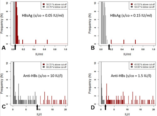
Figure 2. HBsAg and anti-HBs testing in DBS eluates (modified from17). The ROC-guided increase of the cut-off value for HBsAg quantification from 0.05 IU/ml (A) to 0.15 IU/ml (B) reduced the rate of false-positive results from 14.7 % to 0 %. Similarly, ROC analysis of anti-HBs concentrations suggested a decrease of the respective threshold from 10 IU/l (C) to 1.5 IU/l (D), thereby completely eliminating false-negatives, which initially accounted for 14.2 % of all determinations. Please click here to view a larger version of this figure.
The results of the coupled serum/DBS analyses are summarized in Table 117.No non-specificity was recorded in the course of DBS testing for any of the seven analytes.
When DBS eluates were investigated for the presence of HBsAg, i. e. the serological key parameter of both acute and chronic infection with HBV, an analytical sensitivity of 98.6 % was determined. HBsAg concentrations in the two falsely-negative sera were 749 IU/ml and 6 IU/ml, respectively. Anti-HBc antibodies were successfully detected in 176 out of 204 (i. e 86.3 %) DBS eluates. Twenty-two out of the 28 specimens which were not detected by the measurements originated from individuals co-infected with HIV. Thus, if HIV-positive individuals were excluded from the calculation, a sensitivity of 97.1 % was achieved for retrieving anti-HBc antibodies from eluted dried whole venous blood. Similar observations held true for the determination of anti-HBs antibodies. Even after adjustment of the cut-off value of the assay to 1.5 IU/l, nine discrepant serum DBS results occurred. In the patients not co-infected with HIV, serum anti-HBs concentrations of 11 – 26 IU/l were found, which were too low to allow for detection after elution of the dried blood.
Anti-HCV antibodies are indicative of either an acute or chronic or resolved infection. Only four (i. e. 2.2 %) falsely-negative anti-HCV results were determined from DBS eluates and further serological and molecular analyses clearly demonstrated that the respective sera were very probably taken from patients, whose infections had long since been resolved.
The detection of anti-HIV antibodies in DBS eluates with the fully automated system was a completely smooth process which yielded an analytical sensitivity of 100 %.
“Mean HBV DNA concentration from whole blood eluates was performed on 100 specimens (mean DNA concentration: 1,573,898 IU/ml, range: < 357 - > 17,860,000 IU/ml) and yielded a value of 93.0 %. Thus, seven samples with low serum HBV DNA concentrations between 409 and 3,643 IU/ml were not detected by the DBS testing. Of 100 sera which had proven to be HCV RNA positive (mean HCV RNA concentration: 1,415,944 IU/ml, range: 2,479 - >7,692,000 IU/ml), corresponding whole blood aliquots were available. The comparative investigation resulted in an analytical sensitivity of 100 % for HCV RNA determinations from DBS eluates.”17
To test the outlined protocol for preparing and processing DBS under field conditions, it was used in close collaboration with the Robert Koch-Institute (Berlin, Germany) during the pilot phase of the study “Drugs and Chronic Infectious Diseases”, which was conducted on active drug users in the German cities of Berlin and Essen18. All 534 participants enrolled (433 men and 101 women) underwent capillary blood sampling as described in detail in sections 2.2.1 – 2.2.3 of the preceding protocol, and DBS were, again, eluted by addition of 1,000 µl of PBS-based buffer.
Two individuals were diagnosed to be chronically infected with HBV, and 39 showed the serological pattern of a resolved HBV infection. Furthermore, fifteen participants were positive for anti-HBs (status after vaccination) and eight were found to be positive for anti-HBc alone. The anti-HCV prevalence among the participants was 57.3 % (N = 193) in Berlin and 73.0 % (N = 144) in Essen. From the antibody-positive samples 65 % (N = 125) and 62 % (N = 89) were also viremic. Twenty-five out of the 534 individuals tested positive for anti-HIV. Nineteen of the infections were already known and 24 of these individuals were co-infected with HCV.
The results obtained by DBS testing were carefully compared with the study participants’ anamnestic data regarding the self-reported HBV and HCV status. This comparison in conjunction with the results of testing coupled serum/DBS pairs led to minor modifications of the testing algorithm for the main phase of the “DRUCK Study”, which will enrol active drug users in six additional large German cities: “Individuals infected with HIV will be always tested for the presence of HBV DNA. Participants whose DBS eluates are positive for anti-HBc or anti-HBs should be subjected to a venipuncture during a second consultation in order to definitely clarify their anti-HBc/anti-HBs status and, finally, all DBS eluates will be screened for HCV RNA regardless of the results of anti-HCV testing.”17
| Parameters | Coupled sera/DBS eluates (N) | Discrepant results (N) | Reason for discrepancies between testing serum and DBS eluates |
| HBV | |||
| HBsAg | 299 | 2 | Two patients with chronic HBV infection. Both were under antiviral treatment and one of them was co-infected with HIV. HBsAg concentrations in serum: 749 IU/ml and 6 IU/ml, respectively. |
| Anti-HBc | 305 | 28 | In serum, twenty-two patients had a resolved HBV infection. Six individuals showed the serological finding “anti-HBc alone”. Twenty-two of the patients were co-infected with HIV. From the “HBV resolvers”, twenty were assessed as “anti-HBs positive alone” by DBS testing and, thus, were falsely classified as “condition after vaccination”. |
| Anti-HBs | 310 | 9 | The four patients not co-infected with HIV had serum anti-HBs concentrations of 11 ‑ 26 IU/l, which were too low to allow for antibody detection after elution of the dried blood spots. |
| HBV DNA | 150 | 7 | The respective sera had been obtained from seven patients with low serum HBV DNA concentrations ranging from 409 IU/ml to 3,643 IU/ml. |
| HCV | |||
| Anti-HCV | 339 | 4 | These four sera had been taken from patients, with long since resolved HCV infections. |
| HCV RNA | 150 | 0 | – |
| HIV | |||
| HIV 1-p24/anti-HIV 1/2 | 209 | 0 | – |
Table 1: Results obtained from 1,762 DBS, which were prepared and processed from whole venous blood following the protocol proposed in this communication, compared to the findings in coupled serum samples as a reference.
Discussion
DBS have been used for 100 years2 but, surprisingly, there is still no general consensus about their preparation and processing. To date, a sufficient standardization of this important pre-analytical phase has only been achieved in the field of newborn screening14, whereas a variety of different protocols exists for all other applications of DBS testing5, 16, 23. To overcome this remarkable heterogeneity, a comprehensive step-by-step instruction for preparing and processing DBS to be utilized in immunoassays and by molecular techniques is presented in this communication and evaluated with regard to its effectiveness for detecting markers of HBV, HCV and HIV infections. The focus of the following discussion is primarily placed on the different steps of the suggested protocol.
In the history of DBS testing many different filter paper cards have been used2 but today only two commercial sources are approved by the FDA as class II medical devices for blood collection5, 16. These filter card systems are highly uniform and have very similar absorption characteristics so that analytical results obtained from capillary blood prepared on either of them do not differ by more than 4% – 5%28. Not surprisingly, Masciotra and co-workers29 therefore detected HIV-1 RNA equally well with a qualitative assay after elution from blood collection cards from different sources. Given these data, it can be virtually excluded that any of the discrepancies observed when testing coupled serum/DBS pairs for markers of HBV, HCV and HIV infections17 was caused by the choice of the filter card alone. However, when accurate quantification of an analyte is indispensable, source-to-source variation may no longer be negligible and more sophisticated techniques, e.g., perforated DBS (PDBS) as a method for microsampling30 or the preparation of dried serum spots (DSS)31 may have to be applied instead of conventional DBS testing10.
Since only small quantities of blood are used for DBS testing (one drop of capillary blood consists of approximately 50 µl)5, 16, variations in the sample volume are crucial and, admittedly, at least some of the discrepancies between serological results and participant’s self-reported HBV and HCV status recorded during the piloting of the “DRUCK Study” are attributable to this variable. The single most important factor for minimizing “fluctuations” of the sample volume is undoubtedly a correct technique of capillary blood collection, which can only be achieved by careful and ongoing training of the technical personnel22. Furthermore, as a measure of quality control, all laboratories working with DBS should have already initiated procedures for identifying and subsequently excluding DBS specimens that have to be considered as unsatisfactory or invalid16.
Studies conducted primarily in the context of HIV32 and newborn screening33 have shown that high humidity may lead to a degradation of analytes, but hitherto no consensus has been reached regarding the question of how long DBS should be air-dried. An interval of at least 4 hr or preferably O/N is proposed in this communication, which therefore adopted conditions that were used in the vast majority of all relevant publications32, 34 Data on storage of DBS to be subsequently used for HBV and HCV testing are conflicting. In 1981, Villa and co-workers35, who applied contemporary analytical techniques, reported that storage at RT did not affect the results of HBV analyses during the entire observation period of 180 days if antibody titers were > 1/1,000, but that DBS became borderline-positive or even negative after 15 days when titers in serum were only 1/100. Storage at -20 °C or 4 °C did not result in a substantial improvement. In contrast, a study conducted thirty years later36 tested replicates of a HBV-positive sample, and the authors found that anti-HBc as well as anti-HBs antibodies were stable for up to 183 days at RT, whereas HBsAg under the same conditions became false-negative already after 63 days. With regard to HBV DNA detection from DBS eluates, the concentration of the viral nucleic acid was stable for at least seven days at 37 °C37 or proved to be “resistant” to storage at RT for up to three weeks38. Testing for anti-HCV antibodies using two commercially available third generation immunoassays39 provided accurate results for a period of 117 days using DBS samples stored at -20 °C, 2 – 8 °C, and 20 – 25 °C, respectively. Storage at -20 °C, however, resulted in the lowest variation of optical densities. Applying a fourth generation anti-HCV ELISA, i.e., Monolisa HCV-Ag-Ab-ULTRA, in the context of DBS testing, Larrat et al.40 observed a sharp decrease in analytical specificity after storing the DBS specimens for more than three days at RT. On the other hand, precise testing results were obtained with the same kit utilizing samples deposited for 60 days under various conditions (-20 °C, 2 – 8 °C, and 22 – 26 °C) by Brandao and co-workers41. Observations on the decline of HCV RNA concentrations in DBS under various storage conditions range from no significant alteration at RT for up to one year42 to a tenfold change after four weeks at ambient temperature43. Taking this rather conflicting data on the stability of HBV and HCV antigens, nucleic acids and antibodies into account, it seemed reasonable to retract in the proposed protocol to a consensus, which was defined earlier for the storage of DBS specimens to be used for HIV testing15, 30: For short-term deposition (up to two weeks) antigens, viral nucleic acid, and antibodies are regarded as stable at RT, whereas optimal storage for longer periods is at frozen conditions.
As a rule, three parameters should be considered when designing an elution protocol: (1) the elution buffer; (2) the duration and temperature of elution; and (3) the elution volume23. In the vast majority of all relevant publications phosphate-buffered saline (PBS) was used for eluting DBS, and most authors added a protein, e.g., bovine serum albumin (BSA) or Tween 20, a surfactant, in order to improve the assay signal by stabilizing proteins, as they go into solution, and simultaneously blocking non-specific binding sites23. Only a few reports, which directly compare different elution buffers in the context of DBS testing, are available. Villar et al.36, e.g., recorded almost equivalent elution capacities for all buffers used, but PBS/BSA 0.5% resulted in the lowest level of non-specific reactivity. A very similar observation was made by Croom and co-workers44 when applying the specimen diluent of the Genetics Systems rLAV EIA for the elution of anti-HCV antibodies from DBS. Since the risk of sample degradation is exceedingly low in the first hours after preparation of DBS (see above), it was decided to incubate the spots O/N at ambient temperature and to support the elution process by gentle end-over-end mixing. This approach has the advantage that specimens punched out the previous day can be directly transferred to routine diagnostics early the next morning23. The volume of the elution buffer should be adapted to the minimal respective requirements of the assays used for subsequent analyses in order to keep the dilution factor as low as possible. However, a careful optimization of the elution conditions for every single analyte was not possible in the preceding evaluation because a high sample throughput had to be guaranteed in a comparatively short time during the “DRUCK Study”17. Consequently, the rather unfavorable volume of 1,000 µl of PBS-based buffer was used in order to complete the whole elution process for all parameters in two separate operations.
This approach one the one hand fails “completely in the anti-HBc/anti-HBs system for those individuals infected with HIV due to the low antibody concentrations; … and it required molecular biological procedures with optimal analytical sensitivity with regard to HBV DNA and HCV RNA tests.”17 On the other hand, the high elution volume proved to be in no way disadvantageous for the determination of HBsAg, anti-HCV, and anti-HIV by the kits used throughout the evaluation. “The detection of HBsAg positive materials from whole blood eluates succeeded with a sensitivity of 98.6% to a similarly high degree as in previous studies, which had in part used a much smaller elution volume of 100 µl, 250 µl, or 600 µl, or 500 µl”17, 36, 45, 46. The sensitivity of 97.8% determined in the investigation of 179 serum/DBS pairs for anti-HCV antibodies corresponded to the results of already existing reports24, 44, 47, 48, 49, which had worked with an elution volume that was lower by a factor of 5 to 10. “In addition, the protocols for anti-HCV detection had been appropriately optimised … by stipulating their own cut-off points”17, 24, 48, 49 “or by increasing the sample volumes from 20 µl to 100 µl.”17, 49 Finally, the analytical specificity and sensitivity (100% each) established for anti-HIV detection were equal or superior to the performance characteristics of other immunoassays specifically adapted to DBS testing50, 51 “or a stepwise procedure with the combined use of several anti-HIV tests”17, 52. “They, indeed, also exceeded the performance record of an assay that had been specially developed and optimised for the detection of anti-HIV antibodies in DBS eluates (Q-Prevent HIV 1 + 2 DBS kit).”17, 53
Taken together, the comprehensive step-by-step protocol presented in this communication proved to be a feasible and user-friendly tool for preparing and processing DBS and can, thus, be used reliably in diagnostic virology. It allows approaches using automation 54 and due to its excellent performance characteristics has the potential to serve as a kind of foundation-stone for a future generally accepted consensus protocol in the global field of DBS testing in laboratory medicine.
Disclosures
In the past, RSR received an honorarium for delivering lectures and financial support for his scientific work from Abbott Diagnostics. NG and OM declare that they have no conflicting interests.
Acknowledgements
This communication is based on the results previously published in Virology Journal17 in open access mode under the terms of the Creative Commons Attribution License (http://creativecommons.org/licenses/by/2.0) with BioMed Central as a licensee. The authors gratefully acknowledge the fruitful collaboration in the framework of piloting the study “Drugs and Chronic Infectious Diseases” (“DRUCK Study”) with U. Marcus, W. Cai, W. Zhang, and R. Zimmermann from the Robert Koch-Institute (Berlin, Germany) and are thankful to S. Moyrer for taking the photographs assembled in Figure 1 as well as to D. F. Whybrew, Ph.D, (Göttingen, Germany) for correcting the the manuscript. They also express their appreciation for skilful technical assistance to J. Ackermann, E. Bayrambasi, U. Büttner, S. Dziubek, I. Jakobsche, B. Krellenberg, H. Krohn, P. Kusimski, A. Metcalf, J. Piejek, S. Saar, M. Schröter and K. Seidel. This study was supported in part by a grant of the German Ministry of Health to the National Reference Centre for Hepatitis C.
Materials
| Name | Company | Catalog Number | Comments |
|---|---|---|---|
| Latex rubber gloves | Hartmann | #4049500041515 | |
| 70% isopropyl alcohol | Bode Chemie | #9768050 | |
| Mulifly safety cannula | Sarstedt | #85.1638 | |
| EDTA blood collection tube | Sarstedt | #02.1066.001 | |
| Adhesive bandage | Hartmann | #2994163 | |
| Dry gauze pad | Hartmann | #143213 | |
| Singel-use safety lancet | Owen Mumford | #3802421 | |
| Filter paper card | Whatman/sigmaaldrich | #10534612 | Filter paper card Grade 903 |
| Disposable pipette tips | Starlab | #S1126-7810, #S1120-8810 | |
| Zipper bag | Flexico | #20219 | |
| Mini desiccant bag | Tropack | #- | |
| Disposable Punch | pfmmedical | #48601 | 6.0 mm Disposable Biopsy Punch |
| Disposable Forceps | Servoprax | #H7301 | |
| 12 Well Cell Culture Plate | Greiner | #665180 | |
| PBS Buffer | Gibco | #14190-136 | |
| Tween 20 | Applichem | #A1389.0500 | |
| Sodium Azide | Merck | #66880250 | |
| Safe-Lock Tubes, 1.5 ml | Eppendorf | #30120086 | |
| ARCHITECT HBsAg (Quantitative) | Abbott | #6C3642 | |
| ARCHITECT anti-HBc II | Abbott | #8L4425 | |
| ARCHITECT anti-HBs | Abbott | #7C1825 | |
| ARCHITECT anti-HCV | Abbott | #6C3725 | |
| ARCHITECT HIV Ag/Ab Combo | Abbott | #4J2727 | |
| MagNA Pure 96 DNA and Viral NA Large Volume Kit | Roche | #6374891001 | |
| artus HBV LC PCR Kit | Qiagen | #4506063 (24 tests), #4506065 (96 tests) | |
| VERSANT HCV TMA Assay IVD | Siemens Healthcare | #2554311 |
References
- Schmidt, V. Ivar Christian Bang (1869 - 1918), founder of modern clinical microchemistry. Clin Chem. 32 (1), 213-215 (1986).
- Hannon, W. H., Therell, B. L. Overview of the history and applications of dried blood spot samples. Dried blood spots. Applications and techniques. Li, W., Lee, M. S. 3, Wiley. Hoboken. 3-15 (2014).
- Bang, I., Bergmann, J. F. Der Blutzucker. , Wiesbaden. Available from: https://archive.org/details/derblutzucker00bang (1913).
- Chapman, O. D. The complement-fixation test for syphilis. Use of patient's whole blood dried on filter paper. Arch Derm Syphilol. 9 (5), Available from: http://archderm.jamanetwork.com/article.aspx?articleid=495323 607-611 (1924).
- Smit, P. W., Elliott, I., Peeling, R. W., Mabey, D., Newton, P. N. An overview of the clinical use of filter paper in the diagnosis of tropical diseases. Am J Trop Med Hyg. 90 (2), 195-210 (2014).
- Kuehn, B. M. After 50 years, newborn screening continues to yield public health gains. JAMA. 309 (12), 1215-1217 (2013).
- Guthrie, R. Blood Screening for Phenylketonuria. JAMA. 178 (8), Available from: http://jama.jamanetwork.com/article.aspx?articleid=332184 863(1961).
- Guthrie, R., Susi, A. A simple phenylalanine method for detecting phenylketonuria in large populations of newborn infants. Pediatrics. 32 (3), 338-343 (1963).
- Snijdewind, I. J., van Kampen, J. J., Fraaij, P. L., vander Ende, M. E., Osterhaus, A. D., Gruters, R. A. Current and future applications of dried blood spots in viral disease management. Antiviral Res. 93 (3), 309-321 (2012).
- Demirev, P. A. Dried blood spots: analysis and applications. Anal Chem. 85 (2), 779-789 (2013).
- Li, W., Lee, M. S. Preface. Dried blood spots. Applications and techniques. Li, W., Lee, M. S. , Wiley. Hoboken. VIII-IX (2014).
- Guder, W. G., Narayanan, S., Wisser, H., Zawta, B. Diagnostic Samples: From the Patient to the Laboratory. The Impact of Preanalytical Variables on the Quality of Laboratory Results. , 4th edition, Wiley-VHC. Weinheim. (2009).
- Mei, J. V., Alexander, J. R., Adam, B. W., Hannon, W. H. Use of filter paper for the collection and analysis of human whole blood specimens. J Nutr. 131 (5), 1632S-1636S (2001).
- NBS01-A6. Blood Collection on Filter Paper for Newborn Screening Programs; Approved Standard-Sixth Edition. , Clinical and Laboratory Standards Institute (CLSI). Wayne, PA. Available at: http://shop.clsi.org/c.1253739/site/Sample_pdf/NBS01A6_sample.pdf (2013).
- WHO manual for HIV drug resistance testing using dried blood spot specimens. , World Health Organisation (WHO). Available at: http://www.who.int/hiv/pub/drugresistance/dried_blood_spots/en/ (2012).
- Mei, J., Lee, M. S. Dried blood spots sample collection, storage, and transportation. Dried blood spots. Applications and techniques. Li, W., Lee, M. S. , Wiley. Hoboken. 21-31 (2014).
- Ross, R. S., et al. Detection of infections with hepatitis B virus, hepatitis C virus, and human immunodeficiency virus by analyses of dried blood spots. Performance characteristics of the ARCHITECT system and two commercial assays for nucleic acid amplification. Virol J. 10 (72), (2013).
- Zimmermann, R. DRUCK-Studie – Drogen und chronische Infektionskrankheiten in Deutschland. Ergebnisse der Pilotierung eines Sero- und Verhaltenssurveys bei i. v. Drogengebrauchern. Epidemiol Bull. 33, Available from: http://www.rki.de/DE/Content/Infekt/EpidBull/Archiv/2012/Ausgaben/33_12.pdf?__blob=publicationFile 335-339 (2012).
- World Medical Association, Declaration of Helsinki. Ethical principles for medical research involving human subjects. JAMA. 310 (20), 2191-2194 (2013).
- Young, D. S., Bermes, E. W., et al. Specimen collection and processing; sources of biological variation. Tietz textbook of clinical chemistry. Burtis, C. A., Ashwood, E. R. 58, 2nd edition, W. B. Saunders Company. Philadelphia. 58-101 (1994).
- H3-A6. Procedures for the collection of diagnostic blood specimens by venipuncture. , Clinical and Laboratory Standards Institute (CLSI). Wayne, PA. Available from: http://shop.clsi.org/site/Sample_pdf/H3A6_sample.pdf (2007).
- H04-A6. Procedures and devices for the collection of diagnostic capillary blood specimens. , Clinical and Laboratory Standards Institute (CLSI). Wayne, PA. Available from: http://shop.clsi.org/site/Sample_pdf/H04-A6.pdf (2008).
- McDade, T. W. Development and validation of assay protocols for use with dried blood spot samples. Am J Hum Biol. 26 (1), 1-9 (2014).
- Judd, A., et al. Evaluation of a modified commercial assay in detecting antibody to hepatitis C virus in oral fluids and dried blood spots. J Med Virol. 71 (1), 49-55 (2003).
- Zweig, M. H., Campbell, G. Receiver-operating characteristic (ROC) plots: A fundamental evaluation tool in clinical medicine. Clin Chem. 39 (4), 561-577 (1993).
- Greiner, M., Pfeiffer, D., Smith, R. D. Principles and practical application of the receiver-operating characteristic analysis for diagnostic tests. Prev Vet Med. 45 (1-2), 23-24 (2000).
- Clopper, C. J., Pearson, E. S. The use of confidence or fiducial limits illustrated in the case of the binominal. Biometrika. 26 (4), Available from: http://www.bios.unc.edu/~mhudgens/bios/662/2007fall/clopper.pdf 404-413 (1934).
- Mei, J. V., Zobel, S. D., Hall, E. M., De Jesús, V. R., Adam, B. W., Hannon, W. H. Performance properties of filter paper devices for whole blood collection. Bioanalysis. 2 (8), 1403-1410 (2010).
- Masciotra, S., et al. Evaluation of blood collection filter papers for HIV-1 DNA PCR. J Clin Virol. 55 (2), 101-106 (2012).
- Li, F., Zulkoski, J., Fast, D., Michael, S. Perforated dried blood spots: a novel format for accurate microsampling. Bioanalysis. 3 (20), 2321-2333 (2011).
- Li, Y., Henion, J., Abbott, R., Wang, P. The use of a membrane filtration device to form dried plasma spots for the quantitative determination of guanfacine in whole blood. Rapid Commun Mass Spectrom. 26 (10), 1208-1212 (2012).
- Basavaraju, S. V., Pitman, J. P. Dried blood spots for use in HIV-related epidemiological studies in resource-limited settings. Dried blood spots. Applications and techniques. Li, W., Lee, M. S. , Wiley. Hoboken. 76-94 (2014).
- Chace, D. H., Spitzer, A. R., De Jesus, V. R. Applications of dried blood spots in newborn and metabolic screening. Dried blood spots for use in HIV-related epidemiological studies in resource-limited settings. Dried blood spots. Applications and techniques. Li, W., Lee, M. S. 3, Wiley. Hoboken. 53-75 (2014).
- Gakhar, H., Holodniy, M. Use of dried blood spot samples in HCV-, HBV-, and influenza-related epidemiological studies. Dried blood spots. Applications and techniques. Li, W., Lee, M. S. 3, Wiley. Hoboken. 95-113 (2014).
- Villa, E., Cartolari, R., Bellentani, S., Rivasi, P., Casolo, G., Manenti, F. Hepatitis B virus markers on dried blood spots. A new tool for epidemiological research. J Clin Pathol. 34 (7), 809-812 (1981).
- Villar, L. M., de Oliveira, J. C., Cruz, H. M., Yoshida, C. F. T., Lampe, E., Lewis-Ximenez, L. L. Assessment of dried blood spot samples as a simple method for detection of hepatitis B virus markers. J Med Virol. 83 (9), 1529-1510 (2011).
- Lira, R., et al. Use of dried blood samples for monitoring hepatitis B virus infection. Virol J. 6 (1), 153(2009).
- Jardi, R., et al. Usefulness of dried blood samples for quantification and molecular characterization of HBV-DNA. Hepatology. 40 (1), 133-139 (2004).
- Marques, B. L., et al. Dried blood spot samples: optimization of commercial EIAs for hepatitis C antibody detection and stability under different storage conditions. J Med Virol. 84 (10), 1600-1607 (2012).
- Larrat, S., et al. Performance of an antigen–antibody combined assay for hepatitis C virus testing without venipuncture. J Clin Virol. 55 (3), 220-225 (2012).
- Brandao, C. P., et al. Simultaneous detection of hepatitis C virus antigen and antibodies in dried blood spots. J Clin Virol. 57 (2), 98-102 (2013).
- Bennett, S., et al. Detection of hepatitis C virus RNA in dried blood spots. J Clin Virol. 54 (2), Available from: http://www.ncbi.nlm.nih.gov/pubmed/22418454 106-109 (2012).
- Abe, K., Konomi, N. Hepatitis C virus RNA in dried serum spotted onto filter paper is stable at room temperature. J Clin Microbiol. 36 (10), 3070-3072 (1998).
- Croom, H. A., et al. Commercial enzyme immunoassay adapted for the detection of antibodies to hepatitis C virus in dried blood spots. J Clin Virol. 36 (1), 68-71 (2006).
- Mendy, M., et al. Hepatitis B surface antigenaemia and alpha-foetoprotein detection from dried blood spots: applications to field-based studies and to clinical care in hepatitis B virus endemic areas. J Viral Hepat. 12 (6), 642-647 (2005).
- Komas, N. P., Baï-Sepou, S., Manirakiza, A., Léal, J., Béré, A., Le Faou, A. The prevalence of hepatitis B virus markers in a cohort of students in Bangui, Central African Republic. BMC Infect Dis. 10, 226(2010).
- Parker, S. P., Cubitt, W. D., Ades, A. E. A method for the detection and confirmation of antibodies to hepatitis C virus in dried blood spots. J Virol Methods. 68 (2), 199-205 (1997).
- McCarron, B., et al. Hepatitis C antibody detection in dried blood spots. J Viral Hepat. 6 (6), Available from: http://www.ncbi.nlm.nih.gov/pubmed/10607263 453-456 (1999).
- Tuaillon, E., et al. Dried blood spot for hepatitis C virus serology and molecular testing. Hepatolog. 51 (3), 752-758 (2010).
- Solomon, S. S., et al. Dried blood spots (DBS): a valuable tool for HIV surveillance in developing/tropical countries. Int J STD AIDS. 13 (1), 25-28 (2002).
- Lakshmi, V., Sudha, T., Bhanurekha, M., Dandona, L. Evaluation of the Murex HIV Ag/Ab Combination assay when used with dried blood spots. Clin Microbiol Infect. 13 (11), 1136-1110 (2007).
- Sarge-Njie, R., et al. Evaluation of the dried blood spot filter paper technology and five testing strategies of HIV-1 and HIV-2 infections in West Africa. Scand J Infect Dis. 38 (11-12), 1050-1056 (2006).
- de Castro, A. C., Borges, L. G. dosA., de Souza, R. daS., Grudzinski, M., D’Azevedo, P. A. Evaluation of the human immunodeficiency virus type 1 and 2 antibodies detection in dried whole blood spots (DBS) samples. Rev Inst Med Trop Sao Paulo. 50 (3), 151-156 (2006).
- Fan, L., Wan, K., Kavetskaia, O., Wu, H. Automation in dried blood spot sample collection, processing, and analysis for quantitative bioanalysis in pharmaceutical industry. Dried blood spots. Applications and techniques. Li, W., Lee, M. S. , Wiley. Hoboken. 229-234 (2014).
Reprints and Permissions
