影象残留的颜色
English
Share
Overview
资料来源: 实验室的乔纳森 Flombaum — — 约翰 · 霍普金斯大学
人类色觉是令人印象深刻。正常色觉的人可以告诉分开数以百万计的个人色彩。最令人惊讶的是,这种能力是相当简单的硬件配置实现的。
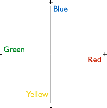
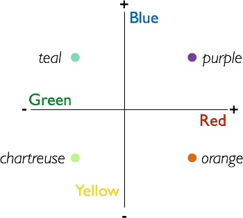
一部分人类色觉的力量来自于一些聪明的人类大脑中的工程。在那里,色觉依赖于所谓的对手的系统。这意味着一种刺激的存在被视为另一,缺乏证据,反之亦然;没有一种刺激的情况下被认为是证据为别人的存在。尤其是,在人类的大脑有火他们接收信号显示蓝色指示灯亮起,或时他们不会收到信号,表示黄灯的细胞。同样,有火在黄色或蓝色的缺席的细胞。蓝色和黄色因此被视为对手值在一维空间,并可以认为的负面与正面的价值观,在笛卡尔平面的一个轴上。如果刺激定性为负值,则在该轴,它也不能有积极的价值。所以,如果它的特点为黄色,它还不能称为蓝色。同样,绿色和红色 (或真的,品红),占据另一个对手维度。有人类大脑中响应出现一个或如果缺少了其他的细胞。图 1 和图 2在笛卡尔术语解释颜色对立特性。

图 1。的对手颜色尺寸。人类大脑处理颜色使用对手尺寸系统。这是蓝色和黄色占领一个轴,可以被认为是只是积极或消极的和红色和绿色占领另一个轴的二维平面。系统的后果是大脑处理的一些颜色作为指示他人,没有存在,反之亦然。所有的感知颜色占据对手空间中的点。

图 2。所有的感知颜色占据对手空间中的点。这里显示的是颜色中的对手空间两个维度的每个包含非零值的示例。
一种方法那颜色对立特性是发现在 1878 由埃瓦尔德赫林,甚至之前科学家成像大脑本身获得的技术-是通过称为一个颜色残像的一种错觉。影象残留的今天仍然使用证明人类的色彩感知的对手属性,研究他们。
该视频演示如何创建一个颜色的残像幻觉和从人类观察员收集主观感知响应的简单方法。
Procedure
Results
For each of the inducer colors in the experiment, identify the most frequently selected after-color. Make a table that visualizes the results, like the one in Figure 8.

Figure 8. Representative result. Most frequently selected after-colors as a function of inducer colors. The most frequently perceived after-colors will be opponent values of the respective inducers.
The most frequently perceived after-colors should be opponent values of the respective inducer colors. The reason is because color-sensitive cells in the human brain are mapped spatially-they respond to specific regions of space dependent on where the subject fixates their eyes. Normally, people move their eyes around, causing different cells to share the burden of responding to regions of external space. By fixating the disc in the inducer images (slide #1 in each pair), the observer causes the same groups of cells to respond in a sustained way to the saturated colors present in a given region of external space. During the fixation period, these cells respond heavily. Blue-sensitive cells produce large blue signals, yellow-sensitive cells produce large yellow signals, and so on. When the black and white image is suddenly shown, and while the observer still fixates, these cells are no longer stimulated-there is no color in the image. But, because they were signaling so strongly a moment before, the rest of the brain interprets their sudden lack of activity as signaling the presence of an opponent color. The sudden lack of signaling in blue-sensitive cells is interpreted as the presence of yellow. The sudden lack of signaling in the yellow-sensitive cells is interpreted as the presence of blue, and so on. The brain interprets the absence of activity in color cells as indicating the presence of opponent colors, when in fact the lack of activity in this case is caused by the absence of color altogether. The brain is effectively tricked, causing people to see colors where there aren't any because of the way it organizes color in terms of opponent dimensions.
Applications and Summary
Color opponency is among the great demonstrations of the scientific method. Researchers in the 1800s were able to infer the nature of color representation in the human brain without any ability to observe brain activity. Today, in fact, color afterimages have become a useful tool for identifying the brain regions involved in processing color. In monkeys, scientists have recorded neurons that fire as though color is present, when it is not, after showing the monkeys sequences of slides that produce afterimages in human observers.1,2 Similarly, with fMRI, scientists have found regions of visual cortex that respond selectively to presence of a color, and that also respond when that color is perceived as an illusion, induced by an afterimage pair of slides.
References
- Zeki, S. Colour coding in the cerebral cortex: the reaction of cells in monkey visual cortex to wavelengths and colours. Neuroscience, 9(4), 741-765 (1983).
- Conway, B. R., & Tsao, D. Y. Color architecture in alert macaque cortex revealed by FMRI. Cerebral Cortex, 16(11), 1604-1613 (2006).
Transcript
Human color vision is impressive, and can help us see—and distinguish between—over a million distinct hues. Psychologists use a visual illusion called the color afterimage to study our perception of color.
What a person perceives as the hue of objects they encounter, like an orange, starts with sensory information received by the eyes.
When this light enters an individual’s eyes, the cornea and lens focus it onto photoreceptor cells in the retina. In response, these cells generate signals that are relayed to the visual cortex, where the colors of objects are identified.
Along this pathway, individual neurons exist that respond differently to distinct color signals.
For example, there are certain neurons—called “green on, red off” cells—that are only activated by green light. However, these same cells are inhibited by red light.
Similarly, there are neurons called “red on, green off” cells that respond to red, but which are inhibited by green. Thus, components of the brain treat these colors as “opponents”: the presence of one is interpreted as the other being absent.
As a result, green and red can be thought of as occupying opposite values on a two-dimensional plane—respectively, negative and positive. In fact, all the specific individual colors that we see are ordered pairs of points in this coordinate system.
This video demonstrates how to investigate such opponency, originally discovered by Ewald Hering in 1878, using the color afterimage illusion—where individuals’ brains are tricked into perceiving an opponent color in a position previously filled by its antagonistic hue.
We not only explain how to generate stimuli, and collect and interpret color perception data, but we also explore how researchers can apply this illusion to study different facets of color vision.
In this experiment, participants are asked to perform several trials in which they must first stare at colored shapes—during what is called the inducer phase—and then while looking at the same shapes in black and white, state what hues they see—referred to as the afterimage phase.
During the first inducer phase, two of the same shape—like a star—are shown side-by-side on a computer screen for 10 s. These shapes are identical in size and orientation, but are filled with different colors, such as blue and yellow, each of which should have a distinct opponent—a stipulation critical for the second phase.
For the duration of each trial, participants are instructed to fixate on a circle centered between the shapes, in order to prevent eye movements that could interfere with the illusion.
In the afterimage phase, the black disc remains onscreen, but the colored pictures on either side of it are replaced by images of the same shapes devoid of color—white stars outlined in black.
Participants are then asked what hue they see in a random position in a star, and the color they report serves as the dependent variable.
The trick here is that during the inducer phase, color-sensitive cells are active—for example, some fire heavily in response to a blue stimulus. However, the switch from the inducer stars to black-and-white shapes results in these cells suddenly ceasing to signal due to the absence of any color.
Yet, participants’ brains interpret this abrupt stoppage as meaning that the opponent colors are present—a lack of signaling from previously active blue-sensitive cells is taken as evidence for yellow.
As a result, participants will fill in the black-and-white shapes with the opponent colors of those shown during the inducer phase—where a blue inducer star was, a yellow afterimage star is perceived, and vice versa.
This is the color afterimage illusion, which quickly fades as soon as participants move their eyes.
Due to how the brain organizes colors, it is expected that participants will predominantly see after-colors that are opponents of the inducer: green for red, for example.
To prepare stimuli for the experiment, first open a blank slide in a slide-editing program. Use the shape tool to generate two equally-sized stars—one on the right side and the other on the left—both centered vertically.
Then, create a small black disc positioned between them to serve as the fixation point.
To generate images for an inducer phase, outline both shapes in black. Then, select the star on the left and fill it uniformly with a bright blue. Similarly, fill the right one with a bright yellow.
Repeat this process, creating additional inducer slides with stars in different colors, like green and red and another set in green and blue.
Now, copy one of the inducer slides. In this replica, keep both stars outlined in black, but change their internal colors to white. This completed slide will be shown in the afterimage phase of all trials.
Finally, arrange them so that each stimulus series consists of two slides: a colored inducer set followed by an afterimage one in black and white for each trial.
When the participant arrives, direct them to a computer monitor and verify that they are not color-blind. Proceed by explaining the instructions for the task that they will be performing.
Emphasize that throughout a trial, the participant should try to avoid moving their eyes, and remain focused on the black disc that will appear in the center of the computer screen.
Once you are confident that the participant understands the afterimage task, have them perform between 10 and 20 trials. For each one, next to inducer-color, record the hue that was selected as the after-color.
To analyze the data, tally the results and construct a table depicting the most frequently selected after-color for each inducer color.
Notice that the selected after-colors are predominantly opponents of the inducer hues, indicating that the participants’ brains were tricked, causing them to see hues that weren’t actually there—color afterimage illusions.
Now that you know how to use visual stimuli to elicit a color afterimage illusion, let’s look at how researchers can use this technique to better understand the anatomical basis behind color vision, and diseases that affect this perception.
Up until now, we’ve focused on normal vision. However, variations of the afterimage test can also be employed to better understand diseases that affect color perception—like types of color blindness in which both hues of an opponent pair appear the same.
For example, researchers can compare how long an individual perceives a color afterimage to times reported by normal, control participants. Illusions persisting for abnormal lengths of time can be indicative of a color vision disease.
Afterimage tests can then be paired with other color perception assessments and imaging techniques to pinpoint whether a defect in the retina, visual cortex, or the visual pathway—which relays signals between these two areas—is responsible.
You’ve just watched JoVE’s video on color afterimages. By now, you should know how to use shapes of different hues to investigate this illusion, and collect and interpret opponent color data. Importantly, you should also have an understanding of how afterimages can help identify brain regions involved in color perception, and diagnose diseases relating to color vision.
Thanks for watching!
