JoVE
JoVE
Faculty Resource Center
Research
Behavior
Biochemistry
Biology
Bioengineering
Cancer Research
Chemistry
Developmental Biology
Engineering
Environment
Genetics
Immunology and Infection
Medicine
Neuroscience
JoVE Journal
JoVE Encyclopedia of Experiments
Education
Biology
Chemistry
Clinical
Engineering
Environmental Sciences
Pharmacology
Physics
Psychology
Statistics
JoVE Core
JoVE Science Education
JoVE Lab Manual
JoVE Quiz
JoVE Business
Videos Mapped to your Course
Authors
Librarians
High Schools
About
Sign-In
Sign In
Contact Us
Research
JoVE Journal
JoVE Encyclopedia of Experiments
Education
JoVE Core
JoVE Science Education
JoVE Lab Manual
High Schools
EN
EN - English
CN - 中文
DE - Deutsch
ES - Español
KR - 한국어
IT - Italiano
FR - Français
PT - Português
EN
EN - English
CN - 中文
DE - Deutsch
ES - Español
KR - 한국어
IT - Italiano
FR - Français
PT - Português
Close
Research
Behavior
Biochemistry
Bioengineering
Biology
Cancer Research
Chemistry
Developmental Biology
Engineering
Environment
Genetics
Immunology and Infection
Medicine
Neuroscience
Products
JoVE Journal
JoVE Encyclopedia of Experiments
Education
Biology
Chemistry
Clinical
Engineering
Environmental Sciences
Pharmacology
Physics
Psychology
Statistics
Products
JoVE Core
JoVE Science Education
JoVE Lab Manual
JoVE Quiz
JoVE Business
Videos Mapped to Your Course
Teacher Resources
Get in Touch
Instant Trial
Log In
EN
EN - English
CN - 中文
DE - Deutsch
ES - Español
KR - 한국어
IT - Italiano
FR - Français
PT - Português
Journal
/
Neuroscience
/
干无创多通道头皮脑电传感器在小鼠视觉诱发电位记录中的应用
JoVE Journal
Neuroscience
A subscription to JoVE is required to view this content.
Sign in or start your free trial.
JoVE Journal
Neuroscience
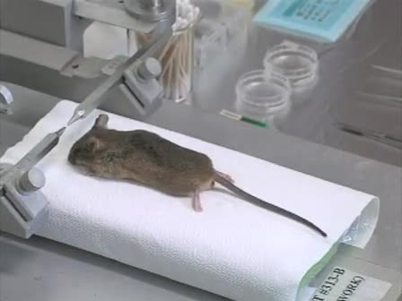
Visual Evoked Potential Recordings in Mice Using a Dry Non-invasive Multi-channel Scalp EEG Sensor
Please note that all translations are automatically generated.
Click here for the English version.
干无创多通道头皮脑电传感器在小鼠视觉诱发电位记录中的应用
DOI:
10.3791/56927-v
•
06:19 min
•
January 12, 2018
•
Chanmi Yeon
,
Donghyeon Kim
,
Kiseon Kim
,
Euiheon Chung
3
1
Department of Biomedical Science and Engineering (BMSE)
,
Gwangju Institute of Science and Technology (GIST)
,
2
School of Electrical Engineering and Computer Science (EECS)
,
Gwangju Institute of Science and Technology (GIST)
,
3
School of Mechanical Engineering (SME)
,
Gwangju Institute of Science and Technology (GIST)
Chapters
00:05
Title
00:53
Sensor Assembly
02:10
Animal Preparation
02:44
Mouse EEG Setup
04:18
Results: Novel Mouse EEG Sensor Shows Repetitive VEP Response and Good Signal Quality
04:49
Conclusion
Summary
Automatic Translation
English (Original)
العربية (Arabic)
中文 (Chinese)
Nederlands (Dutch)
français (French)
Deutsch (German)
עברית (Hebrew)
italiano (Italian)
日本語 (Japanese)
한국어 (Korean)
português (Portuguese)
русский (Russian)
español (Spanish)
Türkçe (Turkish)
Automatic Translation
我们设计了一种干式16通道脑电图传感器, 它是无创、可变形和可重用的。本文介绍了利用干无创多通道脑电传感器对小鼠头皮上的视觉诱发电位 (诱发电位) 信号进行处理的全过程。
Tags
Keywords: Visual Evoked Potential
Non-invasive
Multi-channel
Scalp EEG
Sensor
Mouse
Pre-clinical
Translational Research
Dry EEG
Electrode
Stereotaxic Frame
Impedance
Article
Embed
ADD TO PLAYLIST
Usage Statistics
Related Videos
check_url/56927?article_type=v
电极的制作及植入
海兔californica
完整的,自由的行为的动物多通道的神经和肌肉的录音
check_url/56927?article_type=v
高密度的自由移动,使用基于聚酰亚胺的微电极的小鼠的脑电图记录
check_url/56927?article_type=v
transretinal ERG的录音,从小鼠视网膜杆和锥光反应
check_url/56927?article_type=v
双突触诱发的星形胶质细胞和神经元的反应,在急性海马脑片电生理记录
check_url/56927?article_type=v
提取脑电数据视觉诱发电位记录在功能磁共振成像引导下经颅磁刺激
check_url/56927?article_type=v
在实验视神经脱髓鞘模型大鼠视觉诱发电位记录
check_url/56927?article_type=v
电图同时记录和麻醉大鼠视觉诱发电位
check_url/56927?article_type=v
可视化指南排序的电生理记录使用“SpikeSorter”
check_url/56927?article_type=v
用单单元记录法研究猕猴背视流中的对象表示
check_url/56927?article_type=v
c57bl6 小鼠耳蜗植入术与电唤起脑干反应记录
Read Article