1. Preparation of the placenta at delivery
- Label the umbilical cords of the twins with one (for the first-born) or two (for the second-born) clamps.
- Inspect the maternal and fetal surface of the placenta for completeness or disruption.
- Record the following data: type of cord insertion (central, eccentric, marginal or velamentous), number of blood vessels in the umbilical cord (usually one vein and two arteries, sometimes only one artery) and color difference between both placental shares. A section of the dividing membranes can be sent to Pathology to confirm the type of chorionicity.
- The placenta can then be placed in a plastic bowl and refrigerated until the final examination (best within one week) and color dye injection.
- The placenta must not be frozen or fixed (do not use formalin).
2. Catheterization of the umbilical vessels
- Wash the placenta with warm water or saline.
- Trim the peripheral membranes, remove the inter-twin dividing membrane and peel off the amnions (for better visualization of the vascular anastomoses and better quality of the placental pictures).
- Transect each umbilical cord at approximately 5 cm distance from the cord insertion.
- Gently squeeze out blood clots from the umbilical vessels and placental vessels.
- The umbilical vein is usually easy to identify due to its larger diameter, compared to the smaller diameter of the two umbilical arteries.
- Cannulate the umbilical vein with an appropriately sized catheter. Avoid false passages.
- Cannulate one umbilical artery with a smaller catheter. Use tweezers to widen the lumen of the umbilical artery. Avoid false passages. Only one of the 2 umbilical arteries needs to be catheterized since an anastomosis (of Hyrtl) connects the 2 arteries near the cord insertion.
- Repeat both steps for the other umbilical cord.
- Placement of the catheter can be facilitated by gentle back and forth massaging of the umbilical vessels. Any type of catheter can be used for this procedure. We choose to use (and recycle) the catheters used at our neonatology ward for umbilical catheterization in neonates.
- Tie a piece of tape around both cords to avoid back flow of the colored dye during dye injection.
3. Injection with colored dye
- Connect a 20 ml syringe filled with colored dye to each catheter.
- Any viscous colored dye can be used to visualize the placental angio-architecture. Use contrasting colors to allow good visualization of the anastomoses (dark colors for the arteries, bright colors for the veins).
- Gently inject (with low pressure) the colored dye in the vein while an assistant gently pushes the dye to allow the colored dye to fill all placental vessels, also the smallest ones.
- Pay particular attention to the small vessels near the vascular equator (the vascular equator is the place where the anastomoses from either twin connect with each other).
- Repeat the previous steps to inject colored dye into the artery. Of note: arteries may be more difficult to inject and require more patience.
- Repeat both steps for the other umbilical cord.
4. Evaluation and documentation of the placenta after colored dye injection
- Carefully examine the vascular equator and record the number and types of anastomoses.
- Place a measuring tape on the placenta to measure the diameters and placental shares on the digital picture.
- Use a high-resolution digital camera and take pictures of the injected placenta. Make sure that the pictures are taken perpendicular to the placenta.
5. Representative Results:
The placental angio-architecture in monochorionic twins varies according to the type of monochorionic twin pregnancy. Injection studies have demonstrated that AA, AV and VV anastomoses are present in respectively 80%, 95% and 20% of uncomplicated monochorionic twin pregnancies1 (Figure 1). AA anastomoses are considered to protect against the development of TTTS and TAPS6. Injections studies have shown that AA anastomoses occur in only 20% of TTTS placentas and 10% of TAPS placentas1,2,7 (Figure 2). In TTTS placentas treated with fetoscopic laser surgery, the scars caused by laser coagulation of the vascular anastomoses can be seen on the placental surface (Figure 3 and 4). TAPS placentas are characterized by the presence of only a few minuscule AV anastomoses2 (Figure 5). The placental share of the TAPS recipient is often plethoric, whereas the placental share of the donor is pale (Figure 6). In monochorionic twin pregnancies with birth weight discordance, the growth restricted fetus often has a velamentous cord insertion and a much smaller placental share (Figure 7). AA anastomoses are often present in monochorionic placentas from twins with birth weight discordance8. Monochorionic-monoamniotic placentas have a characteristic angio-architecture with a short distance between both cord insertions (Figure 8). The incidence of AA anastomoses in monoamniotic-monochorionic placentas is virtually 100% and prevents the development of TTTS in monoamniotic twins9. The figures in this article show the characteristic findings of monochorionic placentas injected at our center with color dye. We routinely use darker colors (blue or green) for arteries and lighter colors (yellow, pink or orange) for veins.

Figure 1. Monochorionic placenta from a normal, uncomplicated monochorionic twin pregnancy showing several AV anastomoses from green arteries to pink veins (white stars), several VA anastomoses from blue arteries to yellow veins (green stars) and 1 large AA anastomosis (identified by the mixing of the blue and green dye; blue star).

Figure 2. Monochorionic placenta in a TTTS pregnancy treated with serial amnioreduction showing the presence of only AV (white stars) and VA (green stars) anastomoses without an AA anastomosis.

Figure 3. TTTS placenta after fetoscopic laser coagulation of the vascular anastomoses using the selective laser technique in which a small residual anastomosis was inadvertently left patent (white star). With the selective laser technique, the vascular anastomoses are first identified and subsequently coagulated one by one.
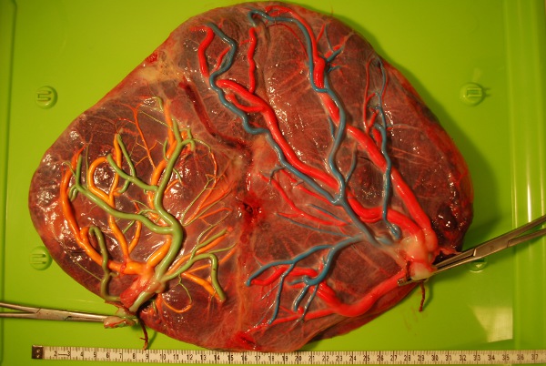
Figure 4. TTTS placenta after fetoscopic laser coagulation using the Solomon technique in which, after identification and coagulation of each individual anastomosis, the complete vascular equator is coagulated from one placental margin to the other.

Figure 5. TAPS placenta in which only a few, minuscule VA anastomoses (white stars) are visible along the vascular equator.

Figure 6. The maternal side of the TAPS placenta (shown in Figure 5) demonstrates the characteristic color difference between both placental shares.

Figure 7. Monochorionic placenta of a twin pregnancy with selective intrauterine growth restriction of one twin. The growth restricted fetus has a velamentous cord insertion and a much smaller placental share. The white star indicates an AA anastomosis.

Figure 8. Monoamniotic placenta: Note the various AV (green star), VA (blue star) and the 2 AA anastomoses (white stars), and the short distance between both cord insertions.
