We developed an easily customized strand-specific fluorescent in situ hybridization (FISH) protocol combined with immunofluorescence. This allows for a detailed examination of RNA dynamics with simultaneous insight into the chromatin structure, nuclear organization, and transcriptional regulation at the single cell level.
Combining RNA fluorescent in situ hybridization (FISH) with immunofluorescence (immuno-FISH) creates a technique that can be employed at the single cell level to detect the spatial dynamics of RNA localization with simultaneous insight into the localization of proteins, epigenetic modifications and other details which can be highlighted by immunofluorescence. X-chromosome inactivation is a paradigm for long non-coding RNA (lncRNA)-mediated gene silencing. X-inactive specific transcript (Xist) lncRNA accumulation (called an Xist cloud) on one of the two X-chromosomes in mammalian females is a critical step to initiate X-chromosome inactivation. Xist RNA directly or indirectly interacts with various chromatin-modifying enzymes and introduces distinct epigenetic landscapes to the inactive X-chromosome (Xi). One known epigenetic hallmark of the Xi is the Histone H3 trimethyl-lysine 27 (H3K27me3) modification. Here, we describe a simple and quick immuno-FISH protocol for detecting Xist RNA using RNA FISH with multiple oligonucleotide probes coupled with immunofluorescence of H3K27me3 to examine the localization of Xist RNA and associated epigenetic modifications. Using oligonucleotide probes results in a shorter incubation time and more sensitive detection of Xist RNA compared to in vitro transcribed RNA probes (riboprobes). This protocol provides a powerful tool for understanding the dynamics of lncRNAs and its associated epigenetic modification, chromatin structure, nuclear organization and transcriptional regulation.
Mammalian X-chromosome inactivation (XCI) is a strategy to compensate for the imbalance in X-linked gene dosage between XX and XY, wherein one of the two X-chromosomes in females is transcriptionally inactivated1. X-chromosome inactivation is a great model system for long non-coding RNA (lncRNA) research. X-chromosome inactivation is regulated by multiple lncRNAs, and has been extensively studied over the past few decades to uncover the crosstalk mechanisms between lncRNAs, transcription, chromatin structure and nuclear organization2,3.
The X inactivation center (XIC) located on the X-chromosome is a complex genetic locus comprised of a number of genes producing non-coding RNAs4. X-inactive specific transcript (Xist) lncRNA in eutherian mammals is one such lncRNA which plays a crucial role in X-chromosome inactivation5,6. Xist transcripts surround the location of the future Xi to initiate X-chromosome inactivation, and appear as a cloud when visualized using RNA FISH; this formation is referred to as the “Xist Cloud”7. Since Xist RNA interacts with various chromatin-modifying enzymes, co-localization of the Xist clouds with different epigenetic modifications for silent chromatin and repressive transcription is observed during X-chromosome inactivation8. For example, Xist RNA interacts with polycomb repressive complex 2 (PRC2) which is responsible for H3K27me3 and induces a repressive chromatin state9. The occurrence of the Xist cloud on the Xi and its co-localization with the intensive H3K27me3 modification represents a facultative heterochromatin landscape of the Xi10,11.
Cytogenetic techniques, such as DNA/RNA FISH have come a long way from the traditional method using radiolabelled probes12 to recent and advanced techniques with enhanced sensitivity and fluorescent imaging using multiple oligonucleotide probes13,14. DNA/RNA FISH coupled with immunofluorescence has been routinely used as a cytological tool to understand spatiotemporal nuclear organization, RNA localization, chromatin structure and modifications. The most standard probe preparation for RNA FISH involves the use of plasmid or bacterial artificial chromosome (BAC) clones and their subsequent labeling either with nick translation or random priming15. However, nearly 70% of genes in mice and 40% of genes in humans show an overlap of sense and antisense transcripts16, hence requiring a strand-specific FISH method in order to distinguish sense and antisense transcripts. In vitro transcribed RNA probes (riboprobe) are often used for strand-specific RNA FISH17,18; however, this involves preparing a plasmid clone or PCR product with T7, SP6, or T3 promoter and synthesizing riboprobes. Furthermore, riboprobes derived from genomic DNA or cDNA often contain non-specific regions and repetitive elements, which result in high background noise. Another issue is that riboprobes, which are a few hundred nucleotides in length and contain multiple fluorophores, cannot efficiently penetrate into the nucleus. To circumvent this, multiple shorter oligonucleotide probes labeled with a single fluorophore at the end have been developed that have good sensitivity, uniform signal strength, and ease of purification and handling14. In addition, DNA oligonucleotides are generally more stable than RNA. We applied a similar strategy of using oligonucleotides in Xist RNA FISH19 with immunofluorescence to understand the epigenetic dynamics of the Xi induced by Xist lncRNA during the process of X-chromosome inactivation. This protocol describes the creation of oligonucleotide probes and proper preparation of cells, as well as utilization of immunofluorescence and RNA FISH. Xist RNA FISH using multiple oligonucleotides is cost effective approach in the long-term if Xist RNA FISH is performed routinely in one’s laboratory. This technique can be used to identify lncRNAs in cells while simultaneously mapping its co-localization with epigenetic modifications or factors. One major advantage of the protocol is the ability to easily modify it to suit one’s research interests.
1. Probe Preparation
2. Slide Preparation
NOTE: Slides can be prepared for immuno-FISH using mouse embryonic stem (ES) or embryoid body (EB) differentiating cells20,21 by either growing them directly on a poly-lysine-treated slide or using cytospin with cell suspension. Here, slide preparation by cytospin is shown.
3. Immunofluorescence
4. RNA FISH with Oligonucleotide Probes
Representative images of quick immuno-FISH are shown in Figure 1A. Co-localization of the Xist RNA cloud and H3K27me3 signal on the Xi was detected in differentiating female cells. At day 12 upon differentiation, more than 90% of EB cells had an Xist cloud (Figure 1B). Short oligonucleotide probes efficiently penetrated into the nuclei, leading to a visualization of almost all H3K27me3 signals co-localized with Xist RNA (Figure 1C; 97%, n = 150).
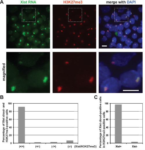
Figure 1: Xist RNA and H3K27me3 co-localize on the Xi in differentiating mouse EB. (A) Immuno-FISH for Xist RNA with H3K27me3 modification in differentiating female EB cells at day 12 upon differentiation. Xist probes were labeled with Alexa Fluor 488, and anti-H3K27me3 primary antibody was detected by anti-mouse IgG Alexa Fluor 555-conjugated secondary antibody. Nuclei were counterstained with DAPI. The boxed region is magnified in the bottom panels. Scale bars: 10 µm. (B) Frequency of Xist cloud- and H3K27me3-positive cells in EB at day 12 upon differentiation. (C) Frequency of co-localization of Xist cloud with H3K27me3 signal in EB at day 12 upon differentiation. Please click here to view a larger version of this figure.
Xp1 | ggtaagtatccaaaaccccgttgg |
Xp2 | cgatcagcagcaacagtacacg |
Xp3 | gcaattggttgcttttatccagtcc |
Xp4 | cgcaacaccgcacactaatacg |
Xp5 | gccatcttagacacattcaagagcat |
Xp6 | cacacgtgaagtaccaagcgaaac |
Xp7 | gccacgtatagagcactgtaagagactatg |
Xp8 | caaagcacactatcagacgtgtcg |
Xp9 | ggagaatctagatgccataaaggcaag |
Xp10 | tgatggacactgcattttagcactg |
Xp11 | ggacactgcattttagcaatacgattc |
Xp12 | gtgatgggcactgcattttagc |
Xp13 | agatgggctatctcagtcttataggct |
Xp14 | ggaagtcagtatggagggggtatg |
Xp15 | tgggactgtgactactacagcaatga |
Xp16 | aagactcaattcctagtcaggattatccac |
Xp17 | gggctgtagctctatgacagtgcttt |
Xp18 | gtcttcaccagatgcagattactacagtg |
Xp19 | aatagtttgaggaaggggtttcaagtg |
Xp20 | gctgttcaggtttccttctgtagtga |
Xp21 | gcaagagatacaatggtccgaaaagt |
Xp22 | agcacttcgtacaaccctctttctg |
Xp23 | gaagagagcaggtcattcgtcagag |
Xp24 | gcaactgagacactgtagccatatgaag |
Xp25 | ttcctggaggaagaacggaaaga |
Xp26 | tgattagaaggcttaggtcatcttcca |
Xp27 | ttttgttcagagtagcgaggacttga |
Xp28 | aatagagcagaatggcttcctcgaa |
Xp29 | acattgcttgatcacgctgaagac |
Xp30 | gcaaggaagaaatagacacacaaagc |
Xp31 | ggaagaaatagatgtaacaaagaattagacaca |
Xp32 | cacttcagagccacttgaatcctg |
Xp33 | agtcacaggtgtcctgtagaaacagttc |
Xp34 | cctttatgggcaatggcaacaat |
Xp35 | ggcacatctgcatattgcttgtcta |
Xp36 | gcaactaagaccatgaacccacaa |
Xp37 | aaacacactggccttaagtatatggactg |
Xp38 | cattcatttgcacacatggaacaat |
Xp39 | tgggagacaatatttagcctccaggt |
Xp40 | cctagcaagggcactgttttgtaataa |
Xp41 | taacatttagcacactgccttgcac |
Xp42 | cagtgatctacactaggtccacctcaca |
Xp43 | ttatgttgaaggaatcttggccttg |
Xp44 | aagtgagagctgtagtctcaaggtgtga |
Xp45 | gtattcaacctctgaggcaaactgtg |
Xp46 | agattgtggaacttagatggctgtca |
Xp47 | tggaactgcattaaagtcccaacttag |
Xp48 | gaactcccagacctcttcaacctg |
Table 1: Sequence of fluorescently labeled oligonucleotides for Xist RNA FISH. The 48 oligonucleotides were synthesized with a 5’-amino modification to prepare fluorescently labeled oligonucleotide probes for Xist RNA FISH.
In this paper, we have presented a quick immuno-FISH protocol that takes less than 5 hr to complete slide preparation, Xist RNA FISH, and immunofluorescence for H3K27me3. In comparison with the general immuno-FISH approach for Xist RNA detection, which usually needs overnight incubation for RNA FISH, this protocol not only significantly reduces time spent but also improves the sensitivity of immune-FISH using oligonucleotide probes.
Since Xist is highly transcribed during X-chromosome inactivation and its transcript is found to intensely accumulate on the Xi in cis, it is easy to detect Xist RNA signal in differentiating and differentiated cells by RNA FISH utilizing a relatively brief incubation time. This immuno-FISH protocol could be applied for other proteins of interest and abundant RNA, although optimization in immunofluorescence and RNA FISH may be required. In order to apply the protocol to other RNA, the number of possible oligonucleotide probes would need to be considered, followed by trial-and-error optimization of the probe’s concentration, incubation time, and temperature for RNA FISH. One example of other applications is our previous success in detecting telomeric RNA using single LNA probes22. For probe design and preparation for the detection of low abundant RNA targets, refer to the article by Raj and Tyagi23. Another issue that must be considered is the permeabilization step. Careful consideration regarding different permeabilization condition is needed; for example, milder effects than what is described in our protocol can occur with permeabilization after fixation15. Various permeabilization procedures also need to be considered when carrying out immuno-FISH using different cell types. The order of immunofluorescence and RNA FISH may also affect the detection of target proteins, modifications, and RNA by immuno-FISH. If RNA FISH is performed prior to immunofluorescence, one hour incubation during the RNA FISH segment is enough to detect Xist RNA signal. However, since the formamide contained in the hybridization buffer and FISH wash buffer negatively affects H3K27me3 immunofluorescence, we always carry out H3K27me3 immunofluorescence prior to Xist RNA FISH in this protocol. For other applications, the order of procedures would depend on the target proteins and epigenetic modifications to be observed by immunofluorescence. As the primary antibody we used for H3K27me3 immunofluorescence has a high titer and specificity to H3K27me324, we are able to use a shorter incubation time for immunofluorescence. However, reaction time and temperature for immunostaining are dependent on the antibody used and thus optimization is needed for each antibody.
Using oligonucleotide probes, Xist RNA detection by RNA FISH was significantly improved. In our previous studies, we found that a subset of differentiating cells with H3K27me3 staining on the Xi are not associated with Xist RNA signal25. Since H3K27me3 modification on the Xi is associated with PRC2 recruitment by Xist RNA9–11,26, we speculated that this was due to the inefficient penetration of riboprobes into the nuclei compared to the antibody for immunofluorescence. By using oligonucleotide probes in this protocol, we observed that H3K27me3 signal in differentiating female cells nearly always co-localized with Xist RNA (Figure 1).
This quick immuno-FISH protocol allows us to gain a snapshot of the dynamic changes revolving around Xist lncRNA and offers a new tool to help gain a better understanding of the way lncRNA works. IncRNAs affect various cellular functions such as genomic imprinting, gene regulation, RNA translation, and RNA maturation27. Developing an increased understanding of the diverse roles played by lncRNA would help to propel the field forward and allow for future discoveries in the regulation of vital cellular processes within the human body, as well as contribute to the development of potential targets for disease treatments. Since this protocol can be easily optimized with respect to other markers and lncRNAs of interest, it is a great tool to help tackle the mysteries of various lncRNAs in cells.
The authors have nothing to disclose.
We thank Hongjae Sunwoo for helpful advice for oligonucleotide design in RNA FISH. We also thank Serenity Curtis for editing the manuscript. N.Y. was supported by a Postdoctoral Fellowship for Research Abroad of the Japan Society for the Promotion of Science (JSPS). This work was supported by the NIH (RO1-GM102184), the March of Dimes Research Foundation (#6-FY12-337), and the Developmental Fund and Trustee Grant at Cincinnati Children’s Hospital Medical Center to Y.O.
oligonucleotides with 5’-amine modification | IDT | |
Alexa Fluor 488 Reactive Dye | Life Technologies | A32750 |
MicroSpin G-25 columns | GE Healthcare | 27-5325-01 |
Cytospin 2 | Shandon | 59900102 |
Bovine Serum Albumin, Molecular Biology Grade (for hybridization buffer) | Roche | 10715859103 |
Histone H3K27me3 antibody | Active Motif | 61017 |
Accutase | Innovative Cell Technologies | AT104 |
Alexa Fluor 555 Goat Anti-Mouse, highly cross-adsorbed | Life Technologies | A21424 |
ProLong Gold antifade reagent | Life Technologies | P36931 |