Fast Enzymatic Processing of Proteins for MS Detection with a Flow-through Microreactor
Summary
A quick protocol for proteolytic digestion with an in-house built flow-through tryptic microreactor coupled to an electrospray ionization (ESI) mass spectrometer is presented. The fabrication of the microreactor, the experimental setup and the data acquisition process are described.
Abstract
The vast majority of mass spectrometry (MS)-based protein analysis methods involve an enzymatic digestion step prior to detection, typically with trypsin. This step is necessary for the generation of small molecular weight peptides, generally with MW < 3,000-4,000 Da, that fall within the effective scan range of mass spectrometry instrumentation. Conventional protocols involve O/N enzymatic digestion at 37 ºC. Recent advances have led to the development of a variety of strategies, typically involving the use of a microreactor with immobilized enzymes or of a range of complementary physical processes that reduce the time necessary for proteolytic digestion to a few minutes (e.g., microwave or high-pressure). In this work, we describe a simple and cost-effective approach that can be implemented in any laboratory for achieving fast enzymatic digestion of a protein. The protein (or protein mixture) is adsorbed on C18-bonded reversed-phase high performance liquid chromatography (HPLC) silica particles preloaded in a capillary column, and trypsin in aqueous buffer is infused over the particles for a short period of time. To enable on-line MS detection, the tryptic peptides are eluted with a solvent system with increased organic content directly in the MS ion source. This approach avoids the use of high-priced immobilized enzyme particles and does not necessitate any aid for completing the process. Protein digestion and complete sample analysis can be accomplished in less than ~3 min and ~30 min, respectively.
Introduction
The identification and characterization of purified proteins is frequently achieved by using MS techniques. The protein is digested with an enzyme and its peptides are further analyzed by MS by using a simple infusion experimental setup. Proteolytic digestion is necessary for generating small peptide fragments that fall in the useful mass range of most MS analyzers, and that can be easily fragmented through low energy collision induced dissociation to generate amino acid sequence information. For isolated proteins or simple protein mixtures, there is no further need for chromatographic separation of peptides prior to MS detection. A mixture of 25-50 peptides can be easily analyzed by infusing the sample with a syringe pump directly in the MS ion source.
The mass spectrometer can perform the analysis and confirm the sequence of a protein within a short time-frame. With modern data acquisition methods, this process can be accomplished within a few minutes or even seconds. The limiting factor in completing the entire process on a short time-scale is the proteolytic digestion step. Typically, this is performed over a few hours (or O/N), in solution, at 37 ºC, using substrate:enzyme ratios of (50-100):1. To reduce the enzymatic digestion time to minutes or seconds, immobilized enzyme microreactors, in the form of microfluidic reactors or commercially available cartridges, have been described.1-6 Typically, the enzyme is immobilized by covalent, non-covalent/physical adsorption, complex formation or encapsulation,3,6 the enhanced efficiency of the enzymatic process being enabled by the large surface-to-volume and enzyme-to-substrate ratios. Additional advantages of immobilized reactors include reduced autolysis and interference from the enzyme in MS analysis, improved enzyme stability and reusability. A variety of approaches, using glass or polymeric microfabricated devices have been described, using enzymes immobilized on magnetic beads by antibody-antigen interactions,7,8 entrapped in gold nanoparticle networks,9 encapsulated in titania-alumina sol-gels10 and nanozeolites,11 or captured through Ni-NTA or His-Tag complex formation.6 Alternatively, open-tubular capillaries with immobilized enzymes have been developed, as well.12 Moreover, enhanced proteolytic cleavage has been demonstrated by using controlled microwave irradiation13 or pressure-assisted or pressure cycling technology (PCT) for reducing the reaction times to 30-120 min.14
Despite the multiple advantages of immobilized enzyme reactors, the costs of commercial cartridges is high, the availability of microfluidic devices for routine use is limited, and the use of microwave or PCT technologies results in need for additional instrumentation. The goal of this work was to develop a method that circumvents these disadvantages, and that can be easily implemented in every laboratory to empower researchers with a simple and effective approach for performing enzymatic cleavage of proteins in preparation for MS analysis within minutes. The approach relies on the use of hydrophobic, C18-particles which are pre-loaded in a capillary or microfluidic device, and the adsorption of the protein(s) of interest on these particles followed by enzymatic digestion during the infusion of the enzyme over the packed bed and captured protein(s). In this approach, the substrate is immobilized through non-covalent interactions, and the enzyme is infused over the immobilized protein. The proteolytic digestion efficiency is increased by the large particle surface areas that expose the protein for enzymatic processing, reduced distances and diffusion times to and from the surface of particles, improved mass transfer, no covalent attachment that may affect the activity of the enzyme, ability to quickly evaluate combinations of different enzymes, disposability, and multiplexing if the process is executed in a microfluidic format. This approach is demonstrated with the use of a mixture of standard proteins and trypsin-the most commonly used enzyme for proteolytic digestion prior to ESI-MS detection. The mass spectrometer used for detection in this study was a linear trap quadrupole (LTQ) instrument.
Protocol
1. Preparation of the Capillary Microreactor
- Cut the 100 µm internal diameter (ID) x 360 µm outer diameter (OD) capillary to a length of 7-8 cm, and the 20 µm ID x 90 µm OD capillary to 3-5 cm with the glass capillary cleaver; verify under the microscope that both capillary ends have a clean, straight cut, without any protruding burrs.
- Insert the 20 µm ID x 90 µm OD capillary into one end of the 100 µm ID x 360 µm OD capillary, for a length of ~6 mm; in case of need, observe this operation under a microscope.
- Apply a small droplet of glue E6000 around the junction of the 90 µm OD/100 µm ID capillaries with the end of a Q-tip, and let the glue cure O/N at RT.
- Cut the inserted 20 µm ID x 90 µm OD capillary to a length of ~10-15 mm; this will be the electrospray ionization emitter.
- Pre-cut the 1/32" OD PEEK, 1/16" OD PEEK and 1/16" OD PTFE tubing in pieces of 4-5 cm in length; these will be the sleeves used in capillary unions for providing a leak-free connection.
- Connect the opposite end of the 100 µm ID x 360 µm OD capillary to a PEEK union; use a 1/32" PEEK sleeve for providing a leak-free connection.
- Insert/tighten a piece of ~5 cm long 1/16" PTFE tubing into the opposite end of the union.
- Weigh ~4 mg of C18 (5 µm) particles in a pre-cleaned/dried 2 ml glass vial; add 0.5 ml isopropanol, close the vial and disperse the slurry in the ultrasonicator bath.
- Withdraw with a 250 µl syringe approximately 200 µl slurry, insert the syringe needle in the 1/16" PTFE tubing of the PEEK union, and dispense slowly the slurry into the 100 µm ID x 360 µm OD capillary; observe under the microscope as the capillary fills with particles until a length of 2-3 mm packing is achieved; this will be the microreactor (Figure 1). The 20 µm ID capillary retains the particles in the microreactor through a Keystone effect.15
- Rinse the microreactor with ~50 µl solution of H2O/CH3CN 50:50 v/v, and then with ~50 µl solution of H2O/CH3CN 98:2 v/v.
2. Preparation of Sample Solutions
Note: Operations that involve handling of organic solvents and acids and solution preparation are to be performed in a fume hood. Wear goggles, gloves and protective clothing.
- Rinse with CH3OH and dry a few glass vials (4 ml) and polypropylene tubes (15 ml).
- Prepare 10 ml of a solution of H2O/CH3CN (98:2 v/v) in a 15 ml polypropylene tube by mixing 9.8 ml H2O with 200 µl CH3CN; mix by sonication.
- Prepare 10 ml of a solution of H2O/CH3CN (50:50 v/v) in a 15 ml polypropylene tube by mixing 5 ml H2O with 5 ml CH3CN; mix by sonication.
- Prepare 4 ml of an acidified aqueous solution (TFA 0.01% v/v) by adding 4 µl TFA(10%) to 4 ml H2O/CH3CN (98:2 v/v) in a 4 ml glass vial; mix by sonication.
- Prepare 4 ml of an acidified organic solution (TFA 0.01% v/v) by adding 4 µl TFA(10%) to 4 ml H2O/CH3CN (50:50 v/v) in a 4 ml glass vial; mix by sonication.
- Weigh 16 mg NH4HCO3 with an analytical microbalance in a 4 ml glass vial; add 4 ml of the H2O/CH3CN (98:2 v/v) solution and disperse by sonication; this results in a 50 mM solution of NH4HCO3 (pH~7.8) in aqueous buffer.
- Weigh 5 mg of a standard protein (e.g., bovine serum albumin, hemoglobin α/β, cytochrome C, carbonic anhydrase, α-2-HS-glycoprotein, etc.) with an analytical microbalance in a 4 ml glass vial; add 4 ml DI water and disperse by sonication; this typically results in a high µM-level concentration solution.
- Prepare 1 ml protein solution (1-2 µM) by diluting the high µM-level concentration solution with an appropriate volume of NH4HCO3 (50 mM) aqueous buffer; mix by sonication.
- Prepare the trypsin solution (5 µM) by dissolving 20 µg sequencing-grade trypsin in 170 µl NH4HCO3 (50 mM) aqueous buffer; disperse by sonication.
3. Experimental Setup
Note: The LTQ-MS system is fitted with a modified ESI source that includes a home-built XYZ-stage which enables interfacing of the mass spectrometer to various sample input approaches.
- Disconnect the capillary microreactor from the PEEK union and connect to the PEEK Tee.
- Insert a ~2 cm long Pt wire in the side-arm of the PEEK Tee; use a 1/32" PEEK sleeve for providing a leak-free connection and for insulating the Pt wire.
- Connect a 50 µm ID x 360 µm OD (~0.5 m long) sample transfer capillary to the opposite end of the PEEK Tee; use a 1/32" PEEK sleeve for providing a leak-free connection.
- Secure the PEEK Tee on the XYZ-stage and position the ESI emitter ~2 mm away from the mass spectrometer inlet capillary.
- Connect the Pt wire to the ESI power supply source of the mass spectrometer.
- Connect the opposite end of the sample transfer capillary to the stainless steel union; use a 1/16" PEEK sleeve for providing a leak-free connection.
- Connect a 4-5 cm long 1/16" PTFE tubing to the opposite end of the stainless steel union and insert the 250 µl syringe needle in the PTFE tubing; turn on the pump and set the flow rate at the desired value (e.g., 2 µl/min).
- Input tandem MS data acquisition parameters into the software package that controls the MS instrument and save as a method file to ready the setup for performing the analysis; for the LTQ-MS system, use the following data dependent analysis parameters: dynamic exclusion enabled for 180 sec with exclusion mass width ±1.5 m/z; one MS scan followed by one zoom and MS2 scans on the top 5 most intense peaks, zoom scan width ±5 m/z; collision induced dissociation (CID) parameters set at isolation width 3 m/z, normalized collision energy 35%, activation Q 0.25, and activation time 30 msec.
4. Microfluidic Setup
- Prepare a microfluidic device from glass or polymeric substrates;16-18 the layout of the device is provided in Figure 2A, consisting of a microreactor (1) (0.13 mm width x 2 mm length x 50 µm deep) connected to an inlet (2), outlet (3) and a side port (4); the interconnecting channels for fluidic manipulations are ~110 µm wide and ~50 µm deep; a multichannel structure (5), consisting of ~1.5-2 µm deep x 10 μm wide x 100 μm long microchannels placed 25 µm apart, will act as a filter for retaining the particles in the microreactor.
- Enable fluid-delivery connections to the chip by gluing specialized connectors to the inlet ports, or by devising a polymeric stand that accommodates fittings for fluid delivery (Figure 2B).16
- Prepare a slurry of C18 particles (5 µm) as described in step 1.8; deliver the slurry to the microreactor with a syringe by using 1/16" PTFE tubing connected to the chip side port (4); seal the port after particle loading with epoxy glue and cure for 1 hr in an oven at 90 ºC.16
- Insert a capillary (20 µm ID x 90 µm OD x 10 mm length) in the outlet port, seal with E6000 glue, and let cure O/N; this will become the ESI emitter (6).
- Connect a 50 µm ID x 360 µm OD (~0.5 m long) sample transfer capillary to the chip inlet port (2); connect the opposite end of the capillary to a 250 µl syringe and the syringe pump, as described in 3.6-3.7.
- Place a light-weight PEEK Tee connector on the sample transfer line just prior to the inlet port (2) and insert a Pt wire on the side-arm of the Tee, as described in 3.2; this will be the ESI electrode.
- Secure the microfluidic device on the XYZ stage with the ESI emitter positioned ~2 mm away from the mass spectrometer inlet capillary; prepare the MS for analysis as described in 3.8.
5. Sample Loading, Proteolytic Digestion and Elution for MS Analysis
Note: All solution/sample transfer steps are performed with the aid of a syringe pump and should allow for a few additional minutes to complete, to compensate for the dead-volumes associated with the transfer lines to and from the microreactor; the necessary time will depend on the flow rates involved.
- Rinse the microreactor and prepare it for analysis by infusing an aqueous solution of NH4HCO3 (50 mM) in H2O/CH3CN (98:2 v/v) at 2 µl/min for 5 min; use a 250 µl syringe.
- Load the protein sample (1 µM) on the microreactor by infusing the sample solution at 2 µl/min for 5 min; use a 250 µl syringe.
- Infuse the trypsin solution (5 µM) over the adsorbed protein on the microreactor at 2 µl/min for 1-3 min.
- Quench the proteolytic digestion process by rinsing the microreactor with an acidified aqueous solution of TFA (0.01% v/v) in H2O/CH3CN (98:2 v/v) at 2 µl/min for 5 min; use a 250 µl syringe.
- Start eluting the protein digest from the microreactor by infusing an acidified organic solution of TFA (0.01% v/v) in H2O/CH3CN (50:50 v/v) at 300 nl/min.
- Turn on the MS data acquisition process and the ESI voltage (~2,000 V); acquire MS data using the data acquisition parameters provided in step 3.8; observe the elution of the tryptic peptides.
6. Data Processing
- Process the tandem MS data with a search engine compatible with the raw MS data format.
- Process the LTQ-MS raw files using an organism specific minimally redundant protein database; upload the raw data in the search engine, set the parent and fragment ion mass tolerances to 2 and 1 Da, respectively, and use only fully tryptic fragments with up to two missed cleavages for the search; do not allow for posttranslational modifications; set the high and medium-confidence false discovery rates (FDR) to 1% and 3%, respectively, and do not allow for posttranslational modifications.
- Filter the data to select only high-confidence peptide matches to the proteins in the database; evaluate the protein and peptide-level results.
Representative Results
A representative result of a proteolytic digestion process performed simultaneously on a mixture of proteins, with the above-described microreactors (Figures 1 or 2), is provided in Table 1. The table comprises the unique peptide sequences that identify a particular protein, the cross correlation score (Xcorr) (i.e., a score that characterizes the quality of the experimental-to-theoretical match for the corresponding tandem mass spectrum), the number of missed cleavages, the peptide charge state (z), and the mass/charge (m/z) of the protonated peptides. At the protein level, the table provides the UniProt protein ID, the description (name) of the protein, the protein score and the protein coverage. All proteins were identifiable by multiple peptides, after a proteolytic digestion performed for 90 sec.
The time necessary for the elution of all peptides was dependent on the length of the transfer capillary that delivered the acidified organic solution at 300 nl/min, a flow rate compatible with nano-ESI optimal operation. In-capillary mixing of the aqueous and organic acidified solutions resulted in the early elution of a few peptides, but the great majority eluted over several minutes only in the organic solution. A mass spectrum displaying the co-elution of several peptides representative of the five proteins, generated with the capillary microreactor, is provided in Figure 3.
Table 1. List of tryptic peptides generated through the proteolytic digestion of five proteins using the capillary microreactor and O/N digestion protocols. Only unique sequences, with the highest Xcorr, are shown. Please click here to download this file.

Figure 1. Capillary microreactor. (A) Experimental setup for the fabrication of the microreactor. (B) Capillary microreactor positioned in the MS ion source. The microreactor is loaded manually with C18-bonded silica particles and placed on an XYZ-stage that facilitates proper positioning in the ESI-MS source. Please click here to view a larger version of this figure.

Figure 2. Microfluidic microreactor. (A) Schematic diagram of the microfluidic reactor. (B) Microfluidic reactor secured in a PEEK stand. The microfluidic reactor and transfer lines are fabricated in a glass substrate which can be secured in a home-made polymeric PEEK stand for further positioning in the MS source with the XYZ stage. Please click here to view a larger version of this figure.
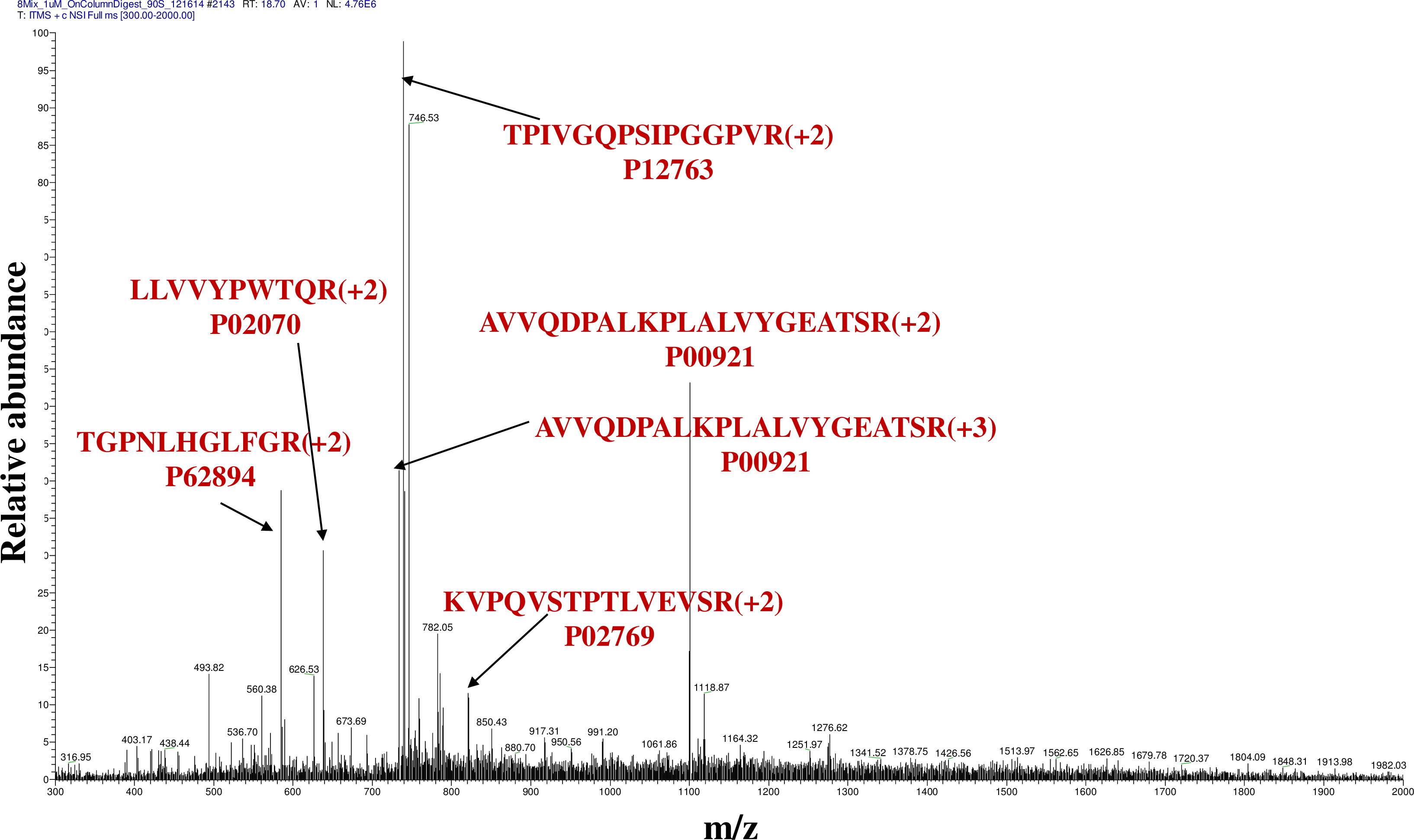
Figure 3. Mass spectrum comprising tryptic peptides generated from the protein mixture subjected to proteolysis in the microreactor. The most intense tryptic peptide ions are labeled with the sequence of the amino acids and the corresponding protein identifier. Please click here to view a larger version of this figure.
Discussion
The microreactor described in this work provides an easy-to-implement experimental setup for performing enzymatic digestion of proteins to enable MS analysis and identification in less than 30 min. The distinct advantages of this system, in comparison to conventional approaches, include simplicity, speed, low reagent consumption and low costs. In particular, there is no need for expensive immobilized trypsin beads and cartridges. The preparation of the capillary microreactor is straightforward (Figure 1A), and can be accomplished within minutes from standard supplies, commonly found in a chromatography laboratory. While the reversed phase C18 particles can be rinsed with high organic-content solvent (CH3CN or CH3OH) for complete removal of adsorbed proteins or peptides, and the microreactor can be re-used, the easy and cost-effective fabrication process invites for the manufacturing of disposable units for single-use applications. The capillary microreactor can be mounted in a home-built ESI source, as shown in Figure 1B, or can be inserted in a standard commercial ion source. While the particle bed for sample capture and digestion does not need to be longer than ~1-2 mm, just enough to capture sufficient material for optimal detection by MS, the length of the capillary itself can be adjusted to fit the size of any particular ion source.
For laboratories that have capabilities to fabricate microfluidic devices, a simple design (Figure 2A) that can be secured into a stand for facilitating interfacing to MS detection (Figure 2B) is provided. The dimensions of the microreactor and transfer channels can be adjusted to the requirements of a particular application, and the device can be developed into a single or multiplexed format. For most commercial mass spectrometers, however, the ion source would need modifications to permit the placement of the device in the source. The photomask drawing for chip fabrication was executed with AutoCAD software and the photomask were prepared by HTA Photomask. The fabrication of the microchips was performed according to protocols described earlier:17,18 alignment of the substrate with the photomask, exposure to UV light (360 nm), removal of the irradiated photoresist and underlying chrome layer, wet chemical etching of channels with buffer oxide etch, removal of the remaining chrome layer, drilling of access holes, cleaning, hydrolysis of the microchip surface (NH4OH + H2O2), and thermal bonding of the substrate to the cover plate by gradual heating to 550 ºC. Both substrate and cover plate were etched, one for deep channels for sample handling (~20-50 µm), and the other for shallow micro-channel filter elements (1.5-2 µm deep).16
The results generated with the above-described microreactor are comparable with the results obtained from O/N,19 conventional digestion protocols that use substrate:enzyme ratios of (50-100):1, followed by a peptide infusion experiment with MS detection (300 nl/min, peptides dissolved in H2O/CH3CN (50:50 v/v) acidified with CH3COOH, 0.1 % v/v). Both microreactor and conventional protocols enabled the identification of all proteins by multiple unique peptides (Table 1). Typically, a somewhat larger number of unique peptides per protein were observable with the microreactor than with the conventional setup. This was a result of incomplete enzymatic cleavage at certain sites. A few additional minutes of proteolytic digestion reduced or eliminated this outcome. Also, some enzymatic peptides generated with the microreactor differed from the ones generated in solution, due to the fact that for the proteins adsorbed on the C18 particles different sites were exposed to the enzyme than in homogenous solutions.20 The Xcorr scores demonstrate, however, that the quality of the tandem mass spectra generated with the microreactor was of high-quality, and that a sufficient protein sequence coverage (11-75%) is achievable for unambiguous protein identifications.
The technique, as described, is limited to the analysis of rather simple protein mixtures that generate at most ~25-50 peptides that can be analyzed by simple infusion experiments and that do not necessitate liquid chromatographic separation prior to MS analysis. Critical to success is the use of freshly thawed and prepared trypsin solutions, to not reduce or lose the activity of the proteolytic enzyme at the operational pH of the microreactor (i.e., pH~8). In addition, a proper balance must be found between the optimal flow rates for electrospray ionization (150-300 nl/min) and the maximal flow rates that can be tolerated by the experimental setup and that enable fast peptide elution from the microreactor (2-3 µl/min). Higher than optimal ESI flow rates result in sensitivity losses and poor detection limits. As a result, the length of the transfer capillaries should be maintained as short as possible to reduce the time necessary for the elution of peptides from the microreactor when operating at ≤300 nl/min. All components that were used for the experimental setup can be replaced by components from other manufacturers that perform similar functions. If the dimensional characteristics of these components differ (length, diameter, volume), the optimization of eluent flow rates, sample concentration, time for performing certain steps and of the ESI voltage may be necessary to obtain similar results.
A unique advantage enabled by the microfluidic setup is the ability to further incorporate on the chip a pumping system, e.g., an electroosmotic flow driven pump,21,22 for enabling the stand-alone operation of the platform and the integration of an additional separation step. The ability to perform on-chip separations of the peptides generated through proteolytic digestion will result in improved detection limits, and will facilitate the analysis of complex protein mixtures from cellular extracts. This will further enable the integration of the technique in workflows that make use of microfabricated platforms and high-speed MS detection for biomedical applications.23
Disclosures
The authors have nothing to disclose.
Acknowledgements
This work was supported by NSF/DBI-1255991 grant to IML.
Materials
| Ion trap ESI-MS | Thermo Electron | LTQ | The LTQ mass spectrometer is used for acquiring tandem MS data |
| XYZ stage | Newport | Multiple parts | The home-built XYZ stage is used to adapt the commercial LTQ nano-ESI source to receive input from various sample delivery systems |
| Stereo microscope | Edmund optics | G81-278 | The microscope is used to observe the microreactor packing process |
| Analytical balance/Metler | VWR | 46600-204 | The balance is used to weigh the protein samples |
| Ultrasonic bath/Branson | VWR | 33995-540 | The sonic bath is used for mixing/homogenizing the samples and dispersing the C18 particle slurry |
| Syringe pump 22 | Harvard Apparatus | 552222 | The micropump is used for loading, rinsing and eluting the sample and the enzyme on and from the packed capillary microreactor |
| Milli-Q ultrapure water system | EMD Millipore | ZD5311595 | The MilliQ water system is used to prepare purified DI water |
| Pipettor/Eppendorf (1000 µL) | VWR | 53513-410 | The pipettor is used to measure small volumes of sample solutions |
| Pipettor/Eppendorf (100 µL) | VWR | 53513-406 | The pipettor is used to measure small volumes of sample solutions |
| Pipettor/Eppendorf (10 µL) | VWR | 53513-402 | The pipettor is used to measure small volumes of sample solutions |
| Fused silica capillary (100 µm ID x 360 µm OD) | Polymicro Technologies | TSP100375 | This capillary is used for the fabrication of the microreactor |
| Fused silica capillary (20 µm ID x 100 µm OD) | Polymicro Technologies | TSP020090 | This capillary is used for the fabrication of the ESI emitter |
| Fused silica capillary (50 µm ID x 360 µm OD) | Polymicro Technologies | TSP050375 | This capillary is used to transfer the samples and the eluent from the syringe pump to the capillary microreactor |
| Glass capillary cleaver | Supelco | 23740-U | This is a tool for cutting fused silica capillaries at the desired length |
| Glue | Eclectic Products | E6000 Craft | This glue is used for securing the ESI emitter into the capillary microreactor or the microfluidic chip |
| Epoxy glue | Epo-Tek | 353NDT | This glue is used to seal the microfluidic inlet hole through which the C18 particles are loaded |
| Reversed phase C18 particles (5 µm) | Agilent Technologies | Zorbax 300SB-C18 | These are C18 particles on which the proteins are adsorbed; the particles were extracted from a 4 mm x 20 cm C18 LC column from Agilent |
| Syringe/glass (250 µL) | Hamilton | 81130-1725RN | The glass syringes are used to load the C18 particle slurry in the capillary microreactor and to deliver the sample and eluents to the microreactor |
| Internal reducing PEEK Union (1/16” to 1/32”) | Valco | ZRU1.5FPK | This union is used to connect the 250 µL syringe to the microreactor for loading the 5 µm particle slurry |
| Stainless steel union (1/16”) | Valco | ZU1XC | The stainless steel union is used to connect the glass syringe needle to the infusion capillary |
| Microvolume PEEK Tee connector (1/32”) | Valco | MT.5XCPK | The Peek tee is used to connect the sample transfer capillary to the capillary microreactor; on its side arm, it enables the insertion of the Pt wire |
| Tee connector (light weight) | Valco | C-NTXFPK | This Tee connector is used to apply ESI voltage to the microfluidic chip through the sample transfer line |
| Pt wire (0.404 mm) | VWR | 66260-126 | The Pt wire provides electrical connection for ESI generation and is connected to the mass spectrometer ESI power supply |
| PTFE tubing (1/16” OD) | Valco | TTF115-10FT | The Teflon tubing is used to enable an air-tight connection between the syringe needle and the stainless steel union |
| PEEK tubing (0.015“ ID x 1/16” OD) | Upchurch Scientific | 1565 | The Peek tubing is used as a sleeve to enable an air-tight connection between the stainless steel union and the 50 µm ID transfer capillary |
| PEEK tubing (0.015” ID x 1/32” OD) | Valco | TPK.515-25 | The Peek tubing is used as a sleeve to enable a leak-free connection between the fused silica capillaries and the Peek Tee |
| Clean-cut polymer tubing cutter | Valco | JR-797 | This cutter is used to pre-cut the 1/16” and 1/32’ Peek polymer tubing that is used as sleeve for leak-free connections in pieces of ~4-5 cm in length |
| Amber vial (2 mL) | Agilent | HP-5183-2069 | The vials are used to prepare sample solutions and the C18 particle slurry |
| Amber vial (4 mL) | VWR | 66011-948 | The vials are used to prepare sample solutions |
| Polypropylene tube (15 mL) | Fisher | 12-565-286D | The vials are used to prepare buffer solutions |
| Cylinder (100 mL) | VWR | 24710-463 | The cylinder is used to measure volumes of solvent |
| Cylinder (10 mL) | VWR | 24710-441 | The cylinder is used to measure volumes of solvent |
| Pipette tips (1000 µL) | VWR | 83007-386 | The pipette tips are used to measure small volumes of sample solutions |
| Pipette tips (100 µL) | VWR | 53503-781 | The pipette tips are used to measure small volumes of sample solutions |
| Pipette tips (10 µL) | VWR | 53511-681 | The pipette tips are used to measure small volumes of sample solutions |
| Glass substrates | Nanofilm | B270 white crown, 3” x 3” | These are glass substrates for microchip fabrication |
| Male nut fitting (1/16”) | Upchurch | P203X | This fitting is used for connecting transfer capillaries to the microfluidic chip |
| Nanoport assembly | Upchurch | N-122H | This fitting is used for connecting transfer capillaries to the microfluidic chip |
| Reagents | |||
| Protein standards | Sigma | Multiple # | |
| Acetonitrile, HPLC grade | Fisher | A955 | |
| Methanol, HPLC grade | Fisher | A452 | |
| Isopropanol, HPLC grade | Sigma | 650447 | |
| Trifluoroacetic acid | Sigma | 302031 | |
| Ammonium bicarbonate | Aldrich | A6141 | |
| Trypsin, sequencing grade | Promega | V5111 |
References
- Petersen, D. H. Microfluidic Bioreactors. Encyclopedia of Microfluidics and Nanofluidics. , (2014).
- Matosevic, S., Szita, N., Baganz, F. Fundamentals and applications of immobilized microfluidic enzymatic reactors. J. Chem. Technol. Biotechnol. 86 (3), 325-334 (2011).
- Liu, Y., Liu, B., Yang, P., Girault, H. H. Microfluidic enzymatic reactors for proteome research. Anal. Bioanal. Chem. 390 (1), 227-229 (2008).
- Wu, H., Zhai, J., Tian, Y., Lu, H., Wang, X., Jia, W., Liu, B., Yang, P., Xu, Y., Wang, H. Microfluidic enzymatic-reactors for peptide mapping: strategy, characterization, and performance. LabChip. 4 (6), 588-597 (2004).
- Jin, L. J., Ferrance, J., Sanders, J. C., Landers, J. P. A microchip-based proteolytic digestion system driven by electroosmotic pumping. LabChip. 3 (1), 11-18 (2003).
- Asanomi, Y., Yamaguchi, H., Miyazaki, M., Maeda, H. Enzyme-immobilized microfluidic process reactors. Molecules. 7 (16), 6041-6059 (2011).
- Aravamudhan, S., Joseph, P. J., Kuklenyik, Z., Boyer, A. E., Barr, J. R. Integrated microfluidic enzyme reactor mass spectrometry platform for detection of anthrax lethal factor. , 1071-1074 (2009).
- Liu, X., Lo, R. C., Gomez, F. A. Fabrication of a microfluidic enzyme reactor utilizing magnetic beads. Electrophoresis. 30 (12), 2129-2133 (2009).
- Liu, Y., Xue, Y., Ji, J., Chen, X., Kong, J., Yang, P., Girault, H. H., Liu, B. Gold nanoparticle assembly microfluidic reactor for efficient on-line proteolysis. Mol. Cell. Proteomics. 6 (8), 1428-1436 (2007).
- Wu, H., Tian, Y., Liu, B., Lu, H., Wang, X., Zhai, J., Jin, H., Yang, P., Xu, Y., Wang, H. Titania and alumina sol-gel-derived microfluidics enzymatic-reactors for peptide mapping: design, characterization, and performance. J. Proteome Res. 3 (6), 1201-1209 (2004).
- Ji, J., Zhang, Y., Zhou, X., Kong, J., Tang, Y., Liu, B. Enhanced protein digestion through the confinement of nanozeolite-assembled microchip reactors. Anal. Chem. 80 (7), 2457-2463 (2008).
- Hustoft, H. K., Brandtzaeg, O. K., Rogeberg, M., Misaghian, D., Torsetnes, S. B., Greibrokk, T., Reubsaet, L., Wilson, S. R., Lundanes, E. Integrated enzyme reactor and high resolving chromatography in "sub-chip" dimensions for sensitive protein mass spectrometry. Scientific Reports. 3 (3511), 1-7 (2013).
- Pramanik, B. N., Mirza, U. A., Ing, Y. H., Liu, Y. H., Bartner, P. L., Weber, P. C., Bose, A. K. Microwave-enhanced enzyme reaction for protein mapping by mass spectrometry: A new approach to protein digestion in minutes. Protein Sci. 11 (11), 2676-2687 (2002).
- Olszowy, P. P., Burns, A., Ciborowski, P. S. Pressure-assisted sample preparation for proteomic analysis. Anal. Biochem. 438 (1), 67-72 (2013).
- Lord, G. A., Gordon, D. B., Myers, P., King, B. W. Tapers and restrictors for capillary electrochromatography and capillary electrochromatography-mass spectrometry. J. Chromatogr. A. 768, 9-16 (1997).
- Lazar, I. M., Kabulski, J. L. Microfluidic LC Device with Orthogonal Sample Extraction for On-Chip MALDI-MS Detection. Lab Chip. 13 (11), 2055-2065 (2013).
- Harrison, D. J., Manz, A., Fan, Z. H., Ludi, H., Widmer, H. M. Capillary electrophoresis and sample injection systems on a planar glass chip. Anal. Chem. 64 (17), 1926-1932 (1992).
- Jacobson, S. C., Hergenroder, R., Koutny, L. B., Warmack, R. J., Ramsey, J. M. Effects of Injection Schemes and Column Geometry on the Performance of Microchip Electrophoresis Devices. Anal. Chem. 66 (7), 1107-1113 (1994).
- Armenta, J. M., Perez, M. J., Yang, X., Shapiro, D., Reed, D., Tuli, L., Finkielstein, C. V., Lazar, I. M. Fast Proteomic Protocol for Biomarker Fingerprinting in Cancerous Cells. J. Chromatogr. A. 1217, 2862-2870 (2010).
- Doucette, A., Craft, D., Li, L. Mass Spectrometric Study of the Effects of Hydrophobic Surface Chemistry and Morphology on the Digestion of Surface-Bound Proteins. J. Am. Soc. Mass Spectrom. 14, 203-214 (2003).
- Lazar, I. M., Trisiripisal, P., Sarvaiya, H. A. Microfluidic Liquid Chromatography System for Proteomic Applications and Biomarker Screening. Anal. Chem. 78 (15), 5513-5524 (2006).
- Lazar, I. M., Karger, B. L. Multiple Open-Channel Electroosmotic Pumping System for Microfluidic Sample Handling,". Anal. Chem. 74 (24), 6259-6268 (2002).
- Lazar, I. M., Rockwood, A. L., Lee, E. D., Sin, J. C. H., Lee, M. L. High-speed TOFMS Detection for Capillary Electrophoresis. Anal. Chem. 71 (13), 2578-2581 (1999).
