We describe three methods of bone marrow transplantation (BMT): BMT with total-body irradiation, BMT with shielded irradiation, and BMT method with no pre-conditioning (adoptive BMT) for the study of clonal hematopoiesis in mouse models.
A subscription to JoVE is required to view this content. Sign in or start your free trial.
Method Article
Bone Marrow Transplantation Procedures in Mice to Study Clonal Hematopoiesis
In This Article
Summary
Abstract
Clonal hematopoiesis is a prevalent age-associated condition that results from the accumulation of somatic mutations in hematopoietic stem and progenitor cells (HSPCs). Mutations in driver genes, that confer cellular fitness, can lead to the development of expanding HSPC clones that increasingly give rise to progeny leukocytes harboring the somatic mutation. Because clonal hematopoiesis has been associated with heart disease, stroke, and mortality, the development of experimental systems that model these processes is key to understanding the mechanisms that underly this new risk factor. Bone marrow transplantation procedures involving myeloablative conditioning in mice, such as total-body irradiation (TBI), are commonly employed to study the role of immune cells in cardiovascular diseases. However, simultaneous damage to the bone marrow niche and other sites of interest, such as the heart and brain, is unavoidable with these procedures. Thus, our lab has developed two alternative methods to minimize or avoid possible side effects caused by TBI: 1) bone marrow transplantation with irradiation shielding and 2) adoptive BMT to non-conditioned mice. In shielded organs, the local environment is preserved allowing for the analysis of clonal hematopoiesis while the function of resident immune cells is unperturbed. In contrast, the adoptive BMT to non-conditioned mice has the additional advantage that both the local environments of the organs and the hematopoietic niche are preserved. Here, we compare three different hematopoietic cell reconstitution approaches and discuss their strengths and limitations for studies of clonal hematopoiesis in cardiovascular disease.
Introduction
Clonal hematopoiesis (CH) is a condition which is frequently observed in elderly individuals and occurs as a result of an expanded hematopoietic stem and progenitor cell (HSPC) clone carrying a genetic mutation1. It has been suggested that by the age of 50, most individuals will have acquired an average of five exonic mutations in each HSPC2, but most of these mutations will result in little or no phenotypic consequences to the individual. However, if by chance one of these mutations confers a competitive advantage to the HSPC—such as by promoting it’s proliferation, self-renewal, survival, or some combination of these—this may lead to the preferential expansion of the mutant clone relative to the other HSPCs. As a result, the mutation will increasingly spread through the hematopoietic system as the mutated HSPC gives rise to mature blood cells, leading to a distinct population of mutated cells within the peripheral blood. While mutations in dozens of different candidate driver genes have been associated with clonal events within the hematopoietic system, among these, mutations in DNA methyltransferase 3 alpha (DNMT3A) and ten eleven translocation 2 (TET2) are the most prevalent3. Several epidemiological studies have found that individuals who carry these genetic mutations have a significantly higher risk of cardiovascular disease (CVD), stroke, and all-causal mortality3,4,5,6,7. While these studies have identified that an association exists between CH and increased incidence of CVD and stroke, we do not know whether this relationship is causal or a shared epiphenomenon with the aging process. To gain a better understanding of this association, proper animal models that correctly recapitulate the human condition of CH are required.
Several CH animal models have been established by our group and others using zebrafish, mice, and non-human primates8,9,10,11,12,13,14. These models often use hematopoietic reconstitution methods by transplantation of genetically modified cells, sometimes using Cre-lox recombination or the CRISPR system. This approach allows for the analysis of a specific gene mutation in hematopoietic cells to assess how it contributes to disease development. In addition, these models often employ congenic or reporter cells to distinguish the effects of mutant cells from normal or wild-type cells. In many cases, a pre-conditioning regimen is required to successfully engraft donor hematopoietic stem cells.
Currently, the transplantation of bone marrow to recipient mice can be divided into two main categories: 1) myeloablative conditioning and 2) non-conditioned transplantation. Myeloablative conditioning can be achieved by one of two methods, namely, total body irradiation (TBI) or chemotherapy15. TBI is carried out by subjecting the recipient to a lethal dose of gamma or X-ray irradiation, generating DNA breaks or cross-links within rapidly dividing cells, rendering them irreparable16. Busulfan and cyclophosphamide are two commonly used chemotherapy drugs that disrupt the hematopoietic niche and similarly cause DNA damage to rapidly dividing cells. The net result of myeloablative preconditioning is apoptosis of hematopoietic cells, which destroys the recipient’s hematopoietic system. This strategy not only allows for the successful engraftment of the donor HSPCs, but can also prevent graft rejection by suppressing the recipient’s immune system. However, myeloablative preconditioning has severe side effects such as damage to tissues and organs and their resident immune cells as well as destruction of the native bone marrow niche17. Therefore, alternative methods have been proposed to overcome these undesirable side effects, particularly in regard to damage to the organs of interest. These methods include shielded irradiation of recipient mice and the adoptive BMT to non-conditioned mice9,17. Shielding the thorax, abdominal cavity, head or other regions from irradiation by the placement of a lead barriers keeps tissues of interest protected from the damaging effects of irradiation and maintains their resident immune cell population. On the other hand, the adoptive BMT of HSPCs to non-conditioned mice has an additional advantage because it preserves the native hematopoietic niche. In this manuscript, we describe the protocols and results of HSPC engraftment after several transplantation regimens in mice, specifically the delivery of HSPC to TBI mice, to mice partially shielded from irradiation, and to non-conditioned mice. The overall goal is to help researchers understand the different physiological effects of each method as well as how they affect experimental outcomes in the setting of CH and cardiovascular disease.
Access restricted. Please log in or start a trial to view this content.
Protocol
All procedures involving animal subjects have been approved by the Institutional Animal Care and Use Committee (IACUC) at the University of Virginia.
1. Prior to preconditioning
- Place the recipient mice on antibiotic-supplemented water (5 mM sulfamethoxazole, 0.86 mM trimethoprim) ~24 h prior to irradiation. This is necessary to prevent infection, as the immune system will be suppressed following irradiation, and maintained for 2 weeks following irradiation. At this point, supplement mice with a nutritional/hydration gel to encourage feeding and to prevent weight loss and dehydration after irradiation.
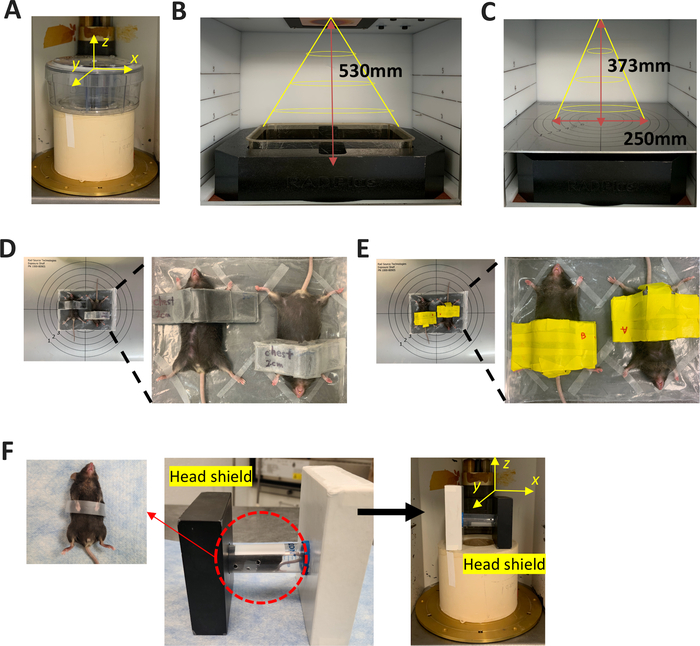
Figure 1: Images showing various preconditioning setups. (A) Pie-cage total body irradiation setup using gamma-ray (Cesium-137): The radiation beam comes from the back of the irradiator in the y-axis direction (horizontal radiation). (B) Mouse cage total body irradiation setup using X-ray: The mouse cage is placed in the reflective chamber. The radiation beam comes from the top of the irradiator in the shape of a cone (vertical radiation). The distance from the radiation source to the cage is 530 mm. (C) Adjustable tray in X-ray irradiator: This setup is used for partially shielded irradiation using X-ray. The radiation beam comes from the top of the irradiator in the shape of a cone (vertical radiation). The distance from the radiation source to the tray is 373 mm, and the radius is 250 mm. (D) Thorax-shielding: Anesthetized mice are placed on a tray. The mice are placed inverted to each other in supine positions with arms and legs fully extended. The lower end of the lead-shield is aligned with the xiphisternum bone and the upper end with the thymus. (E) Abdominal-shielding: Anesthetized mice are placed as in the thorax-shielding set-up with the lower end of the lead-shield aligned with the anus and the upper end below the diaphragm. (F) Head-shielding irradiation setup using gamma-ray (Cesium-137): The anesthetized mouse’s forepaws are taped down and the mouse is placed in a conical restrainer. The black lead-shield (marked) covers the mouse’s head and ears. The radiation beam comes from the back of the irradiator in the direction of the Y-axis (horizontal radiation). Please click here to view a larger version of this figure.
2. Preconditioning of recipient mice (optional)
- Total body irradiation
- Place recipient mice into a uniformly sliced pie-cage, or a mouse cage in the reflective chamber within the calculated radius to receive same irradiation dose; however, a maximum of 8 mice per pie-cage and 5 mice per mouse cage is recommended to ensure uniform irradiation (Figure 1A,B).
- To achieve complete myeloablation, ensure that recipient mice receive a total radiation dose of 11 Gy in two 5.5 Gy fractions separated by a 4–24 h interval.
NOTE: While optimal engraftment can be obtained by implementing a 4 h interval between fractions, this can be extended to a 24 h interval, which can be helpful when labor and/or the irradiator is unavailable.
- Partially shielded irradiation
- Anesthetize the recipient mice by intraperitoneal injection of ketamine (80–100 mg/kg) and xylazine (5–10 mg/kg). The restraint of mouse movement is critical to ensure the uniform irradiation and effective protection of the targeting organs during the shielding process.
- For thorax and abdomen shielding, orient the radiation beam of the X-ray irradiator vertically to the mouse (Figure 1C).
- Position the anesthetized mice onto a flat plate, centering the radiation source from above. Place the mice inverted to each other in a supine position with arms and legs fully extended (Figure 1D,E).
NOTE: X-ray irradiator facility, for this experiment, allows that two mice at a time can be positioned within the effective radius that allows uniform irradiation. While the effective radius is calculated based on the distance between the radiation source and the tray, the number of animals that can be simultaneously irradiated will depend upon the specific irradiator. - Fasten the paws of the mice onto the plate using tape to ensure the mice are immobilized during the irradiation procedure. Place lead shielding so that it covers regions that require protection.
- For thorax shielding, prepare the lead shield by measuring the length from the mouse’s xiphisternum bone to the thymus and calculating the thickness that will provide sufficient protection from the source of irradiation. Place the lead shielding so that the lower end aligns with the xiphisternum bone. The upper end of the lead barrier will fit near the thymus (Figure 1D).
- For abdomen shielding, prepare the lead shield by measuring the length from the mouse’s anus to the diaphragm and calculating the thickness that will provide sufficient protection from the source of irradiation. Place the lead shielding so that the lower end aligns with the anus. The upper end of the lead shield will fit below the diaphragm (Figure 1E).
NOTE: Localizing the lead shield to be consistent among cohorts may reduce some variation with regard to the size of the mice.
- Position the anesthetized mice onto a flat plate, centering the radiation source from above. Place the mice inverted to each other in a supine position with arms and legs fully extended (Figure 1D,E).
- For head shielding, orient the radiation beam of the Cesium irradiator horizontally to the mouse.
- Carefully tape the forepaws of an anesthetized mouse to the abdomen. This ensures that the arms get a full dose of irradiation and are not covered by the shield.
- For head shielding, place the mouse in a conical restrainer, which fits inside a lead shield. Once the mouse is inside the conical restrainer, slide the restrainer into the slot within the lead shield (Figure 1F). The lead shield should completely cover the mouse’s head and ears (~3.2 cm), leaving the rest of the mouse’s body exposed for irradiation. The position of the restrainer inside the shield can be adjusted to fit different sized animals by sliding it further inside or outside the shield.
- Place mice inside the irradiator, perpendicular to the source for irradiation.
- Expose mice to two 5.5 Gy fractions of irradiation (total dose of 11 Gy) separated by a 4–24 h interval.
- After each irradiation fraction, place the cages with anesthetized mice on heated mats or under red heat lamps to prevent hypothermia and aid in the recovery from anesthesia.
NOTE: Caution must be taken to not overheat the anesthetized mouse when using a lamp since they cannot escape the heat. As described above, the positioning of animals and the thickness of lead shield can differ between studies based upon the specific features of the irradiator (radiation type/direction of beam, etc.). Researchers will need to adjust their experiments accordingly.
3. Bone isolation
NOTE: Ideally, donor mice and recipient mice should be similar in age, and within 8–12 weeks old. Using at least 3 mice as donors (rather than single donor) is preferred to minimize for heterogeneity (even when using mice with the same genotype). Approximately, 40 million unfractionated bone marrow cells can be obtained from six bones (two femurs, two tibias, and two humeri) of a single mouse. Transplantation of 5 million bone marrow cells to each recipient mouse will typically ensure engraftment.
- Euthanize donor mice by cervical dislocation without anesthesia (preferred method to avoid chemical contamination of cells) and place each mouse onto an absorbent pad.
- Disinfect the skin using a 70% ethanol spray.
- Make a small transverse cut in the skin below the rib cage and hold the skin tightly at either side of the incision, tear in opposite directions toward the head and feet. Peel off the skin from all the limbs.
- Cut over the shoulders and the elbow joints, and remove the attached muscles and connective tissues with the aid of a Kimwipe to obtain the humeri.
- Carefully dislocate the hip joints between the femur and hip bones. Use blunt scissors to cut along the femur head and detach the legs. Cut over the knee joint to separate the femur and tibia, and carefully remove the attached muscles and connective tissues with the aid of a Kimwipe to harvest the femur and tibia.
NOTE: Pay special attention to keep the bone epiphysis intact during this step. Discard any broken bones due to loss of sterility. Hip bones and spine bones can be collected in addition to the femur, tibia, and humerus. To collect spine bones, a mortar and a pestle can be used to crush the bones into pieces and harvest the bone marrow cells. - Place the isolated bones from mice of the same genotype into correspondingly 50 mL conical tube containing 20 mL ice-cold sterile PBS, and keep it on ice until further use. Pay special attention to correctly place the bones into tubes with matched genotypes.
- Repeat the above steps for each donor animal changing gloves in between each mouse. Also, clean scissors and other instruments with 70% ethanol between each mouse.
4. Bone marrow cell isolation
NOTE: Perform the following steps in a biosafety class II cabinet.
- Preparation of tube sets: Make a small hole in the bottom of a sterile 0.5 mL microcentrifuge tube using an 18 G needle and place it into a sterile 1.5 mL microcentrifuge tube, which contains 100 μL of ice-cold sterile PBS at the bottom.
NOTE: As only six bones can fit into the 0.5 mL microcentrifuge tube, it is recommended to prepare sufficient tube sets to process all the bones at the same time. - Aspirate the PBS and transfer the isolated bones onto a sterile 100 mm cell culture dish. Holding each bone using fine forceps, carefully cut the epiphyses off each end using small scissors that were sterilized in an autoclave. Place the cut bones into the prepared tube sets.
- Centrifuge the tubes at 10,000 x g for 35 s at 4 °C.
- After centrifugation, confirm that the bone marrow has been successfully removed from the bones. Bones should appear white and translucent with a relatively large red pellet at the bottom of the 1.5 mL microcentrifuge tube. Discard the 0.5 mL microcentrifuge tube.
NOTE: If the visual inspection fails to detect bone marrow at the bottom of the 1.5 mL tube, cut the bone again and repeat step 4.3. - Resuspend the bone marrow in 1 mL of ice-cold PBS, then transfer the cell suspension from the same genotype to a matched 50 mL conical tube.
- Dissociate the cells by passing them through an 18 G needle with a 10 mL syringe 10 times.
- Filter single cell suspensions through a 70 μm cell strainer. Add additional ice-cold PBS to a final volume of 10 mL, and resuspend the cells through the gentle use of pipette-aid.
- Centrifuge at 310 x g for 10 min at 4 °C.
- Aspirate the supernatant and resuspend the cell pellet with 10 mL of serum-free RPMI media. Spare 30 μL of this material for cell counting.
- Determine cell concentration with a cell counter, and calculate the volume of cell suspension required for the transplantation. For the example of a 100% BMT, 5 x 10⁶ bone marrow cells are required for each recipient mouse.
NOTE: For a competitive BMT, prepare a total of 5 x 10⁶ bone marrow cells comprising a mixture of donor cells (e.g., CD45.2+) and competitor cells (e.g., CD45.1+). Preparing extra bone marrow cells is highly recommended. For example, if there are 10 recipient mice per experimental group, we typically prepare enough cells for 12 recipient mice. - Transfer the calculated volume of cell suspension into a new 50 mL conical tube. Centrifuge at 310 x g for 10 min at 4 °C.
- Aspirate the supernatant and resuspend the cells using the calculated amount of serum-free RPMI medium to achieve the appropriate cell density and volume. Typically, 200 μL is the optimal volume for a retro-orbital injection.
5. Transplantation of bone marrow cells to irradiated mice
- Anesthetize the recipient mice with 5% isoflurane.
- While mice are anesthetized, slowly inject 200 μL of bone marrow cells into the retro-orbital vein using a 28–30 G needle with an insulin syringe.
- Alternatively, perform the delivery of the donor cells by tail vein intravenous injection and femoral intramedullary injection, with a maximum volume of 0.2 mL and 25 μL, respectively.
- Once the cells are injected, place a drop of proparacaine-containing eye-drops onto the surface of the eye for pain relief. The animal can then be allowed to regain consciousness.
6. Transplantation of bone marrow cells to non-conditioned mice
- Anesthetize the recipient mice by inhalation of 5% isoflurane.
- Inject 5 x 10⁶ unfractionated bone marrow cells from either genotype retro-orbitally into non-irradiated recipient mice with 28–30 G insulin syringe.
- Repeat steps 6.1 and 6.2 over 3 consecutive days, such that the recipient mice will be transplanted with a total of 1.5 x 10⁷ bone marrow cells.
NOTE: Because the adoptive BMT without pre-conditioning procedure requires bone marrow transplantation for 3 consecutive days, one should attempt to alternate eyes for each injection. - Post-injection, administer a drop of proparacaine-containing eye drops to the affected eye.
Access restricted. Please log in or start a trial to view this content.
Results
To compare the effect of three BMT/pre-conditioning methods on donor cell engraftment, the fractions of donor cells in peripheral blood and heart tissue were analyzed by flow cytometry at 1-month post-BMT. Isolated cells were stained for specific leukocyte markers to identify the different subsets of leukocytes. In these experiments, wild-type (WT) C57BL/6 (CD45.2) donor bone marrow cells were delivered to WT B6.SJL-PtprcaPrpcb/BoyJ (CD45.1) reci...
Access restricted. Please log in or start a trial to view this content.
Discussion
For studies of clonal hematopoiesis, we described three methods of BMT: BMT with total-body irradiation, BMT with irradiation with partial shielding, and a less commonly used BMT method that involves no pre-conditioning (adoptive BMT). These methods have been used to assess the impact of clonal hematopoiesis on cardiovascular disease. Researchers can modify these methods accordingly to suit the specific purpose of their study.
Clonal hematopoiesis models
Clonal hematopoi...
Access restricted. Please log in or start a trial to view this content.
Disclosures
The authors have nothing to disclose.
Acknowledgements
This work was supported by US National Institutes of Health grants to K. Walsh (HL131006, HL138014, and HL132564), to S. Sano (HL152174), American Heart Association grant to M. A. Evans (20POST35210098), and a Japan Heart Foundation grant to H. Ogawa.
Access restricted. Please log in or start a trial to view this content.
Materials
| Name | Company | Catalog Number | Comments |
|---|---|---|---|
| 0.5ml microcentrifuge | Fisher Scientific | 05-408-121 | general supply |
| 1.5ml microcentrifuge | Fisher Scientific | 05-408-129 | general supply |
| 1/2 cc LO-DOSE INSULIN SYRINGE | EXELINT | 26028 | general supply |
| Absolute Ethanol (200 prfof) | Fisher chemical | 200559 | general supply |
| BD 1mL Tuberculin Syringes 25G 5/8 Inch Needle | Becton Dickinson | 309626 | general supply |
| BD PrecisionGlide Needle 18G (1.22mm X 25mm) | Becton Dickinson | 395195 | general supply |
| Cesium-137 Irradiator | J. L. Shepherd | Mark IV | equipment |
| DietGel 76A | Clear H2O | 70-01-5022 | general supply |
| Falcon 100 mm TC-Treated Cell Culture Dish | Life Sciences | 353003 | general supply |
| Falcon 50 mL Conical Centrifuge Tubes | Fisher Scientific | 352098 | general supply |
| Fisherbrand sterile cell strainers, 70 μm | Fisher Scientific | 22363548 | general supply |
| Graefe Forceps | Fine Science Tools | 11051-10 | general supply |
| Hardened Fine Scissors | Fine Science Tools | 14090-09 | general supply |
| Isothesia (Isoflurane) solution | Henry Schein | 29404 | Solution |
| Ketamine | Zoetis | 043-304 | injection |
| Kimwipes Delicate Task Wipers | Kimtech Science | KCC34155 | general supply |
| PBS pH7.4 (1X) | Gibco | 10010023 | Solution |
| RadDisk – Rodent Irradiator Disk | Braintree Scientific | IRD-P M | general supply |
| RPMI Medium 1640 (1X) | Gibco | 11875-093 | Medium |
| Sulfamethoxazole and Trimethoprim | TEVA | 0703-9526-01 | injection |
| Xylazine | Akorn | 139-236 | injection |
| X-ray irradiator | Rad source | RS-2000 | equipment |
References
- Evans, M. A., Sano, S., Walsh, K. Cardiovascular disease, aging, and clonal hematopoiesis. Annual Review of Pathology: Mechanisms of Disease. 15 (1), 419-438 (2020).
- Welch, J. S., et al. The origin and evolution of mutations in acute myeloid leukemia. Cell. 150 (2), 264-278 (2012).
- Jaiswal, S., et al. Age-related clonal hematopoiesis associated with adverse outcomes. New England Journal of Medicine. 371 (26), 2488-2498 (2014).
- Dorsheimer, L., et al. Association of mutations contributing to conal hematopoiesis with prognosis in chronic ischemic heart failure. JAMA Cardiology. 4 (1), 25(2019).
- Genovese, G., et al. Clonal hematopoiesis and blood-cancer risk inferred from blood DNA sequence. New England Journal of Medicine. 371 (26), 2477-2487 (2014).
- Jaiswal, S., et al. Clonal hematopoiesis and risk of atherosclerotic cardiovascular disease. New England Journal of Medicine. 377 (2), 111-121 (2017).
- Bick, A. G., et al. Genetic interleukin 6 signaling deficiency attenuates cardiovascular risk in clonal hematopoiesis. Circulation. 141 (2), 124-131 (2020).
- Fuster, J. J., et al. Clonal hematopoiesis associated with TET2 deficiency accelerates atherosclerosis development in mice. Science. 355 (6327), 842-847 (2017).
- Wang, Y., et al. Tet2-mediated clonal hematopoiesis in nonconditioned mice accelerates age-associated cardiac dysfunction. JCI Insight. 5 (6), 135204(2020).
- Sano, S., et al. Tet2-mediated clonal hematopoiesis accelerates heart failure through a mechanism involving the IL-1β/NLRP3 inflammasome. Journal of the American College of Cardiology. 71 (8), 875-886 (2018).
- Sano, S., et al. JAK2-mediated clonal hematopoiesis accelerates pathological remodeling in murine heart failure. JACC: Basic to Translational Science. 4 (6), 684-697 (2019).
- Yu, K. R., et al. The impact of aging on primate hematopoiesis as interrogated by clonal tracking. Blood. 131 (11), 1195-1205 (2018).
- Sano, S., et al. CRISPR-mediated gene editing to assess the roles of Tet2 and Dnmt3a in clonal hematopoiesis and cardiovascular disease. Circulation Research. 123 (3), 335-341 (2018).
- Stachura, D. L., et al. Clonal analysis of hematopoietic progenitor cells in the zebrafish. Blood. 118 (5), 1274-1282 (2011).
- Gyurkocza, B., Sandmaier, B. M. Conditioning regimens for hematopoietic cell transplantation: one size does not fit all. Blood. 124 (3), 344-353 (2014).
- Bacigalupo, A., et al. Defining the intensity of conditioning regimens: working definitions. Biology of Blood and Marrow Transplantation. 15 (12), 1628-1633 (2009).
- Abbuehl, J. P., Tatarova, Z., Held, W., Huelsken, J. Long-term engraftment of primary bone marrow stromal cells repairs niche damage and improves hematopoietic stem cell transplantation. Cell Stem Cell. 21 (2), 241-255 (2017).
- Shao, L., et al. Total body irradiation causes long-term mouse BM injury via induction of HSC premature senescence in an Ink4a- and Arf-independent manner. Blood. 123 (20), 3105-3115 (2014).
- Cui, Y. Z., et al. Optimal protocol for total body irradiation for allogeneic bone marrow transplantation in mice. Bone Marrow Transplantation. 30 (12), 843-849 (2002).
- Koch, A., et al. Establishment of early endpoints in mouse total-body irradiation model. PLOS One. 11 (8), 0161079(2016).
- Ismaiel, A., Dumitraşcu, D. L. Cardiovascular risk in fatty liver disease: the liver-heart axis-literature review. Frontiers in Medicine. 6, 202(2019).
- Amann, K., Wanner, C., Ritz, E. Cross-talk between the kidney and the cardiovascular system. Journal of the American Society of Nephrology. 17 (8), 2112-2119 (2006).
- Liao, X., et al. Distinct roles of resident and nonresident macrophages in nonischemic cardiomyopathy. Proceedings of the National Academy of Sciences. 115 (20), 4661-4669 (2018).
- Honold, L., Nahrendorf, M. Resident and monocyte-derived macrophages in cardiovascular disease. Circulation Research. 122 (1), 113-127 (2018).
- Lavine, K. J., et al. The macrophage in cardiac homeostasis and disease. Journal of the American College of Cardiology. 72 (18), 2213-2230 (2018).
- Ginhoux, F., Guilliams, M. Tissue-resident macrophage ontogeny and homeostasis. Immunity. 44 (3), 439-449 (2016).
- Mildner, A., et al. Microglia in the adult brain arise from Ly-6C hi CCR2+ monocytes only under defined host conditions. Nature Neuroscience. 10 (12), 1544-1553 (2007).
- Cronk, J. C., et al. Peripherally derived macrophages can engraft the brain independent of irradiation and maintain an identity distinct from microglia. Journal of Experimental Medicine. 215 (6), 1627-1647 (2018).
- Lu, R., Czechowicz, A., Seita, J., Jiang, D., Weissman, I. L. Clonal-level lineage commitment pathways of hematopoietic stem cells in vivo. Proceedings of the National Academy of Sciences. 116 (4), 1447-1456 (2019).
- Gibson, B. W., et al. Comparison of cesium-137 and X-ray irradiators by using bone marrow transplant reconstitution in C57BL/6J mice. Comparative Medicine. 65 (3), 165-172 (2015).
- Cui, Y. Z., et al. Optimal protocol for total body irradiation for allogeneic bone marrow transplantation in mice. Bone Marrow Transplantation. 30 (12), 843-849 (2002).
- Kim, C. K., Yang, V. W., Bialkowska, A. B. The role of intestinal stem cells in epithelial regeneration following radiation-induced gut injury. Current Stem Cell Reports. 3 (4), 320-332 (2017).
Access restricted. Please log in or start a trial to view this content.
Reprints and Permissions
