Bioengineering
A subscription to JoVE is required to view this content.
You will only be able to see the first 2 minutes.
The JoVE video player is compatible with HTML5 and Adobe Flash. Older browsers that do not support HTML5 and the H.264 video codec will still use a Flash-based video player. We recommend downloading the newest version of Flash here, but we support all versions 10 and above.
If that doesn't help, please let us know.
Assembly and Characterization of Biomolecular Memristors Consisting of Ion Channel-doped Lipid Membranes
Chapters
Summary March 9th, 2019
Soft, low-power, biomolecular memristors leverage similar composition, structure, and switching mechanisms of bio-synapses. Presented here is a protocol to assemble and characterize biomolecular memristors obtained from insulating lipid bilayers formed between water droplets in oil. The incorporation of voltage-activated alamethicin peptides results in memristive ionic conductance across the membrane.
Transcript
Our protocol describes how to assemble and electrically characterize a peptide-doped biomembrane that closely mimics the composition, structure, and transport properties of biological synapses and which exhibits tunable memory resistance. This technique empowers users to assess activity-dependent, memory resistance and short-term plasticity in engineered systems at time scales and excitation levels relevant to biological synapses and ion channels. This technique provides a framework for characterizing biomimetic membranes containing voltage-activated ion channels, making it applicable to characterization of a variety of cellular transport processes, including those in neurons.
Our suggestion to new researchers is to first become proficient at preparing liposome solutions and assembling a droplet interface bilayer on wire-type electrodes. Seeing firsthand the process for droplet dispensing and positioning on electrodes simplifies this technique for bilayer formation, making it immediately accessible to all. Demonstrating this procedure will be Dr.Joseph Najem, a postdoc from my laboratory.
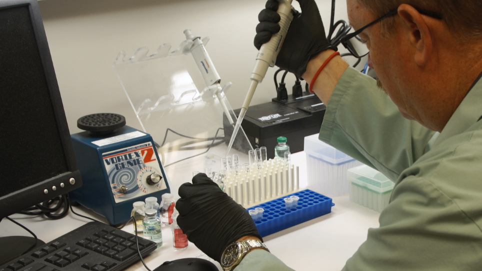
To begin, prepare the alamethicin stock solution in the microcentrifuge tube by dissolving the alamethicin peptides powder in ethanol to a final concentration of 2.5 milligram per milliliter. Vortex the tube briefly to mix well, and store the stock solution in a minus 20 degree Celsius freezer. In a 1.5 milliliter Safe-Lock tube, add one microliter of alamethicin stock solution to 99 microliters of solution A to achieve a final alamethicin concentration of 13 micromolar in the liposome suspension.
Vortex to mix well. The resulting peptide liposome solution is solution B.Mix 117 microliters of solution A with 10 microliters of solution B to achieve a final alamethicin concentration of one micromolar, and then vortex to mix well. Refer to the resulting solution as C.Store the solutions B and C at four degrees Celsius.
Place a one-millimeter thick 25 times 75 millimeters glass side on the stage of an inverted microscope. Dispense a few drops of hexadecane oil onto the center of the glass slide, and then place the oil reservoir directly onto the oil on the glass slide. Completely fill the oil reservoir with hexadecane oil.
Make sure the reservoir is positioned above the objective lens. Then plug the electrode holder to the head stage of a current amplifier mounted on a micromanipulator. The micromanipulator minimizes electrode length and electrical noise.
Then, mount the glass micropipette holder with the second silver-silver-chloride wire onto another micromanipulator. Using the micromanipulators, position the electrodes such that the agarose-coated tips of the silver-silver-chloride wires are fully submerged into the oil reservoir at a similar vertical plane. Align the two electrodes and separate them by a few millimeters.
To form the lipid bilayer, move the electrodes vertically into the oil phase. Use the micropipette to deposit 200 nanoliters of lipid solution A on each of the wires. Wait for three to five minutes to allow for spontaneous lipid monolayer assembly to occur at the water oil interface.
Droplets might sag if the surrounding oil is sufficiently less dense. After that, lower the electrodes to re-submerge until the ends of both electrodes barely touch the bottom of the oil reservoir. Then to form the bilayer, move the electrodes horizontally to bring the droplets into contact.
To obtain the pinched, hysteretic, current-voltage relationship, use a function generator to apply triangular or sinusoidal voltage waveform to an alamethicin-free lipid membrane assembled with droplets of solution A.Record the induced current response across multiple frequencies. To record the size of the interfacial lipid bilayer, either measure the diameter of the lipid membrane on computer or record the peak-to-peak current amplitude resulting from the 10 hertz, 10 millivolts triangular wave to calculate the area of the membrane. Take the wires out of the oil phase to remove the droplets that contain no alamethicin.
Add new aqueous droplets using solution C and form a lipid bilayer. Based on the square wave current amplitude, use the micromanipulators to adjust contact between droplets, such that the bilayer has a similar area as the one formed earlier. Then apply a 10-hertz and 10-millivolts voltage wave form, and record the induced current response as previously.
To conduct pulse experiments using a custom programming software and analog voltage source, generate voltage pulses with specific high and low amplitudes on time and off time. Record the current in response to applied pulses. The plot of current versus voltage shows the non-zero current response upon application of a voltage bias to an alamethicin-free lipid bilayer.
Add 0.017 hertz, a frequency where the impedance is dominated by membrane resistance. A low ohmic current response is shown for the highly insulating membrane. The plot of a lipid bilayer formed between two droplets containing alamethicin peptides shows exponentially increasing currents at voltages higher than the insertion threshold of 100 millivolts.
At high voltage, alamethicin peptides residing at the surface of the lipid bilayer insert into the membrane and aggregate to form conductive pores. The symmetric current responses at both polarities is due to the insertion and aggregation of separate populations of peptides from opposite sides of the membrane. The capacitive current must be subtracted from the total current to obtain only the memristive, pinched hysteresis current-voltage response.
The biomolecular memristor response to subsequent voltage pulses with an increase in conductance during the on time, despite intermittently restoring an insulating state during each off time. Both the present stimulus and prior stimuli contribute to the current increase. Cohesive monolayers on both droplets must form before bringing them together to form the bilayer.
If the droplets are brought together too early, they coalesce and no bilayer is formed. We are now designing and fabricating, microfluidic base neural networks consisting of solid state neurons connected by membrane-based synapses supported by network assimilation optimization of high-performance supercomputers at ORNL. These memristors are the first to have the composition, structure, switching mechanism, and ion transport of biological synapses.
They thus provide a biomolecular basis furnished adding brain-like computing and memory.
Related Videos
You might already have access to this content!
Please enter your Institution or Company email below to check.
has access to
Please create a free JoVE account to get access
Login to access JoVE
Please login to your JoVE account to get access
We use/store this info to ensure you have proper access and that your account is secure. We may use this info to send you notifications about your account, your institutional access, and/or other related products. To learn more about our GDPR policies click here.
If you want more info regarding data storage, please contact gdpr@jove.com.
Please enter your email address so we may send you a link to reset your password.
We use/store this info to ensure you have proper access and that your account is secure. We may use this info to send you notifications about your account, your institutional access, and/or other related products. To learn more about our GDPR policies click here.
If you want more info regarding data storage, please contact gdpr@jove.com.
Your JoVE Unlimited Free Trial
Fill the form to request your free trial.
We use/store this info to ensure you have proper access and that your account is secure. We may use this info to send you notifications about your account, your institutional access, and/or other related products. To learn more about our GDPR policies click here.
If you want more info regarding data storage, please contact gdpr@jove.com.
Thank You!
A JoVE representative will be in touch with you shortly.
Thank You!
You have already requested a trial and a JoVE representative will be in touch with you shortly. If you need immediate assistance, please email us at subscriptions@jove.com.
Thank You!
Please enjoy a free 2-hour trial. In order to begin, please login.
Thank You!
You have unlocked a 2-hour free trial now. All JoVE videos and articles can be accessed for free.
To get started, a verification email has been sent to email@institution.com. Please follow the link in the email to activate your free trial account. If you do not see the message in your inbox, please check your "Spam" folder.