Corneal collagen cross-linking (CXL) is the only conservative treatment currently available to halt keratoconus progression by improving the biomechanical rigidity of the corneal stroma. The aim of this manuscript is to highlight the methods of three different protocols of CXL: conventional CXL (C-CXL), accelerated CXL (A-CXL), and iontophoresis CXL (I-CXL).
Keratoconus is a bilateral and progressive corneal ectasia. In order to slow down its progression, corneal collagen cross-linking (CXL) has recently been introduced as an efficient treatment option. In biological and chemical sciences, crosslinking refers to new chemical bonds formed between reactive molecules. Hence, the aim of corneal collagen CXL is to synthetically increase the formation of crosslinks between collagen fibrils in the corneal stroma. Despite the fact that the efficiency of the conventional CXL (C-CXL) protocol has already been shown in several clinical studies, it might benefit from improvements in duration of the procedure and removal of corneal epithelium. Hence, in order to provide a coherent evaluation of two new and optimized CXL protocols, we studied keratoconus patients who had undergone one of the three CXL treatments: iontophoresis (I-CXL), accelerated CXL (A-CXL), and conventional CXL (C-CXL). A-CXL is a 6 time faster CXL procedure using a ten time higher UVA irradiance but still including an epithelium removal. Iontophoresis is a transepithelial non-invasive technique in which a small electric current is applied to improve riboflavin penetration throughout the cornea. Using anterior segment optical coherence tomography (AS OCT) and in vivo confocal microscopy (IVCM), we conclude that regarding the depth of treatment penetration, conventional CXL protocol remains the standard for treating progressive keratoconus. Accelerated CXL seems to be a quick, effective and safe alternative to treat thin corneas. The use of iontophoresis is still being investigated and should be considered with greater caution.
Keratoconus is a bilateral and progressive corneal ectasia usually reported in 1 in 2,000 in the general population1 resulting in modification of the corneal shape and thus decreased vision2. Keratoconus is usually present in early puberty and progresses until the third to fourth decade of life when the disease typically tends to stabilize, although progression can be variable throughout a patient's life. By halting keratoconus progression, cross linking aims at postpone or avoid keratoplasty.
To date, the only efficient and safe treatment of progressive keratoconus proven in clinical studies is the conventional corneal collagen cross-linking (C-CXL) protocol, which aims to increase stiffness and hence halt keratoconus progression3-8. In order to reduce operation time and other possible risk factors of C-CXL, such as infectious keratitis or stromal haze9, several improved protocols have been described. First, in accelerated CXL (A-CXL), a higher irradiance of UVA is delivered to the cornea over a reduced time10. Secondly, to avoid the necessity for epithelial debridement, transepithelial approaches have been employed. Unfortunately, they have limited success when compared to the conventional protocol11. The most recent transepithelial method for corneal riboflavin delivery during CXL is iontophoresis (I-CXL), but rigorous evaluation of this treatment has not yet been performed12. Iontophoresis is a non-invasive technique in which a small electric current is applied to improve an ionized drug's penetration through a tissue. In CXL by iontophoresis, the riboflavin is ionized to penetrate the cornea through the epithelium.
In vivo confocal microscopy (IVCM) is a method of imaging the cornea that can highlight the cellular changes of abnormal corneas in diseases such as keratoconus13. Indeed, IVCM has demonstrated alterations to all layers of the cornea in keratoconus with a particular reduction in density of the sub-basal nerve plexus and stromal keratocytes13-15. Plus, IVCM has proven to be highly convenient for microstructural analysis of the cornea after C-CXL16.
The corneal demarcation line is described as a hyperreflective line seen in anterior segment optical coherence tomography (AS OCT) 1 month after C-CXL at a depth of 300 µm17,18. IVCM following C-CXL provides information about corneal structural alterations, including the absence of corneal keratocytes to a depth of 300 µm. The depth of this acellular zone, as well as the depth of the demarcation line within the corneal stroma revealed on AS OCT, seems to be associated with the effective depth of CXL treatment19, and measurement of the corneal demarcation line depth in AS OCT 1 month after CXL has been proposed as an efficient clinical method for evaluation of CXL effectiveness18.
In the present study we investigate the efficiency of three different protocols of corneal collagen crosslinking (conventional, accelerated, and iontophoresis) using measurement of the corneal stromal demarcation line by AS OCT and confocal microscopy. We furthermore used IVCM to quantitatively analyze corneal microstructure changes after the three treatments.
These protocols follow the guidelines of our institution's human research ethics committee.
1. Conventional Corneal Collagen CXL (C-CXL)
1. Preparation of the Patient
2. Epithelial Removal
3. Riboflavin Application
4. UVA Irradiation
Figure 1: UVA irradiation in C-CXL. The cornea is irradiated with a 370 nm wavelength UVA light at an irradiance of 3 mW/cm2 (5.4 J/cm2 surface dose) and at a 5 cm working distance for 30 minutes. Please click here to view a larger version of this figure.
5. End of the Surgery
2. Accelerated Corneal Collagen CXL (A-CXL)
1. Preparation of the Patient
2. Epithelial Removal
3. Riboflavin Application
4. UVA Irradiation
5. End of the Surgery
3. Iontophoresis (I-CXL)
1. Preparation of the Patient
2. Position the Iontophoresis Device.
Figure 2. Iontophoresis device. The passive electrode is applied on the forehead under the operative field and the active electrode, a suction ring, is applied to the open eye. Please click here to view a larger version of this figure.
3. Riboflavin Application
Figure 3. Riboflavin application in I-CXL. The suction ring is filled with hypoosmolar 0.1% riboflavine without Dextran. Please click here to view a larger version of this figure.
Figure 4. Iontophoresis device for riboflavin penetration. The electrical current is initially 0.2 mA and gradually increased to 1.0 mA. Total iontophoresis time is 5 minutes. Please click here to view a larger version of this figure.
4. UVA Irradiation
5. End of the Surgery
The corneal demarcation line was visible in AS OCT in 92% of cases at a mean depth of 301.6 µm (SD, 73.6)
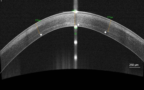
Figure 5. Demarcation line after C-CXL. High-resolution corneal anterior segment optical coherence tomography scan (AS OCT) visualizing the corneal stromal demarcation line at a mean depth of 358 µm (white arrow), 1 month after conventional corneal collagen cross-linking (C-CXL). Scale bar 250 µm. Please click here to view a larger version of this figure.
1 month after C-CXL, whereas after A-CXL it was seen in 85.5% of cases at a mean depth of 183.1 µm (SD, 39.6).
Figure 6. Demarcation line after A-CXL. High-resolution corneal anterior segment optical coherence tomography scan (AS OCT) visualizing the corneal stromal demarcation line at a mean depth of 176 µm (white arrow), 1 month after accelerated corneal collagen cross-linking (A-CXL). Scale bar: 250 µm. Please click here to view a larger version of this figure.
Finally, after I-CXL, the corneal demarcation line was only seen in 46.5% of cases at a mean depth of 214 µm (SD, 37.5). The differences in corneal demarcation line depth following either C-CXL, A-CXL or I-CXL were statistically significant (p <0.001 and p = 0.01). The demarcation line was present significantly more often after C-CXL and A-CXL than after I-CXL (p = 0.005).
Figure 7. Demarcation line after I-CXL. High-resolution corneal anterior segment optical coherence tomography scan (AS OCT) visualizing the corneal stromal demarcation line at a mean depth of 238.5 µm (white arrow), 1 month after iontophoresis (I-CXL). Scale bar: 250 µm. Please click here to view a larger version of this figure.
No intra or postoperative complications were detected in patient follow-ups within 6 months after application of any of the three protocols, including no significant differences in endothelial cell counting. Moreover, the maximum K-value (Kmax) remained stable for each of the protocols after a 6 month follow-up.
Table 1. Efficacy and safety of each protocol of CXL. Evolution of the maximum K-value (dioptry, D) and the endothelial density following conventional (C-CXL), accelerated (A-CXL), and iontophoresis (I-CXL) cross-linking.
For each of the protocols, in the 1-3 month postoperative period, anterior stromal edema with extracellular lacunae and fragmented keratocyte nuclei was observed with IVCM. At 6 months, repopulation of the anterior stroma with keratocyte nuclei was seen and was greater after I-CXL than after the two other protocols. The demarcation between cross-linked and non-cross-linked corneal stroma was seen as a region where keratocytes became elongated and surrounded by large hyper-reflective stromal bands.
Figure 8.: Microstructural corneal changes after C-CXL. In vivo confocal microscopy scans (IVCM) of the corneal stroma obtained 1 month after conventional collagen cross-linking (C-CXL): anterior stromal edema with hyper-reflective cytoplasm (white arrows) and extracellular lacunae (asterisks) are observed. Scale bar: 50 µm.
CXL using UVA irradiation and riboflavin is the standard treatment for arresting the progression of keratoconus. Riboflavin is a photosensitizer which induces chemical covalent bonds (cross-links) when irradiated with UVA3. In the cornea, this phenomenon creates cross-links between collagen fibrils that increase corneal stiffness. Although this phenomenon is well described, until now there has been no direct evidence of intracorneal cross-links. Nonetheless, several studies have reported a stabilization of the Kmax after the procedure thus demonstrating the effectiveness of C-CXL3-8. The question of whether this efficacy is due to cross-links or to other microstructural changes in the corneal stroma remains unanswered.
One of the indirect clinical outcomes of CXL efficacy is the corneal demarcation line detected with AS OCT and IVCM 1 month after CXL. Recently, Kymionis et al. showed that the hyperreflectance evaluated using AS OCT corresponds to the transition area between the acellular and the cellular zone seen on confocal microscopy20. Thus, the corneal demarcation line seen on AS OCT must correspond to a zone of activated keratocytes and fibroblastes under an acellular stroma and above a normal one. Nonetheless, Yam and associates21 failed to demonstrate a correlation between the CXL demarcation line depth with change of visual acuity and of maximum K-value 6 months after C-CXL. The question of whether a greater amount of CXL can result in a greater enhancement in visual acuity and greater reduction of keratometry remains the subject of studies with longer postoperative follow-up. In addition, regarding the mean anterior stromal keratocyte density count on IVCM, a significant reduction was observed during the first 6 months, with normalization at 12 months with C-CXL and A-CXL, and at 6 months with I-CXL22-24. Consequently, varying microstructural corneal changes appear dependent on the type of collagen cross-linking protocol used. This result and the fact that the corneal demarcation line appeared significantly deeper after C-CXL than after A-CXL or I-CXL allows us to discuss the indication and effectiveness of these three protocols.
The conventional protocol has already proved its efficacy and safety with a maximum follow-up of 6 years3-8. C-CXL requires corneal pachymetry of at least 400 µm to protect the endothelial cells25. Its major drawbacks are related to time duration (1 hr) and the necessity to remove the epithelium. Indeed, this leads to patient discomfort and pain and can cause several complications such as infectious keratitis and stromal haze9. Nonetheless, for now, this protocol is still recommended to treat progressive keratoconus, especially when the evolution is aggressive.
Given that one of the major drawbacks of C-CXL was the duration of the procedure, the accelerated protocol initially aimed at reducing operation time by delivering a higher irradiance to the cornea26. However, reducing the soaking time to 10 min may limit the intra-stromal penetration of the riboflavin into the cornea, thus leading to the observed superficial corneal demarcation line. Even if it has not been reported and despite the fact that the same number of photons touch the fibrils, it is possible that the 10 times higher irradiance in A-CXL extends the risk of endothelial injuries27, 28. In this context, it is important to remark that the absence of Dextran in the riboflavin used for A-CXL may explain the absence of endothelial damage despite the higher irradiance. Indeed, Dextran is known to have an osmotic effect that leads to corneal thinning during the procedure and thus to potential endothelial damage29. Consequently, accelerated CXL appears to be a safe CXL modality. Plus, the A-CXL protocol seems to be efficacious; indeed the maximum K-value remained stable at 6 months follow-up. Nonetheless, as for C-CXL, its major limitation is the desepithelialization leading to pain and potential complications such as haze and corneal infection9. Touboul et al. carried out a qualitative study using confocal microscopy of patients treated with A-CXL23. Indeed, when compared to C-CXL, the stiffening effect of the UVA-riboflavin seemed to be most prominent in the anterior 150-200 µm of the cornea with greater keratocyte apoptosis and increased stromal reflectivity. This finding suggests that patients with thin corneas (minimal thickness of 350-400 µm) may benefit from the accelerated CXL. At this time, a hypo-osmolar riboflavin that leads to swelling of thin corneas before C-CXL is used since this protocol still requires corneal pachymetry of at least 400 µm to prevent endothelial damage25. Nonetheless, accelerated CXL may be preferentially used in the future as a faster and less penetrating treatment to stabilize keratoconus for thinner corneas. However, long-term studies are needed to conclusively correlate the depth of the demarcation line with the effect on corneal biomechanics.
Iontophoresis CXL is one transepithelial protocol recently developed to avoid epithelial debridement12, 30. Application of an electric current forces the hypo-osmolar riboflavin to penetrate the corneal stroma. Vinciguerra and co-workers examined 20 eyes which underwent iontophoresis CXL in a prospective study. They showed that the Kmax was stable 1 year after the procedure. However, the demarcation line was not clearly measurable with AS OCT during the follow-up31. Similarly, in our study, the corneal demarcation line assessed with AS OCT was hardly seen at a mean depth of 214 µm in less than half of the patients (46.5%). Plus, confocal microscopy revealed much less keratocyte apoptosis and increased stromal reflectivity after I-CXL than after the two other protocols. Indeed, using confocal microscopy and a modified riboflavin (Ricrolin TE), Caporossi et al. investigated another protocol of transepithelial crosslinking. As for iontophoresis, they find that apoptosis of stromal keratocytes was superficial (mean depth of 140 µm) and unevenly seen in the anterior stroma11. Plus, they confirmed that this Epi-ON protocol resulted in keratoconus evolution after 24 months of follow-up, adding a note of caution to its application in pediatric patients that often suffer from more aggressive forms of the disease32. Indeed, as for other transepithelial protocols, iontophoresis does not seem to ensure an improvement in topographic indices in pediatric patients33. This absence of efficacy can be explained by limited riboflavin and UVA penetration with the epithelium in situ11,34-36. Indeed, the epithelium is a physical barrier for both riboflavin and UVA penetration, limiting the depth of apoptosis and thus of corneal biomechanical effects11. In addition, riboflavin concomitantly serves as a photosensitizer and a UV blocker during UV exposure28. Consequently it is conceivable that, as for other transepithelial protocols, insufficient riboflavin penetration during iontophoresis will not only limit the efficacy of the procedure, but also increase the risk of endothelial cell damage. Nonetheless, no endothelial cell loss was noted as yet after iontophoresis. Finally, in our study, similarly to Vinciguerra et al.31, the highest K-value appeared stable 6 months after I-CXL. However, it remains to be seen from longer follow-ups whether this new procedure remains reliable. Thus, as with other Epi-ON protocols, caution is require when using iontophoresis. Nevertheless, the enthusiasm for transepithelial CXL is understandable, considering the decrease of potential CXL complications. With Epi-OFF CXL, complications occur in approximately 1% of cases essentially caused by temporary haze9. Unfortunately, this haze occasionally leaves corneal scars. Consequently, we believe that currently, iontophoresis CXL should be used with caution on pediatric patients and we would mostly propose this protocol to patients with thin corneas and slowly progressing keratoconus.
Conclusively, regarding penetration, the conventional CXL protocol remains the standard option for treating progressive keratoconus. Accelerated CXL appears to be a quick, effective and safe alternative to treat particularly thin corneas. Iontophoresis is associated with less damage of anterior keratocytes and a less visible demarcation line and should thus be considered with greater prudence.
The authors have nothing to disclose.
The authors have no acknowledgements.
Riboflavin Product number | |||
C-CXL | Sooft SPA, Montegiorgio, Italy | Ricrolin 468465-6 | |
A-CXL | Avedro Inc, Waltham, Massachusetts | VibeX 520-01863-006 | |
I-CXL | Sooft SPA, Montegiorgio, Italy | Ricrolin+ 975481-6 | Passive electrode: PROTENS ELITE 4848LE/ Active electrode: IONTOFOR CXL |
UVA Machine | |||
X-Vega | UVA: 3 mW/cm2 30 min | ||
KXL System | UVA: 30 mW/cm2 10 min | ||
X-Vega | UVA: 10 mW/cm2 9 min |