Repeated blood sampling is necessary and important in animal research. We developed a novel, non-lethal and reliable method for repeated blood collection from adult zebrafish, and applied this method to the study of blood biochemistry, including glucose metabolism.
Repeated blood collection is one of the most common techniques performed on laboratory animals. However, a non-lethal protocol for blood collection from zebrafish has not been established. The previous methods for blood collection from zebrafish are lethal, such as lateral incision, decapitation and tail ablation. Thus we have developed a novel “repeated” blood collection method, and present here a detailed protocol outlining this procedure. This method is minimally invasive and results in a very low mortality rate (2.3%) for zebrafish, thus enabling repeated blood sampling from the same individual. The maximum volume of blood sampling is dependent on body weight of the fish. The volume for repeated blood sampling at intervals should be ≤0.4% of body weight every week or ≤1% every 2 weeks, which were evaluated by measurements of blood hemoglobin. Additionally, hemoglobin, fasting blood glucose, plasma triacylglycerol (TG) and total cholesterol levels in male and female adult zebrafish were measured. We also applied this method to investigate the dysregulation of glucose metabolism in diet-induced obesity. This blood collection method will allow many applications, including glucose and lipid metabolism and hematological studies, which will increase the use of zebrafish as a human disease model organism.
Zebrafish are gaining increasing popularity as a valuable model of human diseases because their organs and genetics are similar to those of humans1,2. In the field of developmental biology, many studies have demonstrated that zebrafish and human show marked similarity in hematopoiesis3, hemostasis4,5, and myelopoiesis6. Adult zebrafish are also used for studying immunological7, neurodegenerative8 and obesity-related diseases9 because this model organism shares common pathways with those disrupted in human diseases. For obesity and obesity-related diseases (diabetes, hepatic steatosis and nonalcoholic steatohepatitis and atherosclerosis), zebrafish blood glucose and lipids levels have been thoroughly investigated in several transgenic and diet-induced obesity models10-13.
Repeated blood sampling from individual animals will reduce animal use and decrease interindividual differences. However, repeated sample collection is technically difficult in small animals such as zebrafish because of their relatively small blood volume and the lack of easily accessible vessels. Several methods for one-time blood collection from zebrafish have been developed, although these methods have their own drawbacks, including lethality, associated tissue damage and limited blood volume. For example, 1–5 µl blood can be harvested from a lateral incision of approximately 0.3 cm in length in the region of the dorsal aorta5. Decapitation with scissors by cutting through the pectoral girdle can collect 5–10 µl blood10. Another convenient blood sampling method is tail ablation14. Cardiac puncture is one potential alternative method for repeated blood collection from the same fish, but the very small amount obtained (approximately 50 nl) with this procedure limits the number of analyses that can be performed11. Accordingly, a new protocol is needed to enable repeated non-lethal blood sampling, which would be a critical advance necessary for this organism to be a standard model organism for human diseases. This technique would allow for testing pharmacological response, discovery of molecular biomarkers for diagnosis, determination of prognosis, and the monitoring of various diseases, such as metabolic diseases, degenerative diseases and several types of malignancies.
We therefore developed a minimally invasive method for obtaining blood from zebrafish serially15. Here we demonstrate the procedure visually and provide a detailed protocol for this technique. Using this method, the normal value based on various parameters, including hemoglobin, fasting blood glucose, and lipids in the blood of healthy adult zebrafish were evaluated. Additionally, we also evaluated whether this method is suitable for studies that require serial samples by monitoring the temporal changes in blood glucose levels during overfeeding experiments.
All animal procedures were approved by the Ethics Committee of Mie University, and were performed according to Japanese animal welfare regulation ‘Act on Welfare and Management of Animals’ (Ministry of Environment of Japan) and complied with international guidelines.
1. Preparation of the Needle
NOTE: All experiments were performed under anesthesia, and all efforts were made to minimize suffering. For euthanasia, fish were immersed in an ice–water bath (5 parts ice/1 part water at ≤4 °C) for ≥20 min.
2. Anesthesia
3. Blood Collection
This blood collection method causes minimal injury to zebrafish (a <1 mm puncture; Figure 1J) and yields a very low mortality rate of 2.3%. We examined the maximum volume of blood that could be collected from a single fish and evaluated the relationship to its body weight (Figure 3). We found that the maximum blood volume collected was linearly correlated with body weight (R = 0.813). The largest volume of blood collected from an individual fish (body weight = 1.071 g) was 25 µl, and the smallest volume was 1.3 µl from a fish weighing 0.115 g. This suggests that the maximum volume of blood collected depends on the body weight of the zebrafish.
Biochemical analysis of hemoglobin, blood glucose, TG and total cholesterol were performed after blood collection (Table 1). Male and female healthy adult zebrafish (4–6 months old) were fasted for 18 hr before blood collection. The biochemical analysis revealed that the normal value of hemoglobin (male 9.91 ± 0.49 g/dl and female 10.02 ± 0.48 g/dl) and TG (male 417 ± 45 mg/dl and female 404 ± 35 mg/dl) did not differ significantly between the two groups. However, the fasting blood glucose and total cholesterol levels of the male group (44 ± 3 mg/dl and 365 ± 18 mg/dl, respectively) were significantly lower (p <0.05) than the female group (69 ± 3mg/dl and 511 ± 52 mg/dl, respectively).
While the minimal trauma to zebrafish using the present method enables repeated blood sampling from the same individual, the effects of repeated blood drawing have not been evaluated. We investigated these effects using measurements of blood hemoglobin level (Figure 4). As we have shown in a previous publication15, adult male fish weighing approximately 0.5 g each were assigned to four groups. Repeated blood sampling (2 µl each time) of the same individual fish once daily for 7 days (for a total of seven blood samples) resulted in significant decrease (p <0.01) in hemoglobin levels from 10.82 ± 0.78 g/dl to 2.38 ± 0.8 g/dl. Removal of 2 µl of blood every 2 days or a single collection of 5 µl per week also yielded a significant decrease (p <0.05) in hemoglobin levels. In addition, one week after a single collection of a 2 µl blood sample, hemoglobin levels were slightly below normal (from 8.11 ± 1.15 g/dl to 7.15 ± 1.17 g/dl). The hemoglobin levels had no effects after a single collection of a 2 or 5 µl blood sample for a 2-week recovery period. Thus, we concluded that repeated collection of 2 µl of blood (0.4% of body weight) per week or 2–5 µl (0.4–1% of body weight) per 2 weeks from individual fish can avoid blood loss anemia.
We further applied this method to the study of glucose metabolism. The changes in blood glucose levels of each individual in the normal diet group (once daily feeding) and overfeeding group (five daily feedings) were monitored over a 5-week period. Normal diet-fed zebrafish (Fish A, B, C) exhibited stable blood sugar levels all the time, while the overfed zebrafish (Fish D, E, F) experienced high blood glucose levels as early as week 1, and maintained this hyperglycemia condition throughout the 5-week study period (Figure 5).
Figure 1: Procedure for Blood Collection From Adult Zebrafish. (A) Glass needles prepared using a needle puller. (B) Cutting the tip of the needle obliquely using a fine scissors. (C) A precut needle with a tip diameter of approximately 135 µm. Scale bar = 1 mm. (D) Blood collection devices: an aspirator tube assembly (left) and a bulb dispenser (right). Arrows indicate the nosepiece to hold the microcapillary needle. The arrowhead shows the mouthpiece of the aspirator tube assembly. The needle is positioned in the end of the nosepiece before sample collection. (E) Heparinizing the needle. (F) An anesthetized fish. (G) Place the fish on a paper towel soaked with the anesthetics. (H) Insert the needle at a 30–45° angle into the blood collection site. (I) Blood rising into the needle. (J) Bleeding has stopped and a <650 µm puncture is circled and shown in a high magnification. (K) Expelling the blood from the needle onto a piece of parafilm. (L) Measurement of blood glucose using a glucose meter.
Figure 2: Schematic Representation of the Anatomic Landmarks for Blood Collection from the Adult Zebrafish. (A) The white line shows the puncture site for blood collection, which is along the body axis and posterior to the anus in the region of the dorsal aorta. (B) The primary vessels are the dorsal aorta and posterior cardinal vein; these are located ventral to the spine. S, spine; DA, dorsal aorta; PCV, posterior cardinal vein.
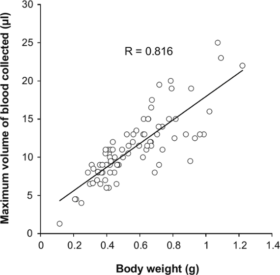
Figure 3: The Relationship of Maximum Volume of Blood Sampling and Body Weight. A total of 83 zebrafish (2–6 months old, 42 male and 41 female) underwent maximal blood collection.
Figure 4: Changes in Hemoglobin Levels Over a 1-week Period with Repeated Blood Sampling.A 2 µl sample of blood was collected from the same individual fish daily, once in 2 days, once weekly or 5 µl once weekly (n = 5). The hemoglobin levels of each group before blood collection (day 0, white bar) and after repeated blood sampling (day 7, gray bar) are shown. Values are means ± standard error of the mean (SEM). *p <0.05, **p <0.01 vs. day 0. Adapted from ref.15.
Figure 5: Changes in the Fasting Blood Glucose Concentrations of Six Individual Male Fish over a 5-week Period. Fish A, B and C were the normal diet group. Fish D, E and F were the overfed group.
Table 1: Hemoglobin, Blood Glucose, TG and Total Cholesterol Levels for Male and Female Zebrafish 4–6 Months of Age.
We present here a detailed protocol for serially obtaining blood from adult zebrafish. This method is straightforward to carry out and we use it in lab on a daily basis. This blood collection method is based on inserting a glass capillary needle into the zebrafish’s dorsal aorta. During this procedure, it is critical to be careful to not ablate the spine because it is the criterion for searching for the dorsal aorta. Reducing the spine injury will improve the survival rate. Although this technique is simple and easy to master, there are best practices that can ensure high success and survival rates. A skillful researcher will take 1–2 min to carry out the blood collection procedure (Protocol 2.3 to 3.6, which is the time that the fish is out of water). To save time in large-scale experiments, a double-team approach is recommended to perform blood collection. For example, one researcher could perform the blood sampling, while a second could handle the fish, perform the anesthesia and analyze the collected blood (measuring blood glucose, recording, or moving the blood to microcentrifuge tube, etc.).
Using this method, we demonstrated that the maximum blood sample volume that could be collected from individual fish is approximately 2% of body weight regardless of sex15, suggesting that the total circulating blood volume of zebrafish is greater than 2% of body weight. Previous studies demonstrated that teleost fish (Osteichthyes spp. and Salmogairdnerigairdneri) possess a total blood volume ranging between 1.8–3.8% of body weight17,18, thus we predict the total circulating blood volume of zebrafish to be 2–3.8% of body weight in zebrafish. For a single blood collection, we strongly recommend that blood sampling should be performed from 3 months old or >0.3 g body weight zebrafish to obtain ≥5 µl of blood. It is noteworthy that approximately half of the zebrafish survived even though 2% of body weight of their blood was removed, which revealed that zebrafish may cope well with blood loss.
The most significant advantage of this method is that it enables repeated blood sampling from the same individual. We determined the optimal volume and frequency of blood sampling by measuring the changes in hemoglobin levels (Figure 4). We recommend that the volume and interval for repeated blood sampling be ≤0.4% of body weight per week and ≤1% of body weight per 2 weeks to avoid blood loss anemia and hemorrhagic death. This conclusion is consistent with the practice guidelines for repeated blood sampling of rodent model animals19,20.
If the experiment allows the sacrifice of zebrafish, the needle can be inserted into a position along the body axis, posterior to the gill in the region of the dorsal aorta as a troubleshooting or an alternative method. This site is near the heart and aorta is relatively big, which can make the blood collection procedure easier.
Zebrafish have been successfully used in modeling the interrelated conditions of metabolic syndromes, including diabetes21,22, obesity23,24, fatty liver disease25 and atherosclerosis26. We further applied this technique to observe glucose metabolism in diet-induced obesity (Figure 5). Similar to mammals, zebrafish also developed abnormal glycolipid metabolism when fed a high-fat diet. The changes in fasting blood glucose of each individual showed individual differences in the response to overfeeding similar to that in humans15,27. This result indicates that an investigation of the temporal changes in blood biochemical parameters of individual fish will provide a good opportunity to determine the individual differences in metabolic disorders such as obesity and type 2 diabetes mellitus, thus further confirming its value as an animal model for human disease.
Overall, we developed a novel method for repeated blood collection from adult zebrafish. This method is essential for zebrafish research requiring repeated blood samples, such as studies of toxicokinetics, pharmacokinetics, and hematology. Furthermore, this repeated blood sampling method can also be applied to other small aquarium fish in biomedical research, for example, medaka (Oryzias latipes) or Xiphophorus (Xiphophorushelleri).
The authors have nothing to disclose.
This work was supported by JSPS KAKENHI Grant Number 25860294 and 25590073. We would like to thank Ms. Yui Namie for the hand-drawn illustration, and Mr. Koshi Kataoka and Ms. Sayuri Ichikawa for assistance with the zebrafish maintenance.
Glass capillaries with filament | Narishige | GD-1 | 1.0-mm-outer-diameter. |
Needle puller | Narishige | PC-10 | To produce the needls |
Heparin | Wako Pure Chemical Industries | 081-00136 | For heparinization |
Aspirator tube assembly | Drummond | 2-040-000 | For blood collection |
Bulb dispenser | Drummond | 1-000-9000 | For blood collection |
2-phenoxyethanol | Wako Pure Chemical Industries | 163-12075 | For anesthetizing the fish |
DRI-CHEM3500V | Fujifilm | – | For hemoglobin measurement |
DRI-CHEM Slides | Fujifilm | Hb-WII | For hemoglobin measurement |
Glutest Neo Super | Sanwa Kagaku Kenkyusho | – | For bood glucose measurement |
Wako L-type TG kit | Wako Pure Chemical Industries | 464-44201 | For TG measurement |
Wako L-type CHO kit | Wako Pure Chemical Industries | 460-44301 | For total cholesterol measurement |
Parafilm M | Alcan Packaging | PM996 | To expel the blood on |