Here, we describe a procedure to visualize and quantify with high sensitivity the endogenous interactions between the endoplasmic reticulum and mitochondria in fixed cells. The protocol features an optimized in situ proximity ligation assay targeting the inositol 1,4,5-triphosphate receptor/glucose-regulated protein 75/voltage-dependent anion channel/cyclophilin D complex at the mitochondria-associated membrane interface.
A subscription to JoVE is required to view this content. Sign in or start your free trial.
Method Article
Study of Endoplasmic Reticulum and Mitochondria Interactions by In Situ Proximity Ligation Assay in Fixed Cells
In This Article
Summary
Abstract
Structural interactions between the endoplasmic reticular (ER) and mitochondrial membranes, in domains known as mitochondria-associated membranes (MAM), are crucial hubs for cellular signaling and cell fate. Particularly, these inter-organelle contact sites allow the transfer of calcium from the ER to mitochondria through the voltage-dependent anion channel (VDAC)/glucose-regulated protein 75 (GRP75)/inositol 1,4,5-triphosphate receptor (IP3R) calcium channeling complex. While this subcellular compartment is under intense investigation in both physiological and pathological conditions, no simple and sensitive method exists to quantify the endogenous amount of ER-mitochondria contact in cells. Similarly, MAMs are highly dynamic structures, and there is no suitable approach to follow modifications of ER-mitochondria interactions without protein overexpression. Here, we report an optimized protocol based on the use of an in situ proximity ligation assay to visualize and quantify endogenous ER-mitochondria interactions in fixed cells by using the close proximity between proteins of the outer mitochondrial membrane (VDAC1) and of the ER membrane (IP3R1) at the MAM interface. Similar in situ proximity ligation experiments can also be performed with the GRP75/IP3R1 and cyclophilin D/IP3R1 pairs of antibodies. This assay provides several advantages over other imaging procedures, as it is highly specific, sensitive, and suitable to multiple-condition testing. Therefore, the use of this in situ proximity ligation assay should be helpful to better understand the physiological regulations of ER-mitochondria interactions, as well as their role in pathological contexts.
Introduction
Mitochondria and endoplasmic reticulum (ER) are not independent organelles in the cell, but they interact structurally and functionally at contact sites defined as mitochondria-associated endoplasmic reticulum membranes (MAM). In fact, MAMs correspond to regions where the membranes of the ER and mitochondria are closely apposed, allowing interactions between proteins from both sides. Nonetheless, the membranes of these organelles do not fuse within these regions, so they maintain their separate entities. The MAMs play a crucial role in calcium (Ca2+) and phospholipid transfer from ER to mitochondria, impacting energy metabolism and cell survival1-3.
The association between the ER and mitochondria was first visualized in the 1970s with electron microscopy. Since then, transmission electron microscopy4,5, electron tomography6,7 or immuno-localization of ER and mitochondria-specific fluorophores/fluorescent proteins8 were classically used to study ER-mitochondria interactions. Another useful tool for the analysis of MAM is based on the use of subcellular fractionation. It allows the isolation of MAM fractions by differential ultracentrifugation coupled to a Percoll gradient9. However, the final product contains enriched MAM fractions, rather than pure fractions. Altogether, these strategies are not particularly sensitive and/or quantitative, and they are not easily amenable to large screening. Alternatively, genetic approaches using drug-inducible fluorescent inter-organelle linkers have emerged, but they do not allow the analysis of organelle interactions at the endogenous expression levels of proteins10.
Based on Szabadkai's discovery of the IP3R/GRP75/VDAC complex at the MAM11, we developed a quantitative method to analyze ER-mitochondria interactions. We used the in situ proximity ligation assay to detect and quantify interactions between VDAC1 and IP3R1, two organelle-surface proteins involved in the Ca2+-channeling complex at the MAM interface in fixed cells12. Briefly, we probed VDAC1 at the outer mitochondrial membrane (mouse anti-VDAC1 primary antibody) and IP3R1 at the ER membrane (rabbit anti-IP3R1 primary antibody) (Figure 1, panel a). Then, according to the assay, we added both anti-mouse and anti-rabbit IgG (mouse and rabbit proximity ligation assay probes), which are conjugated to complementary oligonucleotide extensions. If the two targeted proteins are at a distance below 40 nm, the oligonucleotides can hybridize with the subsequently added connector oligos to allow the formation of a circular DNA template (Figure 1, panel b). This circular DNA molecule is ligated and amplified, creating a single-stranded DNA product covalently attached to one of the proximity probes (Figure 1, panel c). Since the distance between the ER and mitochondria at the MAM interface ranges from 10 nm to 25 nm6, proximity ligation and amplification can be done, leading to subsequent detection due to the hybridization of Texas red-labeled oligonucleotides probes (Figure 1, panel d). Each fluorescent dot represents interactions between VDAC1/IP3R1, thus allowing the quantification of in situ ER-mitochondria interactions in individual cells.
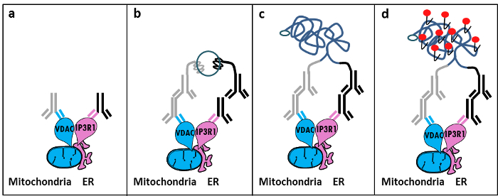
Figure 1: Schematic Illustration of the Detection of the Endoplasmic Reticulum-mitochondria Interactions by In Situ Proximity Ligation Assay. a) A mouse primary antibody directed against VDAC1 and a rabbit primary antibody directed against IP3R1 can bind to their epitopes in proximity at the MAM interface, b) The addition of a pair of proximity ligation probes directed against mouse and rabbit IgG. These probes have attached DNA strands that can form templates for the ligation of connector oligos. c) The circular DNA strand formed after ligation can be amplified and d) visualized by microscopy as a fluorescent dot by using Texas red-labeled oligonucleotides. Please click here to view a larger version of this figure.
Similar in situ proximity ligation assay experiments can be performed with the GRP75/IP3R1 pair of antibodies, as well as cyclophilin D (CypD)/IP3R1 antibodies, considering that CypD was shown to interact with the IP3R/GRP75/VDAC complex at the MAM interface12-14.
Access restricted. Please log in or start a trial to view this content.
Protocol
1. Preparation of Solutions
- Prepare 10% formaldehyde in PBS (low salt) by diluting 5.5 ml of 37% formaldehyde in 14.5 ml of PBS. Prepare 1 M glycine, pH 8.0, by dissolving 3.8 g of glycine in 50 ml of PBS; dilute this solution to obtain 100 mM glycine in 1x PBS.
- Prepare 0.1% Triton-X100 in 1x PBS. Prepare 20x Saline Sodium Citrate (SSC) by using 3.0 M sodium chloride and 0.30 M trisodium citrate prepared in a deionized water buffer with pH 7.0 at 25 °C. Dilute this buffer to 1x and 0.01x using deionized water.
2. Fixation of Cells
NOTE: We used the HuH7 hepatocarcinoma cell line in this study, but this method is applicable to other adherent cell cultures.
- Plate HuH7 cells (cultured in DMEM 1 g/L glucose, supplemented with 10% fetal calf serum and 0.01% penicillin-streptomycin stock) at 150,000 cells per uncoated 35-mm glass-bottom dish. When working on primary cell cultures, use collagen-coated dishes.
- The next day, remove the culture medium. Wash the cells with 1 ml of PBS and aspirate. Fix the cells by adding 1 ml of formaldehyde 10% and incubate for 10 min at room temperature (RT) under agitation.
- Stop the reaction with 1 ml of 1 M glycine and mix by rotation. Remove the stop reaction solution and wash the cells by adding 1 ml of 1x PBS; agitate by rotation and aspirate. Add 1 ml of 100 mM glycine to the cells and incubate them for 15 min at RT under agitation, and then aspirate.
NOTE: The protocol can be stopped here and the following steps can be postponed to another day. In that case, add 1 ml of 100 mM glycine and keep at 4 °C, if necessary.
3. Permeabilization of Cells
- Add 1 ml of 0.1% Triton-X100 in 1x PBS, incubate for 15 min at RT under agitation, and then aspirate. This incubation time could be increased up to 15 - 20 min when working on primary cell cultures (e.g., primary mice hepatocytes). Wash the cells by adding 1 ml of 1x PBS; aspirate.
4. Blocking
- Add 40 µl of blocking solution to each sample (provided by the kit); this volume can be increased in order to cover the sample. Incubate the dishes for 30 min at 37 °C in a humidity chamber.
- Tap the blocking solution off of the dishes. Try to obtain equal residual volumes for each slide, as this will affect reproducibility. Do not let the samples dry!
5. Primary Antibodies
- Dilute the primary antibodies in 1x PBS and add the solution to the dishes (VDAC1 mouse antibody: 1/100, IP3R1 rabbit antibody: 1/500). Alternatively, GRP75 or CypD antibodies (both mouse antibodies used at 1/500) can be used instead of VDAC1.
- Incubate overnight in a humidity chamber at 4 °C. Wash the slides two times using Tris Buffered Saline with 0.01% Tween (TBS-T).
6. Proximity Ligation Assay Probes
- Proximity ligation assay probes are provided by the kit. Choose probes according to the species of primary antibodies.
- Prepare the two proximity ligation assay probes 1:5 in antibody diluent. Allow the mixture to sit for 20 min at RT. Add the diluted proximity ligation assay probe solution. Incubate the dishes in a pre-heated humidity chamber for 1 hr at 37 °C. Wash the dishes two times with TBS-T.
7. Ligation
- Use the in situ detection reagent Texas red kit.
- Dilute the 5x ligation stock (provided by the kit) 1:5 in high-purity water and mix well. Dilute the ligase in the 1x ligation solution (provided by the kit) 1:40 and vortex. Wait to add the ligase until immediately before addition to the samples.
- Add this solution to each sample (40 µl for 35-mm glass-bottom dishes) and incubate the slides in a pre-heated humidity chamber for 30 min at 37 °C. Wash the slides two times with TBS-T.
8. Amplification
NOTE: Be careful, light sensitive reagents.
- Dilute the 5x amplification stock (provided by the kit) 1:5 in high-purity water. Remove the polymerase from the freezer using the freezing block (-20 °C). Dilute the polymerase (provided by the kit) 1:80 in the 1x amplification solution and vortex. Add the polymerase immediately before using the mixture.
- Add this solution to each sample (40 µl for 35-mm glass-bottom dishes). Incubate the slides in a pre-heated humidity chamber for 100 min at 37 °C. Tap the Amplification-Polymerase solution off of the slides.
9. Final Washing
- Wash the slides in 1x SSC washing buffer for 2 min. Wash the slides in 0.01x SSC washing buffer for 2 min. Let the dishes dry at RT in the dark.
10. Preparation for Imaging
- Mount the slides using a minimal volume of mounting medium containing DAPI (aqueous and does not solidify), ensuring that no air bubbles get caught under the cover slip. Nail polish can be used to seal the edges. Wait for approximately 15 min before analyzing using a fluorescence or confocal microscope (excitation: 594 nm, emission: 624 nm, magnification: 63X).
- After imaging, store the slides at -20 °C in the dark.
Access restricted. Please log in or start a trial to view this content.
Results
Based on our experience using this protocol, we can safely recommend this method for the visualization and quantification of ER-mitochondria interactions in fixed cells. Representative images of in situ proximity ligation assay-visualized ER-mitochondria interactions in the HuH7 hepatocarcinoma cell line, using several pairs of antibodies, are shown. As shown in Figure 2 by fluorescent microscopy, each red dot represents an interaction between the two targeted pr...
Access restricted. Please log in or start a trial to view this content.
Discussion
Collectively, our studies indicate that the in situ proximity ligation assay is truly a relevant strategy to follow and quantify endogenous ER-mitochondria interactions in fixed cells, without the need for using organelle-specific fluorophores or fluorescent proteins. The specific use of VDAC1/IP3R1 antibodies has been adapted to study ER-mitochondria interactions in HuH7 cells. However, alternative isoforms of VDAC and IP3R may be used, depending on the cell type. In this case, antibodies need to be validated b...
Access restricted. Please log in or start a trial to view this content.
Disclosures
The authors declare that they have no competing financial interests.
Acknowledgements
We thank all the people in our laboratory who contributed to optimize and validate the protocol. This work was supported by INSERM and the national research agency (ANR-09-JCJC-0116 AND ANR-11-BSV1-033-02). E.T. was supported during her PhD by a research fellowship from the French ministry of higher education and research.
Access restricted. Please log in or start a trial to view this content.
Materials
| Name | Company | Catalog Number | Comments |
|---|---|---|---|
| Formaldehyde | Sigma | F-8775 | |
| Glycine | Sigma | G-8898 | |
| Triton | Sigma | T8532 | |
| 35 mm Glass bottom culture dishes | MatTeK corporation | P35G-0-14-C | |
| Blocking solution | Sigma | DUO-92004 or DUO-92002 | provided in the Duolink PLA probes, Sigma |
| VDAC1 antibody | Abcam | ab14734 | |
| IP3R1-H80 antibody | Santa Cruz | sc28614 | |
| CypD antibody | Abcam | ab110324 | |
| Grp75 antibody | Santa Cruz | sc13967 | |
| TBS 10x | euromedex | ET220 | Dilute to obtain 1x |
| Tween 100x | euromedex | 2001-B | dilute in TBS to obtain 0,01% |
| PLA Probes Mouse MINUS | Sigma | DUO-92004 | Duolink, Sigma |
| PLA Probes Rabbit PLUS | Sigma | DUO-92002 | Duolink, Sigma |
| Duolink detection reagents red | Sigma | DUO-92008 | Duolink, Sigma |
| Ligation solution | Sigma | DUO-92008 | Part of the Duolink detection reagents red, Sigma |
| Ligase | Sigma | DUO-92008 | Part of the Duolink detection reagents red, Sigma |
| Amplification solution | Sigma | DUO-92008 | Part of the Duolink detection reagents red, Sigma |
| Polymerase | Sigma | DUO-92008 | Part of the Duolink detection reagents red, Sigma |
| Duolink Mounting Medium | Sigma | DUO80102 | Duolink, Sigma |
| Softwares | |||
| Blob-finder software | BlobFinder is a freely distributed software that can perform calculations on cells from fluorescence microscopy images. This software can be downloaded for free from The Centre for Image Analysis at Uppsala University who have developed the software and the work was supported by the EU FP6 Project ENLIGHT and Olink Bioscience. http://www.cb.uu.se/~amin/BlobFinder/index_files/Page430.htm | ||
| ImageJ software | Can be downloaded for free from: http://rsb.info.nih.gov/ij/download.html |
References
- Bravo-Sagua, R., et al. Organelle communication: signaling crossroads between homeostasis and disease. The international journal of biochemistry & cell biology. 50, 55-59 (2014).
- Giorgi, C., et al. Mitochondria-associated membranes: composition, molecular mechanisms, and physiopathological implications. Antioxidants & redox signaling. 22, 995-1019 (2015).
- Phillips, M. J., Voeltz, G. K. Structure and function of ER membrane contact sites with other organelles. Nature reviews. Molecular cell biology. 17, 69-82 (2016).
- Cosson, P., et al. The RTM resistance to potyviruses in Arabidopsis thaliana: natural variation of the RTM genes and evidence for the implication of additional genes. PLoS One. 7, 39169(2012).
- Mannella, C. A. Structure and dynamics of the mitochondrial inner membrane cristae. Biochim Biophys Acta. 1763, 542-548 (2006).
- Csordas, G., et al. Structural and functional features and significance of the physical linkage between ER and mitochondria. The Journal of cell biology. 174, 915-921 (2006).
- Mannella, C. A., Buttle, K., Rath, B. K., Marko, M. Electron microscopic tomography of rat-liver mitochondria and their interaction with the endoplasmic reticulum. Biofactors. 8, 225-228 (1998).
- Rizzuto, R., et al. Close contacts with the endoplasmic reticulum as determinants of mitochondrial Ca2+ responses. Science. 280, 1763-1766 (1998).
- Wieckowski, M. R., Giorgi, C., Lebiedzinska, M., Duszynski, J., Pinton, P. Isolation of mitochondria-associated membranes and mitochondria from animal tissues and cells. Nat Protoc. 4, 1582-1590 (2009).
- Csordas, G., et al. Imaging interorganelle contacts and local calcium dynamics at the ER-mitochondrial interface. Mol Cell. 39, 121-132 (2010).
- Szabadkai, G., et al. Chaperone-mediated coupling of endoplasmic reticulum and mitochondrial Ca2+ channels. J Cell Biol. 175, 901-911 (2006).
- Tubbs, E., et al. Mitochondria-associated endoplasmic reticulum membrane (MAM) integrity is required for insulin signaling and is implicated in hepatic insulin resistance. Diabetes. 63, 3279-3294 (2014).
- Paillard, M., et al. Depressing Mitochondria-Reticulum Interactions Protects Cardiomyocytes From Lethal Hypoxia-Reoxygenation Injury. Circulation. 128, 1555-1565 (2013).
- Rieusset, J., et al. Disruption of calcium transfer from ER to mitochondria links alterations of mitochondria-associated ER membrane integrity to hepatic insulin resistance. Diabetologia. 59, 614-623 (2016).
- Allalou, A., Wahlby, C. BlobFinder, a tool for fluorescence microscopy image cytometry. Computer methods and programs in biomedicine. 94, 58-65 (2009).
- Theurey, P., et al. Mitochondria-associated endoplasmic reticulum membranes allow adaptation of mitochondrial metabolism to glucose availability in the liver. Journal of molecular cell biology. , (2016).
- de Brito, O. M., Scorrano, L. Mitofusin 2 tethers endoplasmic reticulum to mitochondria. Nature. 456, 605-610 (2008).
- Soderberg, O., et al. Direct observation of individual endogenous protein complexes in situ by proximity ligation. Nature methods. 3, 995-1000 (2006).
- De Pinto, V., Messina, A., Lane, D. J., Lawen, A. Voltage-dependent anion-selective channel (VDAC) in the plasma membrane. FEBS letters. 584, 1793-1799 (2010).
- Kaul, S. C., Taira, K., Pereira-Smith, O. M., Wadhwa, R. Mortalin: present and prospective. Experimental gerontology. 37, 1157-1164 (2002).
Access restricted. Please log in or start a trial to view this content.
Reprints and Permissions
