티 미 딘 아날로그 듀와 선 충 C. 생식에 세포 주기 분석
Summary
이미징 기반 메서드를 설명 하는 S 단계를 식별 하 고 사용 하는 티 미 딘 C. 선 충 자웅 동체 생식 세포 주기 역학을 분석 하는 데 사용할 수 있는 아날로그 듀. 이 메서드와 필요 없는 transgenes immunofluorescent 얼룩과 호환 됩니다.
Abstract
진핵생물의 세포 주기 분석 염색체 형태, 식 및 세포 주기의 다양 한 단계 또는 nucleoside 아날로그의 설립에 필요한 유전자 제품의 지역화에 자주 활용 합니다. S 단계, DNA polymerases 분석에 대 한 셀을 표시 하는 염색체 DNA에 듀 등 BrdU 티 미 딘 아날로그를 통합 합니다. C. 선 충는 nucleoside 아날로그 듀 일반 문화 중 벌레는 먹이 및 immunofluorescent 기술을와 호환 됩니다. C. 선 충 의 생식은 신호 경로, 줄기 세포, 감수 분열, 세포 주기 때문에 투명 하 고, 유전으로 손쉬운 meiotic 의향 및 세포 분화/gametogenesis 발생의 연구에 대 한 강력한 모델 시스템을 선형 어셈블리 같은 패션. 이러한 기능에 듀 mitotically 자전거 세포 및 생식 개발의 동적 측면을 연구 하는 훌륭한 도구를 확인 합니다. 이 프로토콜 성공적으로 듀 박테리아를 준비, 야생-타입에 피드 하는 방법에 설명 합니다 선 충 C. 광고에 나온 것, 자웅 동체 생식 해 부, DNA에 듀 설립 얼룩 얼룩 다양 한 세포 주기를 검출 하기 위하여 항 체와 고 발달 마커, 생식을 이미지 하 고 결과 분석. 프로토콜은 메서드 및 S 단계 인덱스, M-인덱스, g 2 기간, 세포 주기 기간, meiotic 항목의 위상과 meiotic 의향 진행의 속도의 측정에 대 한 분석에 있는 변이 설명합니다. 이 방법은 세포 주기 또는 다른 조직, 단계, 유전적 배경, 생리 적 조건에 셀 역사 연구에 적응 될 수 있다.
Introduction
동물 개발에 수백, 수천, 수백만, 수십억, 또는 심지어 수조 세포 분열의 성인 유기 체를 형성 하기 위하여 필요 합니다. 세포 주기 G1 세포 이벤트 집합 구성 (gap), S (합성), G2 (gap), M (유사 분열) 정의 일련의 이벤트 실행 각 세포 분열. 세포 주기는 역동적이 고 실시간으로 기술적으로 어려울 수 있는 최고의 평가입니다. 이 프로토콜에 기술 단계 측정 및 스틸 이미지에서 세포 주기의 타이밍을 만드는 수 있습니다.
꼬마 선 충 (C. 선 충) 성인에서 세포 주기 역학의 연구에 S 단계를 식별 하는 금 본 위 제는 nucleoside 아날로그 deoxyuridine (듀)-5-ethynyl-2′ 등 deoxyuridine (BrdU)-5-브로 모-2’로 자웅 동체 생식1,2,3,,45. 로 그들은 어떤 유전자 구조에 의존 하지 않는 거의 모든 유전적 배경에 듀와 BrdU 사용할 수 있습니다. BrdU 시각화 안티 BrdU 항 체 얼룩이 지기은 종종 다른 세포 마커 추가 항 체와 공동 얼룩에 의해 시각의 평가와 호환에 대 한 항 원 노출에 가혹한 화학 치료를 필요 합니다. 대조적으로, 듀 시각화 클릭 화학 가벼운 조건에 의해 발생 하 고 따라서 공동6,7을 얼룩이 지는 항 체와 호환 됩니다.
라벨의 특이성은 분명, 이후 핵만 포함 티 미 딘 (5-ethynyl-2′-deoxyuridine) 유사 체 DNA로 S 단계. 시각화는 고정된 조직에서 일어난다. 듀 라벨 보이지 않으면 저절로 아 지 드를 포함 하는 염료까지 또는 fluorophore 클릭 구리 촉매 화학8듀에 alkyne covalently 반응. 듀 라벨에 핵은 S-위상, 라벨의 짧은 펄스를 사용 하 여 즉각적인 정보를 제공할 수 있습니다. 듀도 정보를 제공할 수 동적, 펄스-체이스 또는 연속 라벨;를 사용 하 여 예를 들어 펄스-체이스 실험 레이블이 각 세포 분열에서 희석 또는 개발을 통해 서 nondividing 세포 진행을 전파.
C. 선 충 자웅 동체 생식 통로, 줄기 세포, 감수 분열, 세포 주기 신호의 연구에 대 한 강력한 모델 시스템입니다. 성인 생식 줄기 세포 항목 및 meiotic 의향, 더 proximally gametogenesis 단계 조정 (그림 1)를 통해 진행 하는 원심 끝에 편광된 조립 라인입니다. 근 위 끝에 oocytes 성숙, ovulated는 수정 고 자 궁9,,1011에 embryogenesis 시작. Meiotic 의향에 mitotically 자전거 생식 줄기, 뿌리 세포와 meiotic S 단계 세포 하지만 되지 셀을 포함 하는 원심 끝 셀 근처 ~ 20 셀-직경 긴 지역 이라고 조상 영역2,4 , 9 , 12. 세포 막 원심 생식에서 핵 사이 불완전 한 분리를 제공 하지만 조상 영역 셀 mitotic 세포 주로 독립적으로 자전거를 받 다. 젊은 성인 광고에 나온 것에 생식 조상 영역 셀의 메디아 mitotic 세포 주기 기간은 ~6.5 h; G 1 단계는 간단한 결 석, 또는 정지1,,213관찰 하지는. 생식 줄기 세포 분화 본질적으로 직접 차별화를 통해 발생 하 고 따라서 부족 교통 증폭 사단4. Pachytene 단계에서 차별화, 동안 약 4 5 점 만점 핵 oocytes를 형성 하지 않습니다 하지만 대신 apoptosis, 세포질 내용을 개발 oocyte12,14 에 기부 하 여 간호사 셀 역할을 받 다 , 15.
Nucleoside 아날로그와 S 단계에서 레이블 셀, 뿐만 아니라 하나의 유사 분열, 감수 분열이 항 체는 얼룩이 지를 사용 하 여 셀을 식별할 수 있습니다. 유사 분열에서 핵은 안티-인-히스톤 H3를 immunoreactive (Ser10) 항 체 (pH3 라고)7,16. 감수 분열에서 핵은 안티-그-3 항 체 (meiotic 염색체 축 단백질)17에 immunoreactive. 조상 영역에서 핵 그-3, nucleoplasmic REC-818의 존재 나 WAPL-119의 존재에 의해 확인할 수 있습니다. WAPL-1 강도 이며 조상 영역에서 높은 체세포 생식에 높은 낮은 초기 meiotic prophases19동안. 여러 가지 세포 주기 측정 프로토콜에 몇 가지 변형 가능 하다: 나) S 단계에서 핵을 식별 하 고 측정 하는 S 단계 인덱스; II) M 단계에서 핵을 식별 하 고 측정 M 단계 인덱스; III) 핵 mitotic 또는 meiotic S 단계;에 있던 여부를 결정합니다 4) g 2;의 기간을 측정 V) 측정 g 2 + M + G1 단계; duartion VI) meiotic 항목;의 속도 측정 VII) meiotic 진행의 속도 추정 합니다.
하나는 몇 가지 유형의 젖은 실험실 실험에서 여러 세포 주기 측정을 만들 수 있습니다. 프로토콜 아래 듀 박테리아와 공동 레이블 M 단계 셀 이라고 표시 된 안티-pH3 항 체와 조상 영역 셀 안티-WAPL-1 항 체와 얼룩에 의해 얼룩이와 선 충 C. 성인 광고에 나온 것을 먹이로 라벨 30 분 펄스에 설명 합니다. 듀 피드 (2.5 단계), 항 체의 종류 기간에 유일한 변화 고용 (5 단계), 그리고 분석 (단계 8.3) 추가 측정을 위해 필요 합니다.
Protocol
Representative Results
Discussion
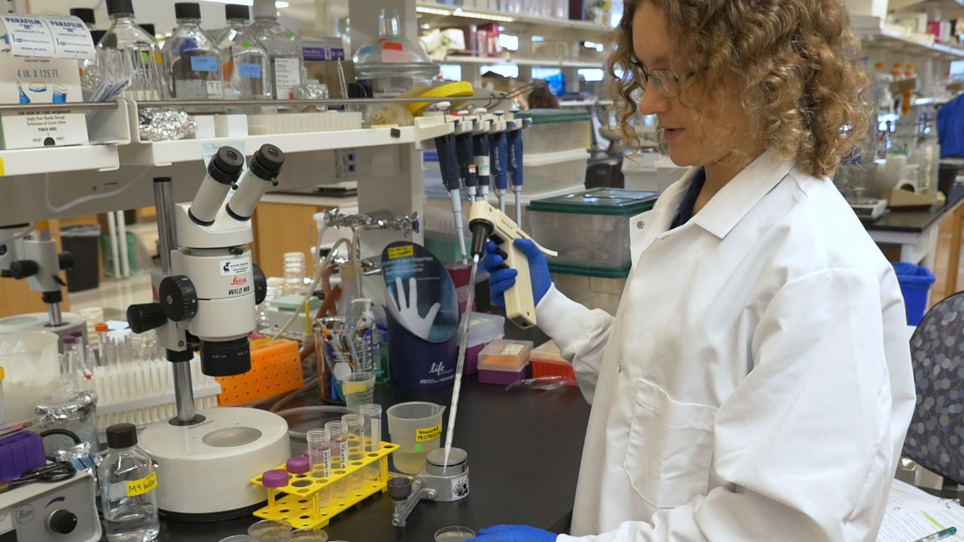
듀 라는 박테리아 (단계 1)의 준비는이 프로토콜 및 문제 해결을 위한 첫 번째 포인트에 대 한 중요 한 이다. 야생-타입 젊은 성인 광고에 나온 것 4 h 듀-펄스, 듀 라는 박테리아의 모든 새로운 배치를 위한 유용한 컨트롤 만들기에 매우 안정적으로 레이블. 또한, 소장 (입력 더 오래 된 동물 또는 특정 인 두/분쇄기 결함이 있는 돌연변이) 그대로 듀 라는 박테리아 클릭 화학 레이블 하 고 용기에 밝은…
Disclosures
The authors have nothing to disclose.
Acknowledgements
우리는 MG1693;에 대 한 대장균 재고 센터에 감사 Wormbase; 프로그램에 의해 국립 연구소의 건강 사무실의 연구 인프라 (P40OD010440) 긴장;에 대 한 투자는 꼬마 유전학 센터 자크 Pincus 통계 조언; Aiping Feng 시 약;에 대 한 루크 슈나이더, 안드레아 Scharf, Sandeep 쿠마, 그리고 교육, 조언, 지원, 및 유용한 토론; 존 브 레너 그리고이 원고에 피드백 Kornfeld Schedl 실험실. 이 작품 부분 건강의 국가 학회에 의해 지원 되었다 주식, TS R01 GM100756 [R01 AG02656106A1]와 [당선-1143954와 ZK DGE-1745038] 국립 과학 재단 predoctoral 친목. 건강의 국가 학회도 국립 과학 재단 연구, 수집, 분석, 및 데이터의 해석의 디자인에도 원고를 쓰기에 어떤 역할을 했다.
Materials
| E. coli MG1693 | Coli Genetic Stock Center | 6411 | grows fine in standard unsupplemented LB |
| E. coli OP50 | Caenorhabditis Genetics Center | OP50 | |
| Click-iT EdU Alexa Fluor 488 Imaging Kit | Thermo Fisher Scientific | C10337 | |
| 5-Ethynyl-2′-deoxyuridine | Sigma | 900584-50MG | or use EdU provided in kit |
| Glucose | Sigma | D9434-500G | D-(+)-Dextrose |
| Thiamine (Vitamin B1) | Sigma | T4625-5G | Reagent Grade |
| Thymidine | Sigma | T1895-1G | BioReagent |
| Magnesium sulfate heptahydrate | Sigma | M1880-1KG | MgSO4, Reagent Grade |
| Sodium Phosphate, dibasic, anhydrous | Fisher | BP332-500G | Na2HPO4 |
| Potassium Phosphate, monobasic | Sigma | P5379-500G | KH2PO4 |
| Ammonium Chloride | Sigma | A4514-500G | NH4Cl, Reagent Plus |
| Bacteriological Agar | US Biological | C13071058 | |
| Calcium Chloride dihydrate | Sigma | C3881-500G | CaCl |
| LB Broth (Miller) | Sigma | L3522-1KG | Used at 25g/L |
| Levamisole | Sigma | L9756-5G | 0.241g/10ml |
| Phosphate buffered saline | Calbiochem Omnipur | 6506 | homemade PBS works just as well |
| Tween-20 | Sigma | P1379-500ML | |
| 16% Paraformaldehyde, EM-grade ampules | Electron Microscopy Sciences | 15710 | 10ml ampules |
| 100% methanol | Thermo Fisher Scientific | A454-1L | Gold-label methanol is critical for proper morphology with certain antibodies |
| Goat Serum | Gibco | 16210-072 | Lot 1671330 |
| rabbit-anti-WAPL-1 | Novus biologicals | 49300002 | Lot G3048-179A02, used at 1:2000 |
| mouse-anti-pH3 clone 3H10 | Millipore | 05-806 | Lot#2680533, used at 1:500 |
| goat-anti-rabbit IgG-conjugated Alexa Fluor 594 | Invitrogen | A11012 | Lot 1256147, used at 1:400 |
| goat-anti-mouse IgG-conjugated Alexa Fluor 647 | Invitrogen | A21236 | Lot 1511347, used at 1:400 |
| Vectashield antifade mounting medium containing 4',6-Diamidino-2-Phenylindole Dihydrochloride (DAPI) | Vector Laboratories | H-1200 | mounting medium without DAPI can be used instead, following a separate DAPI incubation |
| nail polish | Wet n Wild | DTC450B | any clear nail polish should work |
| S-medium | various | see wormbook.org for protocol | |
| M9 buffer | various | see wormbook.org for protocol | |
| M9 agar | various | same recipe as M9 buffer, but add 1.7% agar | |
| Nematode Growth Medium | various | see wormbook.org for protocol | |
| dissecting watch glass | Carolina Biological | 42300 | |
| Parafilm laboratory film | Pechiney Plastic Packaging | PM-996 | 4 inch wide laboratory film |
| petri dishes | 60 mm diameter | ||
| Long glass Pasteur pipettes | |||
| 1ml centrifuge tubes | MidSci Avant | 2926 | |
| Tips | |||
| Serological pipettes | |||
| 500 mL Erlenmyer flask | |||
| Aluminum foil | |||
| 25G 5/8” needles | BD PrecisionGlide | 305122 | |
| 5ml glass centrifuge tube | Pyrex | ||
| Borosilicate glass tubes 1ml | |||
| glass slides | |||
| no 1 coverslips 22 x 40 mm | no 1.5 may work, also | ||
| 37 °C Shaker incubator | |||
| Tabletop Centrifuge | |||
| Clinical Centrifuge | IEC | 428 | with 6 swinging bucket rotor |
| Mini Centrifuge | |||
| 20 °C incubator | |||
| 4 °C refrigerator | |||
| -20 °C freezer | |||
| Observer Z1 microscope | Zeiss | ||
| Plan Apo 63X 1.4 oil-immersion objective lens | Zeiss | ||
| Ultraview Vox spinning disc confocal system | PerkinElmer | Nikon spinning disc confocal system works very well, also, as described here: http://wucci.wustl.edu/Facilities/Light-Microscopy |
References
- Fox, P. M., Vought, V. E., Hanazawa, M., Lee, M. -. H., Maine, E. M., Schedl, T. Cyclin E and CDK-2 regulate proliferative cell fate and cell cycle progression in the C. elegans germline. Development. 138 (11), 2223-2234 (2011).
- Crittenden, S. L., Leonhard, K. A., Byrd, D. T., Kimble, J. Cellular analyses of the mitotic region in the Caenorhabditis elegans adult germ line. Molecular biology of the cell. 17 (7), 3051-3061 (2006).
- Seidel, H. S., Kimble, J. Cell-cycle quiescence maintains Caenorhabditis elegans germline stem cells independent of GLP-1/Notch. eLife. 4, (2015).
- Fox, P. M., Schedl, T. Analysis of Germline Stem Cell Differentiation Following Loss of GLP-1 Notch Activity in Caenorhabditis elegans. Genetics. 201 (9), 167-184 (2015).
- Kocsisova, Z., Kornfeld, K., Schedl, T. Cell cycle accumulation of the proliferating cell nuclear antigen PCN-1 transitions from continuous in the adult germline to intermittent in the early embryo of C. elegans. BMC Developmental Biology. 18 (1), (2018).
- Salic, A., Mitchison, T. J. A chemical method for fast and sensitive detection of DNA synthesis in vivo. Proceedings of the National Academy of Sciences. 105 (7), 2415-2420 (2008).
- vanden Heuvel, S., Kipreos, E. T. C. elegans Cell Cycle Analysis. Methods in Cell Biology. , 265-294 (2012).
- ThermoFisher. . Click-iT EdU Imaging Kits. , (2011).
- Pazdernik, N., Schedl, T. . Germ Cell Development in C. elegans. , 1-16 (2013).
- Hirsh, D., Oppenheim, D., Klass, M. Development of the reproductive system of Caenorhabditis elegans. Developmental Biology. 49 (1), 200-219 (1976).
- Brenner, S. The genetics of Caenorhabditis elegans. Genetics. 77 (1), 71-94 (1974).
- Hansen, D., Schedl, T. Stem cell proliferation versus meiotic fate decision in Caenorhabditis elegans. Advances in Experimental Medicine and Biology. 757, 71-99 (2013).
- Jaramillo-Lambert, A., Ellefson, M., Villeneuve, A. M., Engebrecht, J. Differential timing of S phases, X chromosome replication, and meiotic prophase in the C. elegans germ line. Developmental Biology. 308 (1), 206-221 (2007).
- Agarwal, I., Farnow, C., et al. HOP-1 presenilin deficiency causes a late-onset notch signaling phenotype that affects adult germline function in Caenorhabditis elegans. Genetics. 208 (2), 745-762 (2018).
- Gumienny, T. L., Lambie, E., Hartwieg, E., Horvitz, H. R., Hengartner, M. O. Genetic control of programmed cell death in the Caenorhabditis elegans hermaphrodite germline. Development. 126 (5), 1011-1022 (1999).
- Hendzel, M. J., Wei, Y., et al. Mitosis-specific phosphorylation of histone H3 initiates primarily within pericentromeric heterochromatin during G2 and spreads in an ordered fashion coincident with mitotic chromosome condensation. Chromosoma. 106 (6), 348-360 (1997).
- Zetka, M. C., Kawasaki, I., Strome, S., Mü Ller, F. Synapsis and chiasma formation in Caenorhabditis elegans require HIM-3, a meiotic chromosome core component that functions in chromosome segregation. Genes & development. 13 (17), 2258-2270 (1999).
- Hansen, D., Hubbard, E. J. A., Schedl, T. Multi-pathway control of the proliferation versus meiotic development decision in the Caenorhabditis elegans germline. Developmental Biology. 268 (2), 342-357 (2004).
- Crawley, O., Barroso, C., et al. Cohesin-interacting protein WAPL-1 regulates meiotic chromosome structure and cohesion by antagonizing specific cohesin complexes. eLife. 5 (2), 1-26 (2016).
- Zhao, H., Halicka, H. D., et al. DNA damage signaling, impairment of cell cycle progression, and apoptosis triggered by 5-ethynyl-2′-deoxyuridine incorporated into DNA. Cytometry Part A. 83 (11), 979-988 (2013).
- Stiernagle, T. Maintenance of C. elegans. WormBook. , 1-11 (2006).
- Gervaise, A. L., Arur, S. Spatial and Temporal Analysis of Active ERK. in the C. elegans Germline. Journal of Visualized Experiments. 117 (117), e54901-e54901 (2016).
- Vos, K. . De Cell Counter Plugin. , (2015).
- Schindelin, J., Arganda-Carreras, I., et al. Fiji: an open-source platform for biological-image analysis. Nature Methods. 9 (7), 676-682 (2012).
- Rasband, W. . ImageJ. , (2016).
- Michaelson, D., Korta, D. Z., Capua, Y., Hubbard, E. J. A. Insulin signaling promotes germline proliferation in C. elegans. Development. 137 (4), 671-680 (2010).
- Qin, Z., Jane, E., Hubbard, A., Hubbard, E. J. A. Non-autonomous DAF-16/FOXO activity antagonizes age-related loss of C. elegans germline stem/progenitor cells. Nature communications. 6 (5), 7107 (2015).
- Luo, S., Kleemann, G. A., Ashraf, J. M., Shaw, W. M., Murphy, C. T. TGFB and Insulin Signaling Regulate Reproductive Aging via Oocyte and Germline Quality Maintenance. Cell. 143 (2), 299-312 (2010).
- Narbonne, P., Maddox, P. S., Labbe, J. -. C. daf-18/PTEN locally antagonizes insulin signalling to couple germline stem cell proliferation to oocyte needs in C. elegans. Development. , 4230-4241 (2015).
- Cinquin, A., Chiang, M., et al. Intermittent Stem Cell Cycling Balances Self-Renewal and. Senescence of the C. elegans Germ Line. PLoS Genetics. 12 (4), 1005985 (2016).
- . . Invitrogen EdU (5-ethynyl-2’-deoxyuridine). , 1-7 (2010).
- Tuttle, A. H., Rankin, M. M., et al. Immunofluorescent Detection of Two Thymidine Analogues (CldU and IdU) in Primary Tissue. Journal of Visualized Experiments. 46 (46), e2166-e2166 (2010).