Müller glia primary cultures obtained from mouse retinas represent a very robust and reliable tool to study the glial conversion into retinal progenitor cells after microRNA treatment. Single molecules or combinations can be tested before their subsequent application of in vivo approaches.
Method Article
Primary Cell Cultures to Study the Regeneration Potential of Murine Müller Glia after MicroRNA Treatment
In This Article
Summary
Abstract
Müller glia (MG) are the predominant glia in the neural retina and can function as a regenerative source for retinal neurons. In lower vertebrates such as fish, MG-driven regeneration occurs naturally; in mammals, however, stimulation with certain factors or genetic/epigenetic manipulation is required. Since MG comprise only 5% of the retinal cell population, there is a need for model systems that allow the study of this cell population exclusively. One of these model systems is primary MG cultures that are reproducible and can be used for a variety of applications, including molecule/factor screening and identification, testing of compounds or factors, cell monitoring, and/or functional tests. This model is used to study the potential of murine MG to convert into retinal neurons after supplementation or inhibition of microRNAs (miRNAs) via transfection of artificial miRNAs or their inhibitors. The use of MG-specific reporter mice in combination with immunofluorescent labeling and single-cell RNA sequencing (scRNA-seq) confirmed that 80%-90% of the cells found in these cultures are MG. Using this model, it was discovered that miRNAs can reprogram MG into retinal progenitor cells (RPCs), which subsequently differentiate into neuronal-like cells. The advantages of this technique are that miRNA candidates can be tested for their efficiency and outcome before their usage in in vivo applications.
Introduction
The Müller glia (MG) are the predominant glia in the neural retina. They have similar functions compared to other glia in other parts of the central nervous system such as maintaining the water and ion homeostasis, nourishing neurons, and protecting the tissue. MG have another fascinating feature: although they are mature glia, they still express many genes expressed in retinal progenitor cells (RPCs) during late development1,2. This resemblance is assumed to be the reason for the naturally occurring MG-based neuronal regeneration in the fish retina after retinal damage3,4. During this process, MG re-enter the cell cycle and de-differentiate into RPCs that then differentiate into all six types of retinal neurons. Although this phenomenon occurs naturally in fish, mammalian MG do not convert into neurons5,6. They can, however, be reprogrammed. A variety of factors have been shown to reprogram MG into RPCs/neurons; among these factors is the basic helix-loop-helix (bHLH) transcription factor achaete-scute homolog 1 (Ascl1) that is involved in fish regeneration7,8. In mice, Ascl1 is only expressed in RPCs during retinogenesis but is absent in mature MG or retinal neurons9.
Reprogramming cells directly in vivo is not only methodologically challenging but also requires approval from an institutional animal care and use committee. To receive approval, preliminary data about the factor(s) used or altered, concentrations, off-target effects, underlying mechanisms, toxicity, and efficiency are required. Cell culture systems allow testing for these criteria before usage in in vivo models. Moreover, since MG only comprise about 5% of the entire retinal cell population10, MG cultures allow the study of their function11 as well their behavior, including migration12,13, proliferation14, stress reaction to injury/damage15,16, their interaction with other cell types such as microglia17 or retinal ganglion cells (RGCs)18, or their neurogenic potential19,20,21. Many researchers use immortalized cell lines for their studies since they have an unlimited proliferative potential and can be easily maintained and transfected. Primary cells, however, are preferable for biologically relevant assays than immortalized cells since they have true cell characteristics (gene and protein expression) and, more importantly, they represent a certain stage in development and therefore have an "age". The age of an animal (and consequently of the cells obtained from an animal) is an especially crucial factor in cellular reprogramming since cell plasticity reduces with progressed stage of development22.
This protocol describes in detail how to reprogram primary MG with miRNAs as a current in vitro method for studying regeneration. This MG primary culture model was established in 2012 to evaluate cell proliferation characteristics of MG in P53 knock-out mice (trp53-/- mice)23. It was shown that cultured MG maintain their glial features (i.e., expression of S100β, Pax6, and Sox2 proteins evaluated via immunofluorescent labeling), and that they resemble in vivo MG (microarray of FACS-purified MG)23. Shortly thereafter, glial mRNA and protein expression were validated and confirmed in a different approach using viral vectors20. A few years later, it was confirmed that the vast majority of cells found in these cultures are MG by using the MG-specific Rlbp1CreERT:tdTomatoSTOPfl/fl reporter mouse24. Moreover, quantification of the set of miRNAs in both FACS-purified MG and cultured primary MG showed that the levels of MG miRNAs (mGLiomiRs) do not vary much in cultured MG during the growth phase. Elongated culture periods, however, cause changes in miRNA levels and consequently in mRNA levels and protein expression since miRNAs are translational regulators25.
In 2013, this MG culture model was used to test a variety of transcription factors with respect to their capability to reprogram MG into retinal neurons20. Ascl1 was found to be a very robust and reliable reprogramming factor. Overexpression of Ascl1 via viral vectors induced morphological changes, expression of neuronal markers, and the acquisition of neuronal electrophysiological properties. More importantly, the insights and results obtained from these first in vitro experiments were successfully transferred to in vivo applications22,26 demonstrating that primary MG cultures represent a solid and reliable tool for initial factor screenings and evaluation of glial features prior to in vivo implementation.
A few years ago, it was shown that the brain-enriched miRNA miR-124, which is also highly expressed in retinal neurons, can induce Ascl1 expression in cultured MG21. Ascl1 expression in living cells was visualized via an Ascl1 reporter mouse (Ascl1CreERT:tdTomatoSTOPfl/fl). A reporter mouse is a genetically engineered mouse that has a reporter gene inserted in its DNA. This reporter gene encodes for a reporter protein, which is in this study tdTomato, a red fluorescent protein. This reporter protein reports the expression of a gene of interest, in this case, Ascl1. In other words, cells that express Ascl1 will turn red. Since Ascl1 is only expressed in RPCs9, this Ascl1CreERT:tdTomatoSTOPfl/fl mouse allows tracking of MG conversion into Ascl1 expressing RPCs, meaning converting cells will express red fluorescent tdTomato reporter protein. This is irreversible labeling since the DNA of these cells is altered. Consequently, any subsequent neuronal differentiation will be visualized because the tdTomato label remains in differentiating cells. If Ascl1 expressing MG-derived RPCs (with tdTomato label) differentiate into neurons, these neurons will still have their red label. This mouse, therefore, allows not only the labeling of MG-derived RPCs for live-cell imaging but also allows fate mapping and lineage tracing of these MG-derived (red) RPCs. More recently, the set of miRNAs in RPCs was identified and MG cultures of Ascl1CreERT:tdTomatoSTOPfl/fl RPC-reporter mice were used to screen and test the effect of these miRNAs on reprogramming capacity and efficiency27. One candidate, the RPC-miRNA miR-25, was found capable of reprogramming cultured MG into Ascl1 expressing (Ascl1-Tomato+) cells. These reprogrammed cells adopt neuronal features over time, including neuronal morphology (small somata and either short or long fine processes), expression of neuronal transcripts measured via scRNA-Seq, as well as expression of neuronal proteins validated via immunofluorescent labeling27.
Here, the protocol details how to grow and transfect MG from P12 mice adapted from the previous work21,24,27. Chosen for this protocol is the aforementioned miRNA miR-25, a miRNA highly expressed in RPCs, with low expression levels in MG or retinal neurons. In order to overexpress miR-25, murine miR-25 mimics, i.e., artificial miRNA molecules are used. As a control, mimics of a miRNA from Caenorhabditis elegans are chosen, that have no function in mammalian cells. Visualization of the conversion of MG into RPCs was accomplished via the RPC reporter mouse (Ascl1CreERT:tdTomatoSTOPfl/fl), a mouse with mixed background (C57BL/6, S129, and ICR strains). This culture can, however, be performed with all mouse strains, including wild-type strains. In the past few years, the original protocol has been modified to reduce growth phase duration and the overall culture period and ensure a more robust glia cell status and minimize the degree of cellular degeneration, which occurs in prolonged culture periods. The regular transfection time window was also extended from 3 h to 2 days. As mentioned before, although the current protocol describes MG cultures as a tool for regeneration studies, the method is not only useful for testing reprogramming factors, but can also be adapted for other applications, including studies about MG migratory or proliferative behavior, injury/cell damage related paradigms, and/or the identification of underlying mechanisms and pathways.
Protocol
Procedures involving animal subjects have been approved by the Institutional Animal Care and Use Committee (IACUC) at SUNY College of Optometry.
NOTE: This culture protocol consists of three phases: growth, transfection, and conversion phase. A summary of the overall protocol with the timeline is given in Figure 1.
1. Preparation of media and all required reagents
NOTE: All steps need to be carried out in an A2 or B2 biosafety cabinet (BSC). During the growth phase, a high-serum growth medium is used which consists of a basal neuronal medium supplemented with epidermal growth factor (EGF). For the conversion phase, a low-serum neurophysiological basal medium supplemented with neuronal supplements is used to ensure neuronal differentiation and survival.
- Prepare growth medium (used during growth phase) by supplementing 200 mL of basal neuronal medium with 20 mL of fetal bovine serum (FBS, 10%), 1 mL of 200 mM L-glutamine (0.5%), 2 mL of penicillin/streptomycin (1%), and 2 mL of N2 supplement (1%). Perform sterile filtration (filter units with 0.22 µm pore size). Pre-warm the medium in 37 °C metal bead bath before use.
- Prepare neuronal medium (used during conversion phase) following manufacturer's instructions (see Table of Materials) by supplementing 100 mL of serum-free neurophysiological basal medium with 2 mL of B27 neuronal supplement (2%), 1 mL of N2 supplement (1%), 20 µL of 100 ng/mL brain-derived neurotrophic factor (BDNF, reconstitute in 0.1% bovine serum albumin (BSA) in phosphate buffered saline (PBS, final concentration 20 ng/mL), 20 µL of 100 ng/mL glia cell-derived neurotrophic factor (GDNF, reconstitute in sterile Hanks' Balanced Salt Solution [HBSS], final concentration 20 ng/mL), 500 µL of 100 mg/mL Dibutyryl-cAMP (reconstitute in DMSO), 70 µL of 50 ng/mL ascorbic acid (reconstitute in sterile PBS), and 1.5 mL of penicillin/streptomycin. Perform sterile filtration (filter units with 0.22 µm pore size). Pre-warm medium in 37 °C metal bead bath before use.
NOTE: These media can be stored at 4 °C, for 1 month. - Reconstitute Papain, DNase I, and Ovomucoid reagents required for retinal dissociation following manufacturer's protocol. Aliquot 750 µL of Papain in sterile 1.5 mL tubes, 75 µL of DNase I in sterile 0.6 mL tubes, and 750 µL of Ovomucoid protease inhibitor in 2 mL tubes. Freeze Papain and DNase I aliquots at -20 °C and keep Ovomucoid aliquots at 4 °C to avoid degradation of the reagents. Thaw at room temperature right before use.
NOTE: Papain, DNase I, and Ovomucoid are found in a kit called Papain Dissociation System (see Table of Materials). - Reconstitute poly-L-ornithine (Poly-O) and Laminin for coverslip coating if immunofluorescent labeling is performed following the datasheet instructions (Poly-O: 0.1 mg/mL in sterile water; Laminin: dilute 1.2 mg/mL at 1:50 in DMEM). Aliquot Poly-O and Laminin (2.5 mL) and freeze aliquots at -20 °C. Thaw at room temperature right before use.
NOTE: Step 1.4. is required only if immunofluorescent labeling and laser-scanning microscopy is performed.
2. Mice and tissue extraction
NOTE: For these reprogramming studies, the Ascl1CreERT:tdTomatoSTOPfl/fl mouse was created by crossing an Ascl1CreERT mouse (Ascl1-CreERT: Jax # 012882) with a tdTomatoSTOPfl/fl mouse (B6.Cg-Gt(ROSA)26Sortm14(CAG-tdTomato)Hze/J: Jax # 007914). This mouse has a mixed background (C57BL/6, S129, and ICR strain). The genotype of this mouse is shown in Figure 1A. All strains can be used for this protocol.
- Wear gloves and sanitize workspace, including dissection microscope and all fine tools (Dumont #5 fine and Dumont #7 curved forceps, fine scissors, and Vannas scissors) with 70% ethanol. Sanitize a 10 cm silicone-coated black dissection dish by exposing it to UV light for 20 min.
- Preparation of dishes and plates for tissue extraction
- Prepare a 24-well plate for eye collection and sample separation (two eyes per mouse in one well, labeled with mouse IDs) with ~1 mL of cold HBSS (4 °C) per well. Place the 24-well plate on ice.
- Fill a sterile 10 cm Petri dish (for washing) and the sanitized silicone-coated dissection dish with several mL of cold HBSS (4 °C) to ensure that the tissue is fully covered with HBSS.
- Prepare a sterile 1.5 mL tube with 1 mL of 70% ethanol.
- Prepare a 12 well culture plate by labeling the plate with culture number, date, strain, and all the required information.
- Euthanize P12 mice using any approved method.
- Eye removal
- Gently hold the mouse head with the thumb and index finger around the eye.
- Using Dumont #7 curved forceps, gently go behind the eyeball and clip the optic nerve. Carefully take the eye out.
NOTE: If adult mice are used, do not use curved forceps, and do not pull the eye. Instead cut carefully around the eye globe using fine scissors; cut the optic nerve but do not cut the eye itself. Use forceps to carefully remove the eyeball.
- Eyeball cleaning
- Dip the eyeball briefly into an ethanol-containing tube to avoid carryover of bacteria from the animal.
- Wash the eyeball briefly in the 10 cm Petri dish before placing it into the 24 well plate on ice.
- Repeat steps 2.4 and 2.5 for the other eyes. Keep the well plate on the ice during the process. Place two eyes from one animal in the dissection dish placed under a dissection microscope with a light source.
- Retina extraction
- Fix one eyeball by grabbing the optic nerve and the surrounding connective tissue around the sclera with Dumont #5 fine forceps and press it carefully against the dissection dish (cornea upward).
- Make a hole in the center of the cornea using a 30 G needle to allow easier access for the Vannas scissors.
- Dissect the cornea around the ciliary body using Vannas scissors and remove cornea, lens, iris, and vitreous body carefully with Dumont #5 fine forceps. Figure 2A illustrates an eye cup with the retina inside.
- Dissect the sclera with Vannas scissors until the optic nerve is reached. Clip the optic nerve and carefully extract the retina using Dumont #5 fine forceps.
- Use a second pair of Dumont #5 fine forceps to push against the retina and allow complete removal of the vitreous body. Figure 2B illustrates two extracted retinas.
- Transfer and wash
- Cut about 2.5 cm of the tip of a sterile transfer pipette to enlarge the diameter. Using this tip, pick up (suck in) whole retinas without damaging the tissue.
- Transfer the retinas into a new sterile Petri dish with cold HBSS (4 °C) and rock the dish (back and forth, left, and right).
- Using the transfer pipette tip, carefully push the retinas around to wash off retinal pigment epithelial (RPE) cells.
NOTE: Alternatively, the entire retina with lens and vitreous body can be removed from the eye cup. Then, the lens and vitreous body can be removed from the extracted retina. If the retina is ripped, make sure to collect all pieces for dissociation; otherwise, there will be insufficient amounts of tissue to grow confluent cell layers.
- Immediately place the isolated retina in a new, clean well of the 24-well plate filled with 1 mL of HBSS. Keep the 24-well plate on the ice during the dissection process.
- Repeat steps 2.7-2.9 to isolate the second retina.
3. Retina dissociation
NOTE: All following steps (until cell harvest) need to be carried out in an A2 or B2 biosafety cabinet (BSC).
- Prepare the Papain/DNase I dissociation mixture as follows.
- For six retinas, add 75 µL of DNase I into the tube containing 750 µL of Papain (from step 1.3) and mix carefully (dissociation mixture).
- For individual sample preparation that is required for this protocol, split the total volume of 825 µL into three aliquots: 275 µL of the mixture in one 1.5 mL tube per mouse (two retinas). Calculate the required amounts of DNase I and Papain accordingly.
NOTE: Up to six retinas can be dissociated in one tube of Papain/DNase I mixture. However, individual sample preparation results in fewer clumps and better cell growth than in combined samples.
- Transfer two retinas to Papain/DNase I dissociation mixture. Use a transfer pipette with an enlarged tip diameter (step 2.8.1), pick up the retinas, wait until the retinas settle at the bottom of the tip, and then release the retinas without excessive HBSS into the tube containing Papain/DNase I mixture.
- Place on a nutator and incubate it for 10 min in a cell culture incubator (37 °C, 5% CO2).
- Dissociate the cells by carefully pipetting up and down (about 20-30 times) with a 1 mL pipette. After cells are dissociated (i.e., resulting in a homogenous solution with no chunks), add 275 µL of Ovomucoid protease inhibitor from the Papain Dissociation Kit to neutralize the Papain. Mix gently by pipetting up and down.
NOTE: If six retinas were dissociated in 825 µL of Papain/DNase I mixture, 825 µL of Ovomucoid is required. - Centrifuge the mixture at 300 x g for 10 min at 4 °C.
- Add epidermal growth factor (EGF, 1 µL per 1 mL of the growth medium, reconstituted at 200 µg/mL in PBS) to the calculated volume of growth medium (1 mL per mouse) pre-warmed at 37 °C.
NOTE: Depending on the experimental design, the proliferation marker 5-ethynyl-2'-deoxyuridine (EdU), as well as 4-Hydroxytamoxifen (4-OHT) or other required factors can be added at the beginning of the culture period. - Remove the tubes carefully from the centrifuge. Do not touch the pellet at the bottom of the tube.
- Remove the supernatant carefully and entirely. Resuspend the cell pellet with 500 µL of EGF-supplemented growth medium.
- Transfer the cell suspension into one well of the labeled 12-well plate (Figure 2C). Rinse the tube with another 500 µL of the EGF-supplemented growth medium and add it to the well (total volume of 1 mL per well).
- Repeat steps 3.8 and 3.9 with all other samples.
- Rock the well plate three times carefully (back and forth; left, and right). Place the plate into the incubator (37 °C, CO2).
NOTE: If transgenic mice are used, perform genotyping for every animal (Figure 2D). Identify Cre recombinase positive and negative mice and label the plate accordingly. For this protocol, only cells of Cre recombinase positive reporter mice were used for the next steps. Cells of Cre negative specimens are frozen and used for other applications.
4. Growth phase
NOTE: The growth phase has a duration of about 4-5 days (Figure 1B). For adding liquids to wells containing cells, the pipette needs to point to the wall of the well and the liquid needs to be released slowly to avoid cell detachment. Do not pipette directly on top of the cells.
- One day after dissociation, remove the medium and add 1 mL of fresh EGF-supplemented growth medium.
- On day 3, remove the medium and add 1 mL of HBSS (room temperature) to remove cell debris. Rock gently back and forth left to right. Remove HBSS, repeat the wash step, and add 1 mL of pre-warmed EGF-supplemented growth medium.
- Monitor the cells every day and evaluate their growth status until the cells reach 90%-100% confluency. Figure 3 shows an example of good MG growth over time. Check for possible contamination or cell death (Supplementary Figure 1). Discard contaminated cultures.
NOTE: To monitor and record cell state, take images at various magnifications using a light microscope with an attached camera and 4x, 10x, or 20x objectives. In this study, a fluorescence microscope is used.
5. Preparation of coverslips with poly-L-ornithine (Poly-O) and Laminin coat
NOTE: This step is only necessary if immunofluorescent labeling and confocal laser-scanning microscopy are performed. Round glass coverslips (12 mm diameter) are required for proper imaging. The coating protocol can also be found in the neuronal medium datasheet (see Table of Materials).
- Place sterile coverslips carefully in the center of every well of a 24-well plate using sterile Dumont #2AP forceps.
NOTE: Place coverslips in the center of the well. Placing coverslips close to the wall of a well will cause surface tension issues for the following steps. - Thaw a 2.5 mL Poly-O aliquot at room temperature and place 100 µL of it in the center of each coverslip.
- Incubate the well plate for 30 min in a 37 °C incubator.
- Remove Poly-O and wash the well with the coverslip three times with ~1 mL of sterile water.
- Let the well plate dry overnight in the BSC. Thaw 2.5 mL of Laminin at 4 °C overnight.
- The next morning, add 100 µL of Laminin in the center of each coverslip and incubate for 4 h in a 37 °C incubator.
- Remove the Laminin carefully and entirely.
- Keep the plate at 4 °C if passaging cannot be performed immediately.
NOTE: The coated coverslips in prepared plates can be kept for a few days at 4 °C.
6. Cell passage to remove neuronal survivors
NOTE: Cell passage is required to remove neuronal cells, not to increase the cell population. Glia divide only a few times and will not grow further after passage. Do not dilute cell suspensions. The cells of one confluent well of a 12-well plate can be distributed onto one well of a 12-well plate or two wells of a 24-well plate. When coated coverslips are used, only about one-third of the coverslip is coated. Therefore, six coverslips sitting in a 24-well plate, with confluent cells (~80%-90%) can be obtained from one well of confluent cells of a 12-well plate. Other ratios can be chosen as well to increase or decrease cell density. For this protocol, one Cre+ reporter mouse is used [one experiment, two treatments: miR-25 or control-miR; technical replicates n = 3 (three coverslips per treatment), biological replicate n = 1]. The number of technical and biological replicates can be defined differently depending on the experimental design.
- Check whether the cells are 90%-100% confluent (also at the margin of the well; Figure 3E,F).
- Remove the medium and add 1 mL of HBSS (room temperature) to wash. Gently rock the plate (back and forth, left and right). Remove HBSS completely.
- Add 500 µL of a pre-warmed trypsin-containing solution (pre-warmed at 37 °C in a metal bead bath) to detach the cells from the well. Rock gently (back and forth, left to right) and incubate for 2 min in a 37 °C incubator.
- Move the plate from the incubator to BSC. While tilting, aspirate the trypsin-containing solution and disperse it carefully and slowly over the well several times until the cells detach completely. Hold the plate against the light and make sure no cells are left at the bottom.
- Transfer this cell suspension to a sterile 1.5 mL tube. Centrifuge at 300 x g for 8 min at 4 °C.
- Remove the supernatant and carefully resuspend the cell pellet by adding 600 µL (100 µL per well for 6 wells/coverslips) of pre-warmed growth medium (supplemented with EGF; 1:1000). and pipetting up and down ~30-40 times.
NOTE: The cells can be frozen at this time at -80 °C or in liquid nitrogen and defrosted following standard protocols for thawing cell lines. If cells are frozen, they will not be resuspended in EGF-supplemented medium (growth medium). They will be resuspended in basic medium (without EGF) and freezing solution (1/1 ratio). Steps 6.1.1 to 6.6.2 describe the steps for freezing the cells. If no cells are frozen, continue with step 6.7.- Prepare freezing solution by mixing 100 µL of DMSO and 400 µL of FBS (total volume of 500 µL per well/sample).
- Resuspend the cell pellet in 500 µL of pre-warmed growth medium. Add the cell suspension to 500 µL of DMSO/FBS freezing solution (total volume of 1 mL). Keep tubes on ice until all samples are processed. Freeze cells at -20 °C for 1 h, and then store at -80 °C.
- Seed cells by placing 100 µL of the 600 µL cell/media suspension (step 6.6) in the center of six coated coverslips (see step 5) of the 24-well plate. Place the plate carefully in the incubator and let the cells settle.
NOTE: 100 µL of the 600 µL cell suspension (harvested from one well of a 12-well plate) will result in 90%-100% cell confluency, which is required for transfection. - Check the cells after 3 h to see whether they have settled on the coverslip. Add 400 µL of growth medium supplemented with EGF.
NOTE: Cells are usually ready for transfection the following day (Figure 3G). If not confluent (90%-100%), leave them for another day. If still not confluent, do not use them for transfection. If other downstream applications are conducted, such as miRNA profiling, RNA-Seq, RT-qPCR, or western blot, cells need to be passaged into 12-well plates (1:1 ratio; no plate treatment required) and harvested for RNA/protein extraction.
7. Transfection
NOTE: The transfection phase consists of a 3 h phase in transfection medium only (transfections procedures are described in the transfection manual that comes with the transfection reagent) and an elongated phase in which transfection reagent and miRNAs are still present, but neuronal medium with required supplements is added (total duration is 2 days; Figure 1B). In this protocol, six wells will be transfected: three wells will receive the reprogramming miRNA miR-25 and three wells will receive the control miRNA.
- Check whether the cells reached 90% confluency and record/image the cells before transfection.
- Remove the growth medium and add 500 µL of HBSS (room temperature) to wash the cells.
- Remove HBSS and add 400 µL of reduced serum medium used for transfections. Place the plate back into a 37 °C incubator.
- Prepare the transfection mixture by following the instructions of the manufacturer's transfection reagent protocol. Make two mixtures: transfection reagent mix and miRNA mix (see Figure 1B for illustration).
- Prepare the transfection reagent mixture: For a 24-well plate, 49 µL of reduced serum medium and 1 µL of transfection reagent are required for one well (50 µL in total). For six wells, 294 µL of reduced serum medium and 6 µL of transfection reagent are combined and mixed well by gently pipetting up and down.
- Prepare miRNA mimic mixture: For a 24-well plate, a total volume of 50 µL of the mimic mixture is required for one well. Three wells will receive the control miRNA and the other three wells will be treated with miR-25. For this protocol, a 200 nM final concentration is used.
- For the miR-25 treatment (three wells), 150 µL of reduced serum medium and 3 µL of miR-25 mimics (100 µM stock solution) are combined and mixed well by gently pipetting up and down. Incubate for 5 min.
For the control mimics treatment (three wells), 150 µL of reduced serum medium and 3 µL of control-miRNA mimics (100 µM stock solution) are combined and mixed well by gently pipetting up and down. Incubate for 5 min.
NOTE: The volume of miRNA mimics depends on the dilution factor and concentration needed (20-500 nM). miRNA inhibitors (antagomiRs), or other molecules including plasmids, can be used as well. Also, combinations of molecules are possible to be transfected.
- For the miR-25 treatment (three wells), 150 µL of reduced serum medium and 3 µL of miR-25 mimics (100 µM stock solution) are combined and mixed well by gently pipetting up and down. Incubate for 5 min.
- Combine miRNA mimic mixture and transfection reagent mixture. Mix carefully by pipetting slowly and thoroughly by gently pipetting up and down. Incubate for 20 min at room temperature (per manufacturer's instructions).
- Add the above transfection mixture dropwise and slowly on top of the cells, close to the media surface in the well using a 20 µL pipette. Rock the plate gently (back and forth, left to right).
- Incubate in a 37 °C incubator for 3 h.
- After 3 h, add neuronal medium to the wells (500 µL per well) supplemented with 4-Hydroxytamoxifen (5 mM stock reconstituted with 2.58 mL of ethanol, 250 nM final concentration) to activate the Cre recombinase and 5-ethynyl-2'-deoxyuridine (EdU, 10 mM stock solution reconstituted with 2 mL of DMSO, 1 µM final concentration) to track cell proliferation.
CAUTION: 4-Hydroxytamoxifen and EdU are known to be human carcinogens, teratogens, and mutagens. Read Material Safety Data Sheet before use and wear gloves, goggles, and lab coat. Reconstitute both reagents according to the manufacturer's recommendations. Do not inhale the substance/mixture. Tightly close after use. Waste material must be disposed of following national and local regulations. Wash hands and face after working with the substance. - Incubate in a 37 °C incubator for 2 days (Figure 1B).
8. Cell conversion
NOTE: The cell conversion phase has a duration of about 5-6 days (Figure 1B), but longer periods are possible.
- Check cells daily for successful induction of tdTomato expression, potential cell death, and/or contamination. Cell density does not change (Figure 4). First faint red fluorescent-labeled cells can be observed 1 day after 4-Hydroxytamoxifen treatment. A fluorescent microscope is required to monitor and image the cells.
NOTE: In this study, a fluorescence microscope is used for live imaging. Images are taken with 4x, 10x, or 20x objectives. - Two days after transfection, remove the medium and add 500 µL of pre-warmed neuronal medium supplemented with 4-Hydroxytamoxifen and EdU in each well.
- Replace the medium with a fresh medium every other day until the cells are harvested.
- Take live images for evaluation and assessment of the number of red fluorescent (Ascl1 expressing) cells and their morphological changes (Figure 4 and Figure 5A-C).
9. Cell harvest: fixation for immunofluorescent labeling
NOTE: Cells can be harvested for other downstream applications, including bulk or scRNA-Seq, RT-qPCR, or western blot.
- Remove the medium and add 500 µL of cold HBSS (4 °C) per well and rock the plate gently (back and forth, left to right). Remove HBSS.
- Fix the cells by adding 500 µL of 2% Paraformaldehyde (PFA) and incubate for 20 min at room temperature.
- Perform immunofluorescent labeling according to established protocols.
Results
This protocol describes how to grow MG from P12 mouse retinas and how to reprogram these cells with miR-25 into retinal neurons using the Ascl1CreERT:tdTomatoSTOPfl/fl RPC reporter mouse. This method was used in previous work reporting in detail other suitable miRNAs (mimics or inhibitors, as single molecules or in combination) to reprogram MG into RPC that then adopt neuronal cell characteristics27. This method has been modified to grow cultures faster and thus minimize the cellular alterations caused by the artificial environment over time24,28,29.
Retinal dissociation and growth phase of primary MG cultures
The first MG can be spotted as early as day one in vitro using a light microscope. MG have a tubular, elongated shape and light gray cell bodies (Figure 3A-D, red arrowheads). Most of them are, however, covered by cellular debris at these early stages. MG from two retinas of one mouse, grown in one well of a 12-well plate, reach 90%-95% confluency within 4-5 days (Figure 3E,F). Over time, all neuronal debris will be cleared, and a dense cell layer will be present. Cells are not ready for passage if the bottom of the well is not completely confluent. However, passage should not be done later than 6 days in vitro since the glia lose their glial features in culture over time. Before transfection or any other treatment, cells need to be checked for density and vitality. Transfection should only be performed on 90%-95% confluent cells (Figure 3G). For immunofluorescent labeling and confocal microscopy analysis, cells need to be seeded on coated coverslips.
Early and late phase of the cell conversion period
As early as 15 h post transfection and 4-Hydroxytamoxifen treatment, the first cells start to express faint red fluorescence, i.e., tdTomato red fluorescent reporter protein driven under the (activated) Ascl1 promotor. Ascl1 expressing cells are now further referred to as Ascl1-Tom+ cells. More robust Ascl1-Tom expression can be observed 2 days after transfection with increasing red fluorescence over time (Figure 4). The reprogramming effect becomes visible around 2 days in culture with many Ascl1-Tom+ cells per field found in the reprogramming miRNA treatment: shown here for control-miR and miR-25 (Figure 4B-B''' and Figure 4C-C''', respectively). In both treatments, Ascl1-Tom+ cells are still rather round and relatively big, with a more glia/progenitor cell-like morphology. Moreover, the induction of the red fluorescent reporter does not influence the cell density of the culture (still 90%-95% confluent).
Three to five days after transfection (Figure 4A and Figure 5A), the increase in the number of Ascl1-Tom+ cells becomes more evident in the miR-25 treated wells (Figure 5B-D) showing a four-fold increase in number after miR-25 treatments compared to controls. Moreover, the first morphological changes become visible. These changes include reduction of cell soma size and the development of fine processes. While in control conditions the vast majority of cells are large and flat (glial/progenitor-like, Figure- 5B-B''), many cells with small nuclei and several small processes resembling retinal neurons appear in the miR-25 treatment (Figure- 5C-C''). These neuronal-like cells represent approximately 70% of the total Ascl1-Tom population (15% in control treatment; Figure 5E). Cells are less dense due to cell conversion (smaller cells require less space). Interestingly, these neuronal-like cells even appear to form tiny networks (Figure 5C''). At this time, cells can be harvested for immunofluorescent labeling to confirm neuronal identity.
Confirmation of neuronal identity
Cells are labeled with antibodies against microtubule-associated protein 2 (Map2), a marker for neuron-specific cytoskeletal proteins30,31, and against Orthodenticle homeobox 2 (Otx2) to validate neuronal identity32,33. Both markers were also used in previous reprogramming studies21,27. Otx2 is a transcription factor found in RPCs34,35,36, mature bipolar cells34,35,37,38,39, and photoreceptors35,36,37,38,40. DAPI nuclear labeling is used to counterstain the cultures. Immunofluorescent labeling shows little to absent Map2 or Otx2 expression in control conditions (Figure 6A-A''',C). miR-25 treated samples, however, have many Map2+ and Otx2+ cells (Figure 6B-B'''). Quantification of confocal images shows that after miR-25 overexpression, about 40 neuronal cells per field are present as compared to five neuronal cells per field in controls (Figure 6C, field size: 630 µm x 630 µm). Images were taken with 20x objective. These neuronal cells constitute about 70% of the total Ascl1-Tom+ cell population of the miR-25 treated samples (control ~20%, Figure 6D). All neuronal-like Ascl1-Tom+ cells were Map2+ and Otx2+, confirming neuronal identity. Moreover, the absolute number of Otx2+ and Map2+ cells was higher after miR-25 treatments compared to control-miRNA treatment (miR-25: 60 cells per field; control-miR: 10 cells per field; Figure 6E).
Taken together, results demonstrate that MG cultures can be grown and reprogrammed with miRNAs. miR-25 supplementation induces Ascl1-Tom expression in primary MG. Many MG convert into RPCs that adopt a neuronal morphology and express the neuronal markers Map2 and Otx2 after a few days.
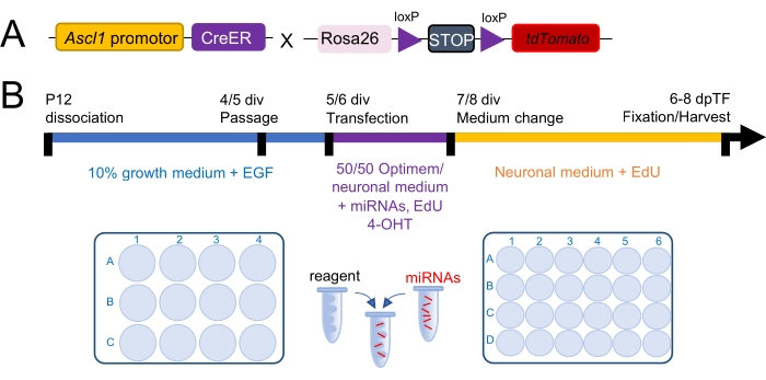
Figure 1: Experimental design and time course of a primary Müller glia culture. (A) Schematic of the Ascl1CreERT:tdTomatoSTOPfl/fl mouse, a retinal progenitor reporter mouse used to track the conversion of Müller glia (MG) into retinal progenitor cells (RPCs). (B) Time course of the culture periods consisting of growth phase (blue, 0-4/5 days in vitro, div), transfection phase (purple, 5/6-7/8 div), and MG conversion phase (yellow, starts 7/8 div). Growth phase: dissociated retinas (two retinas of one mouse) are grown in one well of a 12-well plate in a growth medium. Around day 4/6, cells are passaged into a 24-well plate that contains coated coverslips. Transfection phase: 5-7 div (1 day after passage) cells are transfected with miRNAs for 2 days in transfection medium. Cre recombinase is activated with 4-Hydroxytamoxifen (4-OHT). Cell proliferation is tracked with EdU. MG conversion phase starts 1 day after transfection. Cells are now grown in the neuronal medium until harvest (6-7 days post transfections, dpTF). Please click here to view a larger version of this figure.
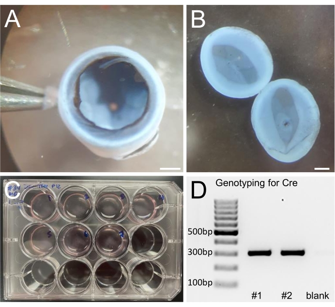
Figure 2: Retinal dissociation and genotyping. (A) Eye cup with removed cornea, lens, iris, and vitreous. (B) Isolated retinas; retinal pigment epithelial (RPE) cells are removed after a thorough wash. (C) 12-well plate with dissociated retinas (two retinas per well). (D) Cutout of a genotyping gel image example. Genotyping is required to identify the mice that have Ascl1 driven Cre recombinase expression and will be used for tracking cell conversion. Scale bars: 1 mm. Please click here to view a larger version of this figure.
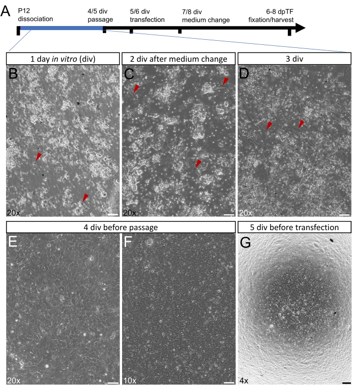
Figure 3: Primary Müller glia during the growth phase. (A) Time course of culture periods during the growth phase (0-4/5 days in vitro (div)) highlighted in blue. (B-G) Live images (phase) of Müller glia (MG) during growth phase after 1 div (B), 2 div after the medium change (C), 3 div (D), 4 div before passage (E,F) and 5 div, 1 day after passage and before transfection (G). MG cell bodies are indicated by red arrowheads. After 3 div, cultures are 60%-80% confluent (D), after 4-5 div, cultures are 90%-100% confluent and ready to be passaged (E-F). After passage on coverslips, cell cultures need to be 80%-90% confluent for subsequent transfection (G). Scale bars: 50 µm (A-D), 100 µm (E), 200 µm (F). Please click here to view a larger version of this figure.
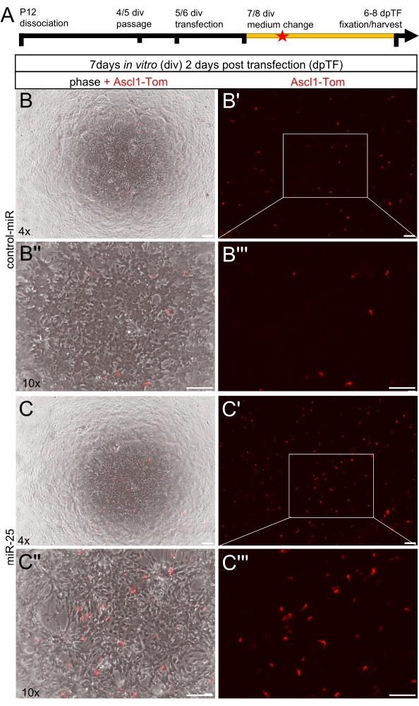
Figure 4: Early stages in the Müller glia conversion phase. (A) Time course of culture periods during conversion phase highlighted in yellow (starts 7/8 days in vitro (div)). The time point of analysis shown in this figure is indicated by the red star: 7 div, 2 days post-transfection (dpTF). (B-C''') Live images of Müller glia (MG) cultures in phase and red fluorescence, combined or single red fluorescence. Red fluorescence visualizes Ascl1 expressing MG (tdTomato+, abbreviated Ascl1-Tom), 2 days after transfection with either control miRNA mimics (control-miR, B-B''') or miR-25 mimics (C-C'''). Areas in B/B' and C/C' are shown in higher magnification in B''/B''', C''/C'''. Scale bars 200 µm. Please click here to view a larger version of this figure.
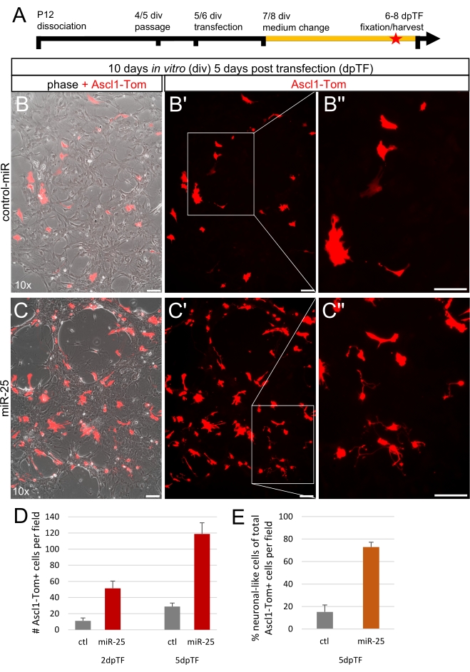
Figure 5: End stages in the Müller glia conversion phase. (A) Time course of culture periods during the conversion phase highlighted in yellow (starts 7/8 days in vitro (div)). The time point of analysis shown in this figure is indicated by the red star: 10 div, 5 days post-transfection (dpTF). (B-C'') Live images of Müller glia (MG) cultures in phase and red fluorescence, combined or single red fluorescence. Red fluorescence visualizes Ascl1 expressing MG (tdTomato+, abbreviated Ascl1-Tom), 5 days after transfection (pTF) with either control miRNA mimics (control-miR, B-B'') or miR-25 mimics (C-C''). Insets in B'/C' are shown in higher magnification in B''/C'', respectively. (D) The absolute number of Ascl1-Tomato+ cells per field (10x; 1440 µm x 1080 µm) after control-miRNA or miR-25 treatment, 2- and 5-days post transfection (dpTF; n =1, five images per well are counted; mean ± S.D.). (E) Percentage of neuronal-like Ascl1-Tomato+ cells of total Ascl1-Tomato+ cells per field (10x; 1440 µm x 1080 µm) after control-miRNA or miR-25 treatment, 5 days post transfection (5dpTF, n = 1, mean ± S.D.). Scale bars: 100 µm. Please click here to view a larger version of this figure.
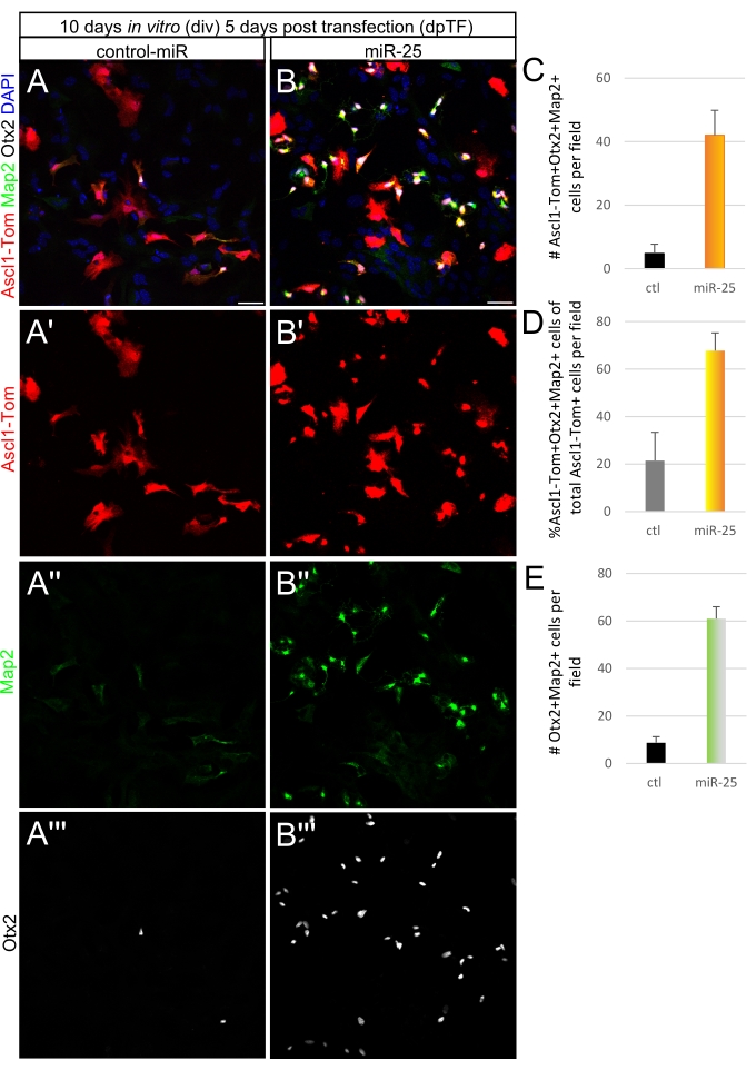
Figure 6: Confirmation of the neuronal identity of reprogrammed cells. (A-A''';B-B''') Immunofluorescent labeling of primary mouse Müller glia (MG) cultures 5 days post transfection treated with control miRNA mimics (control-miR, A-A''') or miR-25 mimics (B-B'''). Cells were fixed with 2% paraformaldehyde (PFA) and incubated with antibodies against RFP to label tdTomato, Map2, and Otx2 to label neurons. DAPI nuclear labeling (blue) was used to stain all cell nuclei. (C-E) Quantification (n = 1; five images per coverslip are counted; mean ± S.D.) of the absolute number of Ascl1-Tom+Otx2+ Map2+ cells per field (C), percentage of Ascl1-Tom+Otx2+Map2+ cells of total Ascl1-Tom+ cells (D), and the absolute number of Otx2+Map2+ cells per field (E). Results show a higher number of neurons in the miR-25 treatment compared to controls. Field size: 630 µm x 630 µm. Scale bars: 50 µm. Please click here to view a larger version of this figure.
Supplementary Figure 1: Cell death and bacterial contamination. (A-A'') Live images of Müller glia (MG) cultures 3 div contaminated with bacteria. Inset 1 in A is shown in higher magnification in A' and displays atrophic, dead glia. Inset 2 in A is shown in A'' with alive glia. Scale bars:100 µm. Please click here to download this File.
Discussion
This protocol describes how to grow MG from dissociated mouse retinas for reprogramming studies using miRNAs. As shown and confirmed in a variety of previous studies, the vast majority (80%-90%) of cells found in these cultures are MG20,23,24. This method is a very robust and reliable technique and results can be easily reproduced if the protocol is followed correctly21,27. The successful growth and reprogramming efficiency of the culture, however, depend on a variety of factors.
First, the age of the mouse plays an important role in successful cell growth. Therefore, if MG cultures are grown from wild-type mice or transgenic mice that resemble wild types such as reporter mice, the latest age to grow confluent MG monolayers is P12. At P12, all retinal neurons and the MG are differentiated, and no RPCs are present in the retina anymore. MG express mature MG markers such as glutamine synthetase or glutamate aspartate transporter (GLAST)20,23,41,42. Cultured P12 MG can still re-enter the cell cycle when exposed to EGF, but the number of cycles is limited albeit enough to form confluent cell layers in vitro. Nevertheless, it can occur that even P12 MG do not grow well because EGF or components in the medium may be degraded. Incomplete dissociation (chunks) can also result in less or delayed MG growth. A major cause for incomplete dissociation is inactivation of the DNase I used for retinal dissociation by harsh handling of the enzyme (fast pipetting, vortexing, etc.). Even if the cells grow well until passage, cells can be less confluent after passage. Reasons for this can be harsh handling during the passage process that leads to increased cell death or because the cells were seeded too sparsely. Since the glia do not grow much, cells should be plated in the same ratio as grown, not diluted (contrary to passaging procedures of immortalized cell lines). Of note, because MG cultures grow from the center to the periphery of a well, higher cell densities will always be found in the center. It is therefore imperative to check the margins of every well before passage to ensure that the whole well is covered by cells. Cell concentration measurements should be performed to ensure the processing of sufficient amounts of cells.
Additionally, primary cultures obtained from mice at P6 or younger will not contain any MG, since MG are just about to mature at this time point. This was shown via single-cell RNA-seq analysis43. Immunofluorescent labeling with mature MG markers at this age will also confirm this. The fraction of RPCs is, however, substantial at P6. Results should, therefore, be interpreted carefully. Moreover, markers such as the intermediate filament markers vimentin and nestin are not appropriate to identify primary MG since these markers are also expressed in astrocytes44,45,46, microglia47,48, endothelial cells, and pericytes49,50,51,52, and are not MG-specific. GFAP, expressed in retinal astrocytes but not in MG of undamaged retinas, is not a good marker for cultured MG. Although it is upregulated in dissociated MG due to the mechanical damage during dissociation, it is downregulated after 3 days in culture28.
Since MG share many RPC markers, the safest procedure to ensure a robust MG culture is to use mice at P12 or older mice. Although this protocol describes cultures obtained from P12 mouse MG, adult mouse MG can be cultured and reprogrammed27. However, more tissue per well needs to be plated (four to six retinas). This is because adult glia do not divide much, even if exposed to EGF and 10% serum. Reduced cell contacts will lead to cell death and cell degeneration (stretched-out cells, enlarged cells). Adult glia are a great tool for co-culture paradigms18 and cell density may not matter as much in these applications. For downstream applications such as transfection or transduction, however, confluent cell layers are a requirement.
Besides impaired growth, cell death can occur. If cell death occurs without any obvious signs, mycoplasma contamination should be considered (mycoplasma size 0.1-0.3 µm) and a mycoplasma detection kit should be used. Keeping every sample separate (not pooling samples) can also reduce the risk of cross-contamination. However, larger bacteria can also be carried over from the animal and can cause contamination even though all work is done in a clean, sterile environment. Dipping the eyeball briefly in 70% ethanol and a thorough wash in an additional 10 cm Petri dish are recommended before dissecting the eye. To achieve a successful culture, it is imperative to work under sterile conditions since contamination can happen quickly. Once bacteria, yeast, or mold/fungus are found in a plate, even if not all wells are affected, the culture is lost. Contaminations will cause cell death (Supplementary Figure 1) and will affect the cells, resulting in non-representative data.
Cell death can also be caused by reagents required for downstream applications such as Poly-O (or Poly-D-Lysine) used to coat coverslips. These substances are toxic and thorough washes are required before Laminin is used. Coverslips should stick to the bottom of the well, not float inside a well. Air bubbles need to be avoided. Moreover, complete coating with Laminin is crucial to ensure cell adhesion to the coverslip. Lack of Laminin will also lead to less cell density and/or cell death.
For identification of true reprogrammed glia, reporter mice and cell proliferation markers that allow cell tracking, such as EdU or BrdU, should be used. Since whole retinas are plated, neuronal survivors are present in all cultures. They are, however, almost completely removed after passage in most cases. Nevertheless, EdU (or BrdU) labeling should be performed to validate that neurons originated from proliferating MG/RPCs and are not neuronal survivors (post-mitotic). The small numbers of neurons found in the controls used here are neuronal survivors.
Interestingly, a few Ascl1-Tom+ cells are also present in the control treatment with slightly increasing numbers over time. These cells could be some rather immature MG express Ascl1 when isolated and cultured. The fraction of this Ascl1-Tom+ cell population is, however, small. Moreover, most of these Ascl1-Tom+ cells found in the control treatment do not express Otx2 and/or Map2 and keep a rather flat cell shape. No neuronal differentiation seems to occur under control conditions. Very delicate neuronal-like cells that appear to form networks are only found after miR-25 treatment.
The protocol described here was used in previous studies to test miRNAs for MG reprogramming21,27 and has been modified slightly in the past years with respect to the well-plates to grow the glia. They are now grown in 12-well plates, instead of 6-well plates. Confluent monolayers can be obtained in 12-well plates after 4/5 days (versus 6/8 days with 6-well plates) and, therefore, the overall culture period that leads to cell alteration/degeneration is reduced. The transfection time window is also elongated. Regular transfection protocols have a 3 h transfection duration after which a complete medium change needs to be performed to ensure cell survival. All molecules are removed after this medium change. In the current protocol, the time window was extended and longer exposure to the miRNAs was allowed by adding 50% neuronal medium (supplemented with the factors required for Cre induction and cell proliferation tracking) to the transfection reagent medium. The cells were healthy and no problems were encountered with this modification. It appears that the transfection results are better with the elongated transfection time, but more data is required to confirm that observation.
Furthermore, all steps required for immunofluorescent labeling to perform confocal laser scanning microscopy are described here. Hence, the MG need to be passaged on glass coverslips. Since the coverslips are smaller than the area of a 24 well-plate, only about one-third of the cells that would cover a full well of a 24-well plate fits on the coated coverslip. For other applications such as qPCR, RNA-Seq, or western blots, cells are passaged in a 1:1 ratio, treated, and harvested at the desired time point.
As mentioned before, this culture system can also be used for other applications, for instance, to study glial behavior, including neuroprotective mechanisms, gliosis and/or to profile mRNA, miRNA, or proteins of cultured glia similar to the studies that used slightly different culture paradigms or species11,28,29,54. These applications would neither require neuronal medium to ensure neuronal survival after reprogramming, nor the addition of EdU to visualize cell proliferation. A low-serum medium without neuronal supplements should be used instead.
Although this method is robust and reliable, similar to all primary culture systems it has limitations; These are limited life span of the cells, progressive cell degeneration over time28, and the fact that it is a 2-dimensional, artificial culture system that cannot mimic the physiological context with regard to both, other retinal cell types and extracellular matrix. Therefore, after identifying top candidates and their most efficient concentrations in MG primary cultures, retinal explant cultures, a 3-dimensional culture system resembling the retinal tissue, should be performed. Explant cultures also have limited culture periods and cells in explants degenerate as well over time. However, they allow the study of MG in their natural 3-dimensional environment and can be performed at various ages reflecting the developmental stage of the animal23,32,55,56.
Taken together, this study reports about MG cultures used to monitor the potential of the RPC miRNA miR-25 to reprogram MG into neuronal-like cells, validated by immunofluorescent labeling. This protocol was used in the previous works21,24,27 and is a current in vitro method to study the regenerative potential of MG. Overall, MG primary cultures are a robust technique and allow reproducible results20,21. Although miRNA overexpression for MG reprogramming is described here exclusively, other factors such as small molecules, DNA (either plasmids or packed into viral vectors), antibodies for protein inhibition32, or specific compounds can be used/tested in MG primary cultures. There is also a broad spectrum of downstream applications, including miRNA profiling24, scRNA-seq27, RT-qPCR20,21, or western blots20. Moreover, MG primary cultures allow daily observation and surveillance of the cells. This can be a substantial advantage for determining time points, for instance, for the onset, duration, and/or end of a certain reaction or cell response. Migratory or proliferative behavior as well as injury/cell damage related paradigms (for example, global miRNA deletion in MG cultures)32 can be studied and analyzed in MG primary cultures to identify underlying mechanisms and pathways. Therefore, MG cultures are a very powerful tool for studying MG at molecular and cellular levels before implementation in in vivo systems.
Disclosures
A patent including some of the findings in this report has been filed for by the University of Washington with inventors Nikolas Jorstad, Stefanie G. Wohl, and Thomas A. Reh. The patent is titled ‘‘Methods and compositions to stimulate retinal regeneration.
Acknowledgements
The authors thank Dr. Ann Beaton and all lab members for their input on the manuscript. Special thanks go to Drs. Tom Reh, Julia Pollak, and Russ Taylor for introducing MG primary cultures as a screening tool to S.G.W. during postdoctoral training at the University of Washington in Seattle. The study was funded by the Empire Innovation Program (EIP) Grant to S.G.W. and start-up funds from SUNY Optometry to S.G.W., as well as the R01EY032532 award from the National Eye Institute (NEI) to S.G.W.
Materials
| Name | Company | Catalog Number | Comments |
|---|---|---|---|
| Animals | |||
| Ascl1-CreERT mouse Ascl1tm1.1(Cre/ERT2)Jejo/J | Jax laboratories | #012882 | Ascl1-CreERT mice were crossed with tdTomato mice |
| tdTomato-STOPfl/fl mouse B6.Cg-Gt(ROSA)26Sortm14(CAG-tdTomato)Hze/J | Jax laboratories | #007914 | Genotyping is requried to identify Ascl1CreER positive mice |
| Reagents | |||
| (Z)-4-Hydroxytamoxifen, ≥98% Z isomer | Sigma-Aldrich | H7904-5MG | reconstituted in ethanol, frozen aliquots |
| 16 % Paraformaldehyde (PFA) aqueous solution | VWR | 100504-782 | 2% PFA made with Phosphate-buffered saline (PBS), frozen aliquots |
| Alexa Fluor 488 - AffiniPure F(ab')2 Fragment Donkey Anti-Rabbit IgG (H+L) | Jackson ImmunoResearch Laboratories | 711-546-152 | dilution 1:500 |
| Alexa Fluor 647 - AffiniPure F(ab')2 Fragment Donkey Anti-Goat IgG (H+L) | Jackson ImmunoResearch Laboratories | 705-606-147 | dilution 1:500 |
| Anti-human Otx2 Antibody, R&D Systems | Fisher Scientific | AF1979 | dilution 1:500 |
| Anti-rabbit MAP2 antibody | Sigma-Aldrich | M9942-200UL | dilution 1:250 |
| Anti-Red Fluorescent Protein (RFP) antibody | Antibodies-Online | ABIN334653 | dilution 1:500 |
| Ascorbic Acid | STEMCELL Technologies | 72132 | reconstituted in PBS, frozen aliquots |
| B-27 Supplement | Fisher Scientific | 17-504-044 | frozen aliquots |
| Brain Phys Neuronal Medium | STEMCELL Technologies | 05790 | used as neuronal medium in section 1.2, store at 4 °C (https://cdn.stemcell.com/media/files/pis/10000000225-PIS_02.pdf?_ga=2.153046205.562651831. 1643231638-1407032920.163831 5521&_gac=1.124727416.1643 231640.Cj0KCQiA_8OPBhDtAR IsAKQu0gbfxhGZMTOU9mHFY dHNsuLirnQzunvMEuS9wA08uY -26yfSbGvNhHEaArodEALw_wcB) |
| Click-iT EdU Alexa Fluor 647 Imaging Kit | Fisher Scientific | C10340 | reconstitute following manual, 4°C |
| Dibutyryl-cAMP | STEMCELL Technologies | 73886 | reconstituted in Dimethyl sulfoxide (DMSO), frozen aliquots |
| Dimethyl Sulfoxide (DMSO) | Fisher Scientific | MT-25950CQC | |
| Fetal Bovine Serum (FBS) | Fisher Scientific | MT35010CV | frozen aliquots |
| Gibco Opti-MEM Reduced Serum Medium, GlutaMAX Supplement | Fisher Scientific | 51-985-034 | store at 4 °C |
| Gibco TrypLE Express Enzyme (1X), phenol red | Fisher Scientific | 12-605-028 | used as solution containing trypsin, store at 4 °C |
| HBSS | Fisher Scientific | 14-025-134 | store at 4 °C |
| Laminin mouse protein, natural | Fisher Scientific | 23-017-015 | frozen aliquots, (https://cdn.stemcell.com/media/files/pis/10000000225-PIS_02.pdf?_ga=2.153046205.562651831. |
| L-Glutamine | Fisher Scientific | 25-030-081 | frozen aliquots |
| miRIDIAN microRNA Mimic Negative Control | Horizon | CN-001000-01-50 | reconstituted in RNase free water (200 µM), frozen aliquots |
| miRIDIAN microRNA Mouse mmu-miR-25-3p mimic | Horizon | C-310564-05-0050 | reconstituted in RNase free water (200 µM), frozen aliquots |
| N-2 Supplement | Fisher Scientific | 17-502-048 | frozen aliquots |
| Neurobasal Medium | Fisher Scientific | 21-103-049 | used for growth medium in section 1.1, store at 4 °C |
| Papain Dissociation System | Worthington Biochemical | LK003153 | reconstituted in Earle's Balanced Salt Solution, frozen aliquots |
| Penicillin Streptomycin | Fisher Scientific | 15-140-122 | frozen aliquots |
| Phosphate-buffered saline (PBS) | Fisher Scientific | 20-012-043 | |
| Poly-L-ornithine hydrobromide | Sigma-Aldrich | P4538-50MG | reconstituted in steriled water, frozen aliquots |
| Recombinant Human BDNF Protein | R&D Systems | 248-BDB-050/CF | reconstituted in steriled PBS and FBS, frozen aliquots |
| Recombinant Mouse EGF Protein | Fisher Scientific | 2028EG200 | reconstituted in steriled PBS, frozen aliquots |
| Recombinant Rat GDNF Protein | Fisher Scientific | 512GF010 | reconstituted in steriled PBS, frozen aliquots |
| Rhodamine Red 570 - AffiniPure F(ab')2 Fragment Donkey Anti-Rat IgG (H+L) | Jackson ImmunoResearch Laboratories | 712-296-150 | dilution 1:1,000 |
| Slide Mounting Medium | Fisher Scientific | OB100-01 | |
| Transfection Reagent (Lipofectamine 3000) | Fisher Scientific | L3000015 | store at 4 °C |
| plasticware/supplies | |||
| 0.6 mL microcentrifuge tube | Fisher Scientific | 50-408-120 | |
| 1.5 mL microcentrifuge tube | Fisher Scientific | 50-408-129 | |
| 10 µL TIP sterile filter Pipette Tips | Fisher Scientific | 02-707-439 | |
| 100 µL TIP sterile filter Pipette Tips | Fisher Scientific | 02-707-431 | |
| 1000 µL TIP sterile filter Pipette Tips | Fisher Scientific | 02-707-404 | |
| 2.0 mL microcentrifuge tube | Fisher Scientific | 50-408-138 | |
| 20 µL TIP sterile filter Pipette Tips | Fisher Scientific | 02-707-432 | |
| Adjustable-Volume Pipettes (2.5, 10, 20, 100, 200, & 1000 µL) | Eppendorf | 2231300008 | |
| Disposable Transfer Pipets | Fisher Scientific | 13-669-12 | |
| Multiwell Flat-Bottom Plates with Lids, No. of Wells=12 | Fisher Scientific | 08-772-29 | |
| Multiwell Flat-Bottom Plates with Lids, No. of Wells=24 | Fisher Scientific | 08-772-1 | |
| PIPET sterile filter 10ML Disposable Serological Pipets | Fisher Scientific | 13-676-10J | |
| PIPET sterile filter 50ML Disposable Serological Pipets | Fisher Scientific | 13-676-10Q | |
| PIPET sterile filter 5ML Disposable Serological Pipets | Fisher Scientific | 13-676-10H | |
| Powder-Free Nitrile Exam Gloves | Fisher Scientific | 19-130-1597B | |
| Round coverslips (12 mm diameter, 0.17 - 0.25 mm thickness) | Fisher Scientific | 22293232 | |
| Vacuum Filter, Pore Size=0.22 µm | Fisher Scientific | 09-761-106 | |
| equipment | |||
| 1300 B2 Biosafety cabinet | Thermo Scientific | 1310 | |
| All-in-one Fluorescence Microscope Keyence BZ-X 810 | Keyence | 9011800000 | |
| Binocular Zoom Stereo Microscope | Vision Scientific | VS-1EZ-IFR07 | |
| Disposable Petri Dishes (100 mm diameter) | VWR | 25384-088 | |
| Dumont #5 Forceps - Biologie/Titanium | Fine Science Tools | 11252-40 | |
| Dumont #55 Forceps - Biologie/Inox | Fine Science Tools | 11255-20 | |
| Dumont #7 curved Forceps - Biologie/Titanium | Fine Science Tools | 11272-40 | |
| Eppendorf Centrifuge 5430 R | Eppendorf | 2231000508 | |
| Fine Scissors-sharp | Fine Science Tools | 14058-11 | |
| McPherson-Vannas Scissors, 8 cm | World Precision Instruments | 14124 | |
| Metal bead bath | Lab Armor | 74309-714 | |
| Nutating Mixer, Electrical=115V, 60Hz, Speed=24 rpm | VWR | 82007-202 | |
| Silicone coated dissection Petri Dish (90 mm diameter) | Living Systems Instrumentation | DD-ECON-90-BLK-5PK | |
| Tweezers, economy #5 | World Precision Instruments | 501979 | |
| Water Jacketed CO2 Incubator | VWR | 10810-744 |
References
- Jadhav, A. P., Roesch, K., Cepko, C. L. Development and neurogenic potential of Müller glial cells in the vertebrate retina. Progress in Retinal and Eye Research. 28 (4), 249-262 (2009).
- Roesch, K., et al. The transcriptome of retinal Müller glial cells. The Journal of Comparative Neurology. 509 (2), 225-238 (2008).
- Goldman, D. Müller glial cell reprogramming and retina regeneration. Nature Reviews. Neuroscience. 15 (7), 431-442 (2014).
- Konar, G. J., Ferguson, C., Flickinger, Z., Kent, M. R., Patton, J. G. miRNAs and Muller Glia Reprogramming During Retina Regeneration. Frontiers in Cell and Developmental Biology. 8, 632632(2020).
- Karl, M. O., Reh, T. A. Regenerative medicine for retinal diseases: activating endogenous repair mechanisms. Trends in Molecular Medicine. 16 (4), 193-202 (2010).
- Wilken, M. S., Reh, T. A. Retinal regeneration in birds and mice. Current Opinion in Genetics and Development. 40, 57-64 (2016).
- Fausett, B. V., Gumerson, J. D., Goldman, D. The proneural basic helix-loop-helix gene ascl1a is required for retina regeneration. The Journal of Neuroscience. 28 (5), 1109-1117 (2008).
- Ramachandran, R., Fausett, B. V., Goldman, D. Ascl1a regulates Müller glia dedifferentiation and retinal regeneration through a Lin-28-dependent, let-7 microRNA signalling pathway. Nature Cell Biology. 12 (11), 1101-1107 (2010).
- Brzezinski, J. A. t, Kim, E. J., Johnson, J. E., Reh, T. A. Ascl1 expression defines a subpopulation of lineage-restricted progenitors in the mammalian retina. Development. 138 (16), 3519-3531 (2011).
- Jeon, C. J., Strettoi, E., Masland, R. H. The major cell populations of the mouse retina. The Journal of Neuroscience. 18 (21), 8936-8946 (1998).
- Del Rio, P., et al. GDNF-induced osteopontin from Muller glial cells promotes photoreceptor survival in the Pde6brd1 mouse model of retinal degeneration. Glia. 59 (5), 821-832 (2011).
- Pena, J., et al. Controlled microenvironments to evaluate chemotactic properties of cultured Muller glia. Experimental Eye Research. 173, 129-137 (2018).
- Pena, J. S., Vazquez, M. VEGF Upregulates EGFR expression to stimulate chemotactic behaviors in the rMC-1 model of Muller glia. Brain Sciences. 10 (6), 330(2020).
- Ueki, Y., Reh, T. A. EGF stimulates Müller glial proliferation via a BMP-dependent mechanism. Glia. 61 (5), 778-789 (2013).
- Zhang, X., Feng, Z., Li, C., Zheng, Y. Morphological and migratory alterations in retinal Muller cells during early stages of hypoxia and oxidative stress. Neural Regeneration Research. 7 (1), 31-35 (2012).
- Sheline, C. T., Zhou, Y., Bai, S. Light-induced photoreceptor and RPE degeneration involve zinc toxicity and are attenuated by pyruvate, nicotinamide, or cyclic light. Molecular Vision. 16, 2639-2652 (2010).
- Wang, M., Ma, W., Zhao, L., Fariss, R. N., Wong, W. T. Adaptive Muller cell responses to microglial activation mediate neuroprotection and coordinate inflammation in the retina. Journal of Neuroinflammation. 8, 173(2011).
- Pereiro, X., Miltner, A. M., La Torre, A., Vecino, E. Effects of adult muller cells and their conditioned media on the survival of stem cell-derived retinal ganglion cells. Cells. 9 (8), 1759(2020).
- Xia, X., Teotia, P., Patel, H., Van Hook, M. J., Ahmad, I. Chemical induction of neurogenic properties in mammalian Muller glia. Stem Cells. 39 (8), 1081-1090 (2021).
- Pollak, J., et al. ASCL1 reprograms mouse Müller glia into neurogenic retinal progenitors. Development. 140 (12), 2619-2631 (2013).
- Wohl, S. G., Reh, T. A. miR-124-9-9* potentiates Ascl1-induced reprogramming of cultured Muller glia. Glia. 64 (5), 743-762 (2016).
- Ueki, Y., et al. Transgenic expression of the proneural transcription factor Ascl1 in Müller glia stimulates retinal regeneration in young mice. Proceedings of the National Academy of Sciences of the United States of America. 112 (44), 13717-13722 (2015).
- Ueki, Y., et al. P53 is required for the developmental restriction in Müller glial proliferation in mouse retina. Glia. 60 (10), 1579-1589 (2012).
- Wohl, S. G., Reh, T. A. The microRNA expression profile of mouse Muller glia in vivo and in vitro. Scientific Reports. 6, 35423(2016).
- Zuzic, M., Rojo Arias, J. E., Wohl, S. G., Busskamp, V. Retinal miRNA functions in health and disease. Genes (Basel). 10 (5), 377(2019).
- Jorstad, N. L., et al. Stimulation of functional neuronal regeneration from Muller glia in adult mice. Nature. 548 (7665), 103-107 (2017).
- Wohl, S. G., Hooper, M. J., Reh, T. A. MicroRNAs miR-25, let-7 and miR-124 regulate the neurogenic potential of Muller glia in mice. Development. 146 (17), 179556(2019).
- Hauck, S. M., Suppmann, S., Ueffing, M. Proteomic profiling of primary retinal Muller glia cells reveals a shift in expression patterns upon adaptation to in vitro conditions. Glia. 44 (3), 251-263 (2003).
- Merl, J., Ueffing, M., Hauck, S. M., von Toerne, C. Direct comparison of MS-based label-free and SILAC quantitative proteome profiling strategies in primary retinal Muller cells. Proteomics. 12 (12), 1902-1911 (2012).
- Buchsbaum, I. Y., et al. ECE2 regulates neurogenesis and neuronal migration during human cortical development. EMBO Reports. 21 (5), 48204(2020).
- Eliscovich, C., Shenoy, S. M., Singer, R. H. Imaging mRNA and protein interactions within neurons. Proceedings of the National Academy of Sciences of the United States of America. 114 (10), 1875-1884 (2017).
- Wohl, S. G., Jorstad, N. L., Levine, E. M., Reh, T. A. Muller glial microRNAs are required for the maintenance of glial homeostasis and retinal architecture. Nature Communications. 8 (1), 1603(2017).
- Yamamoto, H., Kon, T., Omori, Y., Furukawa, T. Functional and evolutionary diversification of Otx2 and Crx in vertebrate retinal photoreceptor and bipolar cell development. Cell Reports. 30 (3), 658-671 (2020).
- Kaufman, M. L., et al. Initiation of Otx2 expression in the developing mouse retina requires a unique enhancer and either Ascl1 or Neurog2 activity. Development. 148 (12), (2021).
- Emerson, M. M., Cepko, C. L. Identification of a retina-specific Otx2 enhancer element active in immature developing photoreceptors. Developmental Biology. 360 (1), 241-255 (2011).
- Ghinia Tegla, M. G., et al. OTX2 represses sister cell fate choices in the developing retina to promote photoreceptor specification. eLife. 9, 54279(2020).
- Brzezinski, J. A. t, Lamba, D. A., Reh, T. A. Blimp1 controls photoreceptor versus bipolar cell fate choice during retinal development. Development. 137 (4), 619-629 (2010).
- Brzezinski, J. A. t, Uoon Park, K., Reh, T. A. Blimp1 (Prdm1) prevents re-specification of photoreceptors into retinal bipolar cells by restricting competence. Developmental Biology. 384 (2), 194-204 (2013).
- Kim, H. T., et al. Mitochondrial protection by exogenous Otx2 in mouse retinal neurons. Cell Reports. 13 (5), 990-1002 (2015).
- Nishida, A., et al. Otx2 homeobox gene controls retinal photoreceptor cell fate and pineal gland development. Nature Neuroscience. 6 (12), 1255-1263 (2003).
- Nelson, B. R., et al. Genome-wide analysis of Muller glial differentiation reveals a requirement for Notch signaling in postmitotic cells to maintain the glial fate. PLoS One. 6 (8), 22817(2011).
- VandenBosch, L. S., et al. Developmental changes in the accessible chromatin, transcriptome and Ascl1-binding correlate with the loss in Muller Glial regenerative potential. Scientific Reports. 10 (1), 13615(2020).
- Clark, B. S., et al. Single-cell RNA-seq analysis of retinal development identifies NFI factors as regulating mitotic exit and late-born cell specification. Neuron. 102 (6), 1111-1126 (2019).
- Lewis, G. P., Fisher, S. K. Up-regulation of glial fibrillary acidic protein in response to retinal injury: its potential role in glial remodeling and a comparison to vimentin expression. International Review of Cytology. 230, 263-290 (2003).
- Luna, G., Lewis, G. P., Banna, C. D., Skalli, O., Fisher, S. K. Expression profiles of nestin and synemin in reactive astrocytes and Muller cells following retinal injury: a comparison with glial fibrillar acidic protein and vimentin. Molecular Vision. 16, 2511-2523 (2010).
- Pogue, A. I., et al. Micro RNA-125b (miRNA-125b) function in astrogliosis and glial cell proliferation. Neuroscience Letters. 476 (1), 18-22 (2010).
- Wohl, S. G., Schmeer, C. W., Friese, T., Witte, O. W., Isenmann, S. In situ dividing and phagocytosing retinal microglia express nestin, vimentin, and NG2 in vivo. PLoS One. 6 (8), 22408(2011).
- Takamori, Y., et al. Nestin-positive microglia in adult rat cerebral cortex. Brain Research. 1270, 10-18 (2009).
- Alliot, F., Rutin, J., Leenen, P. J., Pessac, B. Pericytes and periendothelial cells of brain parenchyma vessels co-express aminopeptidase N, aminopeptidase A, and nestin. Journal of Neuroscience Research. 58 (3), 367-378 (1999).
- Dore-Duffy, P., Katychev, A., Wang, X., Van Buren, E. CNS microvascular pericytes exhibit multipotential stem cell activity. Journal of Cerebral Blood Flow and Metabolism. 26 (5), 613-624 (2006).
- Mokry, J., et al. Expression of intermediate filament nestin in blood vessels of neural and non-neural tissues. Acta Medica (Hradec Kralove). 51 (3), 173-179 (2008).
- Suzuki, S., Namiki, J., Shibata, S., Mastuzaki, Y., Okano, H. The neural stem/progenitor cell marker nestin is expressed in proliferative endothelial cells, but not in mature vasculature. The Journal of Histochemistry and Cytochemistry. 58 (8), 721-730 (2010).
- Wohl, S. G., Schmeer, C. W., Isenmann, S. Neurogenic potential of stem/progenitor-like cells in the adult mammalian eye. Progress in Retinal and Eye Research. 31 (3), 213-242 (2012).
- Eberhardt, C., Amann, B., Stangassinger, M., Hauck, S. M., Deeg, C. A. Isolation, characterization and establishment of an equine retinal glial cell line: a prerequisite to investigate the physiological function of Muller cells in the retina. Journal of Animal Physiology and Animal Nutrition (Berlin). 96 (2), 260-269 (2012).
- Löffler, K., Schäfer, P., Völkner, M., Holdt, T., Karl, M. O. Age-dependent Müller glia neurogenic competence in the mouse retina). Glia. 63 (10), 1809-1824 (2015).
- Schafer, P., Karl, M. O. Prospective purification and characterization of Muller glia in the mouse retina regeneration assay. Glia. 65 (5), 828-847 (2017).
Reprints and Permissions
