The three-dimensional culture method described in this protocol recapitulates pancreas development from dispersed embryonic mouse pancreas progenitors, including their substantial expansion, differentiation and morphogenesis into a branched organ. This method is amenable to imaging, functional interference and manipulation of the niche.
The pancreas is an essential organ that regulates glucose homeostasis and secretes digestive enzymes. Research on pancreas embryogenesis has led to the development of protocols to produce pancreatic cells from stem cells 1. The whole embryonic organ can be cultured at multiple stages of development 2-4. These culture methods have been useful to test drugs and to image developmental processes. However the expansion of the organ is very limited and morphogenesis is not faithfully recapitulated since the organ flattens.
We propose three-dimensional (3D) culture conditions that enable the efficient expansion of dissociated mouse embryonic pancreatic progenitors. By manipulating the composition of the culture medium it is possible to generate either hollow spheres, mainly composed of pancreatic progenitors expanding in their initial state, or, complex organoids which progress to more mature expanding progenitors and differentiate into endocrine, acinar and ductal cells and which spontaneously self-organize to resemble the embryonic pancreas.
We show here that the in vitro process recapitulates many aspects of natural pancreas development. This culture system is suitable to investigate how cells cooperate to form an organ by reducing its initial complexity to few progenitors. It is a model that reproduces the 3D architecture of the pancreas and that is therefore useful to study morphogenesis, including polarization of epithelial structures and branching. It is also appropriate to assess the response to mechanical cues of the niche such as stiffness and the effects on cell´s tensegrity.
Organ culture provides a useful model that bridges the gaps between the complex but highly relevant in vivo investigations and the convenient but approximate simulation of cell line models. In the case of the pancreas, there is no cell line perfectly equivalent to pancreas progenitors although there are transformed cell lines simulating endocrine and exocrine cells. The adult whole pancreas cannot be cultured; isolated endocrine islets can be maintained for few weeks without cell proliferation and tissue slices can be kept in vitro for few hours 5. Embryonic pancreas culture has been widely used not only to study its development, but also to investigate epithelial-mesenchymal interactions 4,6,7, to image processes 8 or to chemically interfere with them 9. Two organ culture methods are mainly used: the first consists in culturing pancreatic buds on fibronectin coated plates 2, which is convenient for imaging purposes; the second option is to culture the organs on filters at the air-liquid interface 3,4 which best preserves morphogenesis. Although very useful, these methods lead to a certain degree of flattening; the expansion of progenitors is very limited as compared to the normal development and the starting population is complex comprising all types of pancreatic cells and mesenchymal cells.
The ability to culture and expand dispersed primary cells is valuable to study lineage relationships and uncover the intrinsic properties of isolated cell types 10. Sugiyama et al. 11 could maintain pancreas progenitors and endocrine progenitors that retained some functional characters for 3-5 days in culture on feeder layers. Pancreatospheres, akin to neurospheres 12 and mammospheres 13, have been expanded from adult islets and ductal cells although the nature of the progenitors/stem cells that generate these spheres is not clear. In addition, in contrast with physiological development, the pancreatospheres contained some neurons 14,15. Spheres were also recently produced from embryonic pancreas progenitors 16,17 and regenerating pancreata18 with good progenitor expansion and subsequent differentiation but failed to recapitulate morphogenesis.
3D models from dispersed and often defined cells that self-organize into miniaturized organs have recently flourished and simulate the development or adult turnover of multiple organs such as the intestine 19,20, the stomach 21, the liver 22, the prostate 23 and the trachea 24. In some instances, developmental morphogenesis and differentiation have been recapitulated in 3D from ES cells, as is the case of optic cups 25, intestine 26 or brain 27.
Here, we describe a method to expand dissociated multipotent pancreatic progenitors in a 3D Matrigel scaffold where they can differentiate and self-organize.
This protocol aims to grow pancreatic organoids derived from murine E10.5 dissociated epithelial pancreatic cells.
The protocol requires ethical approval for animal experimentation.
1. Dissection of Dorsal Pancreatic Bud from E10.5 Mouse Embryos
2. Plating and Culture of Dispersed Cells
Table 1: Organoid medium.
Name of Material | Stock Concentration | Concentration in final medium | Volume of stock |
Penicillin-Streptomycin | 100% | 1% | 50 µl |
KnockOut Serum replacement (supplement) | 100% | 10% | 500 µl |
2-mercaptoethanol | 14.3 M | 0.1 mM | 1 µl |
Phorbol Myristate Acetate (PMA) | 16 µM | 16 nM | 5 µl |
Y-27632 (ROCK inhibitor) | 50 mM | 10 µM | 1 µl |
EGF | 50 µg/ml | 25 ng/ml | 2.5 µl |
Recombinant Human R-spondin 1 | 250 µg/ml | 500 ng/ml | 10 µl |
- or - | |||
Recombinant Mouse R-spondin 1 | 250 µg/ml | 500 ng/ml | 10 µl |
Recombinant Human FGF1 (aFGF) | 100 µg/ml | 25 µg/ml | 1.25 µl |
Heparin (Liquemin) | 2500 U/ml | 2.5 U/ml | 2 µl |
Recombinant Human FGF10 | 100 µg/ml | 100 ng/ml | 5 µl |
DMEM/F-12 | 4,412.25 µl | ||
Total | 5,000 µl |
Table 2: Sphere medium.
Name of Material | Stock Concentration | Concentration in final medium | Volume of stock |
Penicillin-Streptomycin | 100% | 1% | 50 µl |
B27 x50 (supplement) | 100% | 10% | 100 µl |
Recombinant Human FGF2 (bFGF) | 100 µg/ml | 64 ng/ml | 3.2 µl |
Y-27632 (ROCK inhibitor) | 50 mM | 10 µM | 1 µl |
DMEM/F-12 | 4845.8 µl | ||
Total | 5000 µl |
3. Imaging of the Progression of Organoid Development
4. Recovery of Organoids for Histology
5. Recovery of Organoids for PCR and Biochemistry
E10.5 dorsal pancreatic progenitors dissociated and seeded in 3D Matrigel recapitulate pancreas development. Progenitors can be most easily followed with fluorescent reporters. In our case we used a transgenic mouse that expresses a nuclear GFP protein controlled by Pdx1 promoter (Pdx1-Ngn3-ERTM-nGFP) (Movie 1) in the absence of tamoxifen and thus without activating Neurog3 4 (Figure 2).
With the organoid medium, an initial compaction of small clusters of cells occurs in the first hours. Expansion can then be detected by the enlargement of the clusters in the first 4 days (Figure 2A). From day 5, branches form in the 20% largest organoids. Single cells do not expand and lose Pdx1 expression while the large clusters retain Pdx1 expression 28.
In these conditions, progenitors undergo a spectacular morphogenesis with the emergence of branched epithelial structures. This process takes place only when the progenitors are seeded in ≥4-cells clusters, indicating a strong requirement for community signals. Histological analysis reveals that after day 7 of culture, the resulting mini-organs are composed of pancreatic progenitors (SOX9+/HNF1B+/Pdx1+ cells: Figure 3B) and differentiated cells expressing either exocrine (Amylase+) or endocrine (Insulin+ or Glucagon+) markers (Figure 3A,C). The differentiation into endocrine cells is lower than in the endogenous pancreas (around 0.1%) but is increased to 1% when FGF1 is not added to the culture medium 28. Remarkably, not only do the seeded progenitors differentiate into the expected pancreatic lineages, but they also spontaneously adopt the normal pancreatic architecture. Although E10.5 multipotent pancreas progenitors are not polarized, the cells in culture polarize as demonstrated by the segregation of Mucin1 and aPKC in the membrane facing the central lumen and they organize into a branched tubular network. Regionalized “tip and trunk” domains emerge: HNF1B+ progenitors and endocrine cells are localized in the central region, while acinar cells are located at the periphery as a partial or complete crown of cells. The organoids can be maintained in culture for 10 days; after this period, they generally lose their architectural organization and become cystic (not shown). Passaging can be done after partial dissociation but quickly leads to cyst formation, a phenomenon that is reduced by the addition of the BMP inhibitor Noggin 28.
With the sphere medium, expansion is more frequent and is seen from 2% of single cells; nevertheless the efficiency of sphere formation correlates with the size of the seeded clusters 28. At day 2/3, a lumen is detected in the small clusters and expands thereafter, leading to largely mono-layered hollow spheres with occasional local multilayered areas (Figure 2B). These spheres collapse when retrieved from Matrigel (Figure 3D-H). Under these conditions, the resulting structures are mainly composed of pancreatic progenitors, with a small percentage of differentiated exocrine and endocrine cells at day 7 (Figure 3D-H). Progenitors in the spheres also become apically polarized, as demonstrated by the segregation of aPKC at the membrane facing the central lumen of all cells (Figure 3F). Pancreatospheres can be passaged at least twice (not shown).
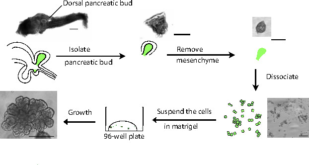
Figure 1. Schematic representation of the procedure. The gastro-intestinal tract is initially dissected from the embryo and subsequently the dorsal pancreatic bud is isolated. The mesenchyme is removed and the pancreatic progenitors are dissociated using trypsin. The resulting partially-dispersed cells are then seeded at low density in growth factor-depleted Matrigel. Scale bars: 1.00 mm except for the resulting organoid picture where the scale bar is 200 µm. Please click here to view a larger version of this figure.
Figure 2. Progress of culture over time. (A) A small cluster of pancreatic progenitors grown with the organoid medium and followed with time-lapse microscopy from 3 hr after plating, for 60 hr. In the bottom panels, an organoid after 7 days of culture. Scale bar: 200 µm; applies to all panels in A. (B) Example of a sphere followed in a 60 hr time-lapse and captured at day 7 of culture. Scale bar: 200 μm; applies to all panels in B. Please click here to view a larger version of this figure.
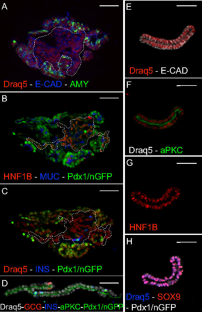
Figure 3. Histology. (A-C) Serial sections of a 7-day organoid stained for progenitor (B – HNF1B) and differentiation (A – amylase, C – insulin) markers. The organoid is composed of epithelial (A – E-cadherin) and apically polarized (B – mucin1) cells. The dashed line corresponds to the non-acinar central region (A), where HNF1B (B) and endocrine (C) cells are detected. (D-H) Sections of 7-day spheres stained for progenitor (G – HNF1B; H – SOX9) and endocrine (D – insulin and glucagon) markers. The spheres are composed of apically polarized (D, F – aPKC) epithelial (E – E-cadherin) cells. Scale bar: 50 µm. Please click here to view a larger version of this figure.
Large-scale production of functional beta cells in vitro is still ineffective 1. In this challenging context, developmental biology studies may help deciphering the exact signals that are required for the differentiation of functional beta cells. This protocol allows for the maintenance, expansion and differentiation of embryonic pancreatic progenitors in vitro. This includes the formation of insulin-producing beta cells that do not co-express other endocrine hormones, have high levels of Pdx1, express the pro-convertases that mature insulin and have the processed insulin 28. Important key factors within the system are the activity of FGF (exogenously stimulated by added FGF potentiated by heparin) and Notch (endogenous) signaling pathways, as well as ROCK-inhibition by Y-27632: in the absence of those factors, no or very limited numbers of organoids and spheres were generated 28. The requirement for FGF and Notch activity is easily understood based on their importance for pancreas development in vivo 28. ROCK inhibitor can be substituted by blebbistatin, thus revealing that hyperactivation of microfilament dynamics upon dissociation leads to both increased cell death, a strong inhibition of the progenitor transcription factor Pdx1 and a lack of expansion. Interestingly, many additional components of the organoid medium were proven to be individually unnecessary, but their combined absence resulted in the loss of epithelial branching 28. In addition to certain essential components of the medium, it is important to control the level of dissociation of progenitors. Indeed, progenitor proliferation and Pdx1 maintenance are significantly promoted in groups of more than 4 cells. A compaction can be observed within the first 12 hr and failure to compact results in failure to maintain Pdx1 and expand. The ROCK inhibitor is essential for this process.
At the moment organoids do not form after FACS sorting but the efficiency of the system could potentially be improved by reaggregating a controlled number of progenitors. Another critical component of this culture system is the 3D matrix. Cells put on Matrigel or in the Matrigel too close to the bottom of the plate spread and lose Pdx1 expression. Matrigel most likely provides biochemical components, notably laminin as well as mechanical cues 28. Indeed, the stiffness of the matrix plays a pivotal role. Stiff hydrogels are not permissive for pancreatic progenitors maintenance and expansion 28 and diluted Matrigel is not either. When Matrigel is diluted 1:10 pancreas progenitor cannot be cultured.
The organoid system can be used to test the effects of small molecules and recombinant proteins on pancreatic progenitors in terms of survival, proliferation, differentiation, polarization and branching 28. It can also be used to test the cooperation of different cell types during pancreas development 28. We are confident that the accessibility of pancreatic progenitor cells will also allow genetic manipulations, such as viral targeting, as seen in other organoid systems 17,27. This could be used for screening 17 with a system that enables morphogenesis in contrast to the spheres described previously as well as here 16,17,28. The culture conditions we developed also present the advantage of being serum-free, feeder-free and devoid of mesenchyme and blood vessels thereby reducing the cellular and biochemical complexity. However, there is a limitation in the ability to passage the organoids and thus to obtain large quantities of progenitors. This could be circumvented in the future by adaptations of the protocol to later stages of development where progenitors are more abundant, to sources of pancreatic progenitors produced from embryonic stem (ES) cells or induced pluripotent stem (iPS) cells. This paves the way for a 3D model of human pancreas development.
This system can also potentially be used for the production of pancreatic cells in the future perspective of therapy. In this context, the production of functional beta cells for transplantation would potentially help in diabetes therapy. The adaptations of the system to human ES or iPS cells would be important for this purpose. It is still unclear whether the organoid or sphere conditions should be used. The organoid conditions allow for the production of cells that have several characteristics of mature beta cells but their function remains to be tested. However, these cells are currently not numerous and are mixed among other cells. It is also likely that the early appearance of heterogeneity in the organoid system leads to uncontrolled signaling between cells and is therefore detrimental to production.
The sphere system that maintains progenitors is in principle preferable for controlled expansion and passaging of progenitors but their subsequent differentiation remains to be controlled. Others have recently produced pancreatospheres that can be efficiently differentiated. It will be important to compare the nature of the spheres obtained in the current protocol devoid of feeders and serum to spheres obtained with the other protocols 16,17. From a therapeutic point of view, the complexity and biological origin of Matrigel may constitute an issue of reproducibility, health and scalability. Preliminary results have shown that soft hydrogels functionalized with laminin are permissive for pancreatic progenitor expansion in vitro. Further optimization is required as these gels are not yet as efficient as Matrigel 28.
The production of beta cells in their natural context could also potentially be useful to test drugs that boost beta cell activity or increase their survival or proliferation but for this purpose it will be important to first test the degree of maturity of the beta cells, to increase the efficiency of their differentiation and to test whether the culture conditions presented here better maintain islets than the current suspension cultures. Producing the exocrine pancreas could also be useful to develop drugs to target pancreatic cancers and pancreatitis. Here again, the degree of maturity of the exocrine cells produced needs to be thoroughly investigated.
The authors have nothing to disclose.
This work was funded sequentially by a NCCR Frontiers in Genetics pilot award, Juvenile Diabetes Research Foundation Grant 41-2009-775 and Grant 12-126875 from Det Frie Forskningsråd/Sundhed og Sygdom. The authors thank the Spagnoli lab for hosting the video shooting.
Penicillin-Streptomycin | Gibco | 15070-063 | Stock keept at -20°C | |
KnockOut Serum replacement (supplement) | Gibco | 10828-028 | Stock keept at -20°C | |
2-mercaptoethanol | Sigma Aldrich | 3148-25ML | Stock keept at 4°C | |
Phorbol Myristate Acetate (PMA) | Calbiotech | 524400-1MG | Stock keept at -20°C | |
Y-27632 (ROCK inhibitor) | Sigma Aldrich | ab120129 | Stock keept at -20°C- Attention! Stability/source is a frequent source of problems | |
EGF | Sigma Aldrich | E9644-2MG | Stock keept at -80°C | |
Recombinant Human R-spondin 1 | R&D | 4645-RS-025/CF | Stock keept at -80°C | |
- or - | ||||
Recombinant Mouse R-spondin 1 | R&D | 3474-RS-050 | Stock keept at -80°C | |
Recombinant Human FGF1 (aFGF) | R&D | 232-FA-025 | Stock keept at -80°C- do not include to increase beta cell production | |
Heparin (Liquemin) | Drossapharm | Stock keept at 4°C | ||
Recombinant Human FGF10 | R&D | 345-FG-025 | Stock keept at -80°C | |
DMEM/F-12 | Gibco | 21331-020 | ||
Penicillin-Streptomycin | Gibco | 15070-063 | Stock keept at -20°C | |
B27 x50 (supplement) | Gibco | 17504-044 | Stock keept at -20°C | |
Recombinant Human FGF2 (bFGF) | R&D | 233-FB-025 | Stock keept at -80°C | |
Y-27632 (ROCK inhibitor) | Sigma Aldrich | ab120129 | Stock keept at -20°C- Attention! Stability/source is a frequent source of problems | |
DMEM/F-12 | Gibco | 21331-020 | ||
Matrigel | Corning | 356231 | Stock keept at -20°C | |
Trypsin 0.05% | Gibco | 25300-054 | Stock keept at 4°C | |
RNAlater – RNA stabilizing reagent | Qiagen | 76104 | Store at room temperature | |
Dispase | Sigma Aldrich | D4818-2MG | Stock keept at -20°C | |
BSA for reconstitution | Milipore | 81-068 | For reconstituition of cytokines – Stock keept at -20°C | |
Fetal calf serum (FCS) | Gibco | 16141079 | Stock keept at -20°C | |
60 well MicroWell trays | Sigma Aldrich | M0815-100EA | ||
4-well plates | Thermo Scientific | 176740 | ||
95-well plates F bottom | Greiner Bio | 6555180 | ||
Glas bottom plates | Ibidi | 81158 | ||
Disposal micropittes | Blaubrand | 708745 |