We present a microfluidic-based electrochemical biochip for DNA hybridization detection. Following ssDNA probe functionalization, the specificity, sensitivity, and detection limit are studied with complementary and non-complementary ssDNA targets. Results illustrate the influence of the DNA hybridization events on the electrochemical system, with a detection limit of 3.8 nM.
Miniaturization of analytical benchtop procedures into the micro-scale provides significant advantages in regards to reaction time, cost, and integration of pre-processing steps. Utilizing these devices towards the analysis of DNA hybridization events is important because it offers a technology for real time assessment of biomarkers at the point-of-care for various diseases. However, when the device footprint decreases the dominance of various physical phenomena increases. These phenomena influence the fabrication precision and operation reliability of the device. Therefore, there is a great need to accurately fabricate and operate these devices in a reproducible manner in order to improve the overall performance. Here, we describe the protocols and the methods used for the fabrication and the operation of a microfluidic-based electrochemical biochip for accurate analysis of DNA hybridization events. The biochip is composed of two parts: a microfluidic chip with three parallel micro-channels made of polydimethylsiloxane (PDMS), and a 3 x 3 arrayed electrochemical micro-chip. The DNA hybridization events are detected using electrochemical impedance spectroscopy (EIS) analysis. The EIS analysis enables monitoring variations of the properties of the electrochemical system that are dominant at these length scales. With the ability to monitor changes of both charge transfer and diffusional resistance with the biosensor, we demonstrate the selectivity to complementary ssDNA targets, a calculated detection limit of 3.8 nM, and a 13% cross-reactivity with other non-complementary ssDNA following 20 min of incubation. This methodology can improve the performance of miniaturized devices by elucidating on the behavior of diffusion at the micro-scale regime and by enabling the study of DNA hybridization events.
Microfluidic lab-on-a-chip (LOC) devices provide numerous advantages in clinical diagnostics, environmental monitoring and biomedical research. These devices utilize microfluidic channels to control fluid flow to regions of the chip where a variety of procedures can take place including reagent mixing, affinity based binding, signal transduction, and cell culturing1-4. Microfluidics provides many advantages over conventional clinical diagnostic tools such as microwell plate readers or electrophoretic gel shift assays. Microfluidic devices require 2 to 3 orders of magnitude (nanoliters as opposed to microliters) fewer reagents to perform similar assays. Also, these devices can increase the speed by which some biological events occur due to the smaller confinement of the species within the channels5,6. Thirdly, sensors can be integrated within microfluidic devices using lithography and etching techniques, which can provide label-free detection. Lastly, these devices are inexpensive to produce and require little work on the part of the technician to operate7-10.
Label-free detection typically is performed using an optical or electrical transducer. Optical devices can present better sensing performance due to lower interference with analytes in the sample. Nevertheless, their performance is compromised in cases where the sample’s background has the same resonating wavelength as the sensor11. There are many advantages to using electrical signals to perform biological and chemical detection in microfluidic systems. The fabrication is inherently less complicated since these sensors typically only require patterned electrodes to operate. In addition, electrical signals can be directly interfaced with most measurement equipment while other signal modalities may require a transducer to convert the signal12-15. Electrical sensors commonly measure changes in impedance16,17, capacitance18, or redox activity19. However, new challenges are presented as these systems are miniaturized. The most important challenges to overcome include: sample preparation and mixing of fluids (due to the low sample volume and Reynolds number), physical and chemical effects (including capillary forces, surface roughness, chemical interactions between construction materials and analytes), low signal-to-noise ratio (produced by the reduced surface area and volume)20-23, and potential interference from electro-active analytes in complex biological samples (e.g., blood and saliva). Further investigation of these effects will result in guidelines for an accurate fabrication and operation of these devices in a reproducible manner that would improve upon their overall performance.
DNA hybridization detection is used extensively to diagnose genetic disorders24,25 and various forms of cancer26. Every year, multiple strains of influenza are identified in patients using results from DNA hybridization techniques27. The influenza virus alone accounts for 36,000 deaths each year in the United States28. Such examples could benefit from a bench-top microfluidic device that can perform the same assay techniques as a plate reader or gel shift assay with low sample volume and at a fraction of the cost without sacrificing sensitivity or specificity. Due to the many advantages of label-free electrochemical sensing, it has been used extensively for detection of DNA hybridization events29,30. A setup where macro-scale electrodes (in the millimeter range) are dipped in beakers with the solution of interest can be used to provide very sensitive data regarding the binding kinetics of single stranded DNA sequences to their matching complementary sequences. Recently, there have been a few advances in incorporating electrochemical sensing in microfluidics for DNA hybridization. Studies have been performed regarding hybridization kinetics31 and integration of sensors for detection15 in microfluidic channels. However, there still exists a need for a rapid high throughput microfluidic device that can analyze DNA hybridization events in parallel without complicated sample preparation steps.
The device presented in this work provides a platform that allows for multiple interactions to be screened in parallel and without complicated sample preparation steps. Our protocol presents how microfluidic-based electrochemical biochips are microfabricated with micro-electromechanical systems (MEMS) technology32,33. We describe the fabrication process of both the microfluidic chip, made of polydimethylsiloxane (PDMS), and the electrochemical chip, comprised of an array of electrodes. The chemical functionalization of the biochip with ssDNA probes is also addressed. Finally, the ability of the biosensor to specifically detect and analyze ssDNA targets is demonstrated. Overall, the microfluidic-based electrochemical biochip is a rapid and high-throughput analysis technique. It can be used to investigate interactions between biological molecules and conducting transducers, and can be utilized in a variety of lab-on-a-chip applications.
1. Microfabrication of the Microfluidic Chip
2. Assemble the Device
3. Analyze DNA Hybridization
NOTE: Introduce each solution slowly into the channel at a flow rate of 200 µl/hr until the whole channel is filled.
A controllable and accurate manufacturing process for the experimental device is essential in research. It allows researchers to obtain reproducible and high throughput experiments. Here we have demonstrated a high yield, high reproducibility microfabrication process of a microfluidic-based electrochemical biochip (Figure 1). With a low failure rate, few devices have shown bonding issues that lead to solution leakage. In order to validate the electrochemical activity of the biochip, cyclic voltammetry measurements are done in the microchannels filled with an electro-active redox couple ferricyanide/ferrocyanide. Figure 2 shows the reproducible electrochemical response of nine different working electrode on the chip. These results show that the electrochemical activity is the same in all nine chambers of the biochip allowing high-throughput measurements.
Most DNA hybridization sensors immobilize ssDNA probes onto a sensor surface. As gold is used to pattern the sensing electrodes for the microfluidic devices, thiols are good candidates to form strong covalent bonds with the sensor surfaces34. One common conjugation technique involved adding a functional thiol (-SH) group at one end of the ssDNA. The S-S disulfide bond protects the free thiol group from oxidation until it is ready to be used. Once the disulfide is reduced by tris(2-carboxyethyl)phosphine (TCEP) addition, the free thiol (-SH) becomes available to bond the DNA to a gold surface. Without TCEP, the disulfide bonds of the ssDNA will also assemble on the electrode, but the bond is much weaker than the thiol-gold bond and will not remain stable throughout the experiment. In this work, a stable ssDNA monolayer for hybridization detection has been achieved with incubation times between one and two hours. Once the ssDNA probes have been assembled onto the electrode, 6-mercapto-1-hexanol (MCH) is used to passivate any exposed regions on the surface to reduce non-specific binding effects35. The MCH’s thiol group allows for self-assembly onto the gold and the hydroxyl group reduces non-specific adsorption of the ssDNA in solution. The high TCEP concentration is used to reduce the thiol groups that may have oxidized to form disulfide bonds. Without the high TCEP content in the buffer, the MCH would form unstable monolayers and the impedance data would vary accordingly due to variations with the surface charge. The MCH has another important function when using it as passivation with ssDNA molecules. The oxidative adsorption process of the MCH injects electrons into the electrode and reduces the surface potential. This causes an electrostatic effect with the anionic probe ssDNA already immobilized and the ssDNA stands upright away from the electrode36. Upright probes greatly increase the hybridization efficiency since the target ssDNA strands have much easier access to the entire length of the probe sequence. This passivation step is crucial for establishing a stable impedance baseline measurement of the sensor, reducing false positive signals and removing any weakly bound molecules from the surface.
The biosensing mechanism of DNA hybridization events is based on repulsion forces between negatively charged DNA and other electro-active molecules. When a hybridization event occurs, stronger repulsion forces between the hybridized DNA and the electro-active molecules make it harder for the electro-active species to diffuse towards the electrode37. Here we use a ferricyanide/ferrocyanide couple as the redox species indicator for these repulsion forces, which are measured using electrochemical impedance spectroscopy (EIS). We utilize this biosensing mechanism in the microfluidic-based electrochemical biochip, and demonstrate its ability to detect DNA hybridization events33. Figure 3 shows the selectivity of the biosensor by illustrating impedance variations as a result of hybridization events between three ssDNA probes and their complementary ssDNA target. A 13% cross-reactivity with other non-complementary ssDNA following 20 min of incubation has been demonstrated. These results show the feasibility of the microfabricated device to measure DNA hybridization events in a reproducible and high-throughput manner. We also have demonstrated the sensitivity of the biosensor by introducing different concentrations of complementary ssDNA target to the sensing probe. Figure 4 shows the functionality of the biosensor with a trend of increasing impedance values for higher ssDNA target concentrations. Following calculations of the restricted diffusional resistance33 for different concentrations of complementary ssDNA target, a linear regression analysis resulted in a theoretical limit of detection of 3.8 nM by the calculation of the corresponding ssDNA target concentration for the background signal. Overall, the biochip shows the ability to rapidly sense the presence of DNA hybridization events.
Figure 1. Photograph of packaged device under test (chip dimensions: 3.5 cm x 3.5 cm; microchannel height is 100 μm and 500 μm in width).
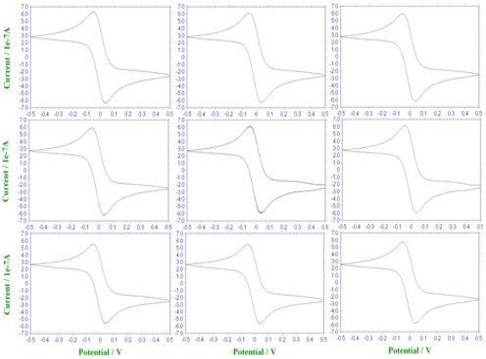
Figure 2. Electrochemical validation. Cyclic voltammograms of 9 (3 x 3 grid) working electrodes in the presence of ferrocyanide/ferricyanide redox couple. The reproducibility among the patterned electrodes is shown by the similar shape, peak heights, and peak separation for all electrodes. Please click here to view a larger version of this figure.
Figure 3. Specificity of the biosensor. The impact of hybridization events between different ssDNA targets and three different ssDNA probes on the charge transfer resistance. Upon DNA hybridization event, the stronger repulsion force between the negatively charged double stranded DNA and the negatively charged ferrocyanide/ferricyanide molecules results in higher charge transfer resistance.
Figure 4. Functionality of the biosensor. Nyquist plot of electrochemical impedance spectroscopy measurements following the introduction of 0.01, 0.1, 1, and 1 μM target ssDNA (Arrow indicates increasing ssDNA target concentrations). The increased impedance values at low frequencies (~15 Hz) for higher target ssDNA concentrations are due to the stronger repulsion forces between the dsDNA and the ferrocyanide/ferricyanide molecules.
Our procedures demonstrate the manufacturing of a microfluidic-based electrochemical biochip and its utilization for DNA hybridization events analysis. Through a high yield controlled microfabrication process we develop a device comprised of microscale channels integrated with an array of electrochemical transducers. We have devised controlled processing parameters for the photolithography procedure of the electrochemical chip and the microfluidic channel mold through an iterative approach. These steps provide guidelines for future creation of other miniaturized analytical devices. Importantly, they provide a method to optimize various biosensing parameters such as signal-to-noise ratio, stability, and sensitivity. Following device fabrication, we functionalize the electrochemical transducers with ssDNA probe as a bioreceptor, providing a unique analytical purpose. Although these procedures demonstrate low device failure rate, solution leakage shows to be a major issue following assembly of the device and during the performance of the DNA hybridization assay. Further investigation of bonding failure in such devices would improve the yield of the process and make it ideal for micro-bio-systems study.
In our studies, we show the ability of the biochip to accurately and specifically sense DNA hybridization events. As part of design parameters for a new analytical micro-device, one should take into consideration the manufacturing of the device, which demands an iterative approach to optimize fabrication parameters (e.g., photolithography parameters, microchannel dimensions, type of the deposited metal). In order to efficiently detect DNA hybridization events, ssDNA probe assembly and hybridization conditions have to be optimized as well (e.g., incubation time, buffer concentration, probe concentration, prevention of non-specific adsorption).
The ability to quickly screen for particular DNA sequences is very beneficial in the fields of cancer research, influenza detection and genetic engineering. Most arrayed detection methods utilize a label to produce the signal and use multiple washing and incubation steps. Utilizing the proposed microfluidic devices, DNA hybridization will be performed with high number of DNA sequences in the same device without the need for labels of any kind. We envision near-term applications integrating more sophisticated capabilities to the biochip to improve its performance and modularity. For example, valve-based microfluidics has the potential to provide the biochip with automatic and controlled analysis abilities. Another example is functionalizing the device with other bioreceptors, providing unique sensing properties that can be used in a wider range of applications.
The authors have nothing to disclose.
The authors acknowledge the Robert W. Deutsch Foundation, the Defense Threat Reduction Agency (DTRA), and the National Science Foundation Emerging Frontiers in Research and Innovation (EFRI) for financial support. The authors also thank the Maryland Nanocenter and its Fablab for cleanroom facility support.
Name of Material/ Equipment | Company | Catalog Number | Comments/Description |
Material | |||
4'' Silicon wafer | Ultrasil | 4-5664 | Single side polished; P-type; Boron doped; Orientation 1-0-0; Thickness 500 micron; Resistance 10-20 ohm*cm |
Shipley 1813 photoresist ("PR1") | Microchem | positive photoresist | |
AZ5214 photoresist ("PR2") | Hoechst Celanse | positive photoresist | |
SU-8 50 photoresist ("PR3") | Microchem | negative photoresist | |
Gold etchant | Transene | TFA | No dilution |
Chromium etchant | Transene | 1020AC | No dilution |
PDMS elastomer | Dow Corning | 3097366 1004 | |
PDMS curing agent | Dow Corning | 3097358 1004 | |
Biopsy punching tool | Healthlink | BP20 | |
Tygon flexible tubing | Cole Parmer | R 3603 | .015" |
Tygon flexible adapter | Cole Parmer | 06417-41 | .0625" |
1 mL syringe | Beckton-Dickenson | 301025 | |
Monobasic potassium phosphate | Fluka | 1551139 | |
Potassium phosphate dibasic anhydrous | Sigma | RES20765-A7 | |
Sodium chloride | Sigma-Aldrich | S7653 | |
tris(2-carboxyethyl)phosphine | Aldrich | C4706 | |
6-mercapto-1-hexanol | Aldrich | 451088 | |
20x concentrated saline-sodium citrate buffer | Sigma | 93017 | |
Potassium hexacyanoferrate(III) | Aldrich | 455946 | |
Sodium ferrocyanide | Aldrich | CDS001589 | |
Target ssDNA #1 | Integrated DNA Technologies (IDT) | 100 nmole DNA oligo | 5’-/5ThioMC6-D/AAAGCTCCGATAGCGCTCCG TGGACGTCCC-3’ |
Complementary ssDNA #1 | Integrated DNA Technologies (IDT) | 100 nmole DNA oligo | 5’-GGGACGTCCACGGAGCGCTA TCGGAGCTTT-3’ |
Target ssDNA #2 | Integrated DNA Technologies (IDT) | 100 nmole DNA oligo | 5’-/5ThioMC6-D/ACGCGTCAGGTCATTGACGA ATCGATGAGT-3’ |
Complementary ssDNA #2 | Integrated DNA Technologies (IDT) | 100 nmole DNA oligo | 5’-ACTCATCGATTCGTCAATGA CCTGACCCGT-3’ |
Target ssDNA #3 | Integrated DNA Technologies (IDT) | 100 nmole DNA oligo | 5’-/5ThioMC6-D/ACCTAGATCCAGTAGTTAGA CCCATGATGA-3’ |
Complementary ssDNA #3 | Integrated DNA Technologies (IDT) | 100 nmole DNA oligo | 5’-TCATCATGGGTCTAACTACT GGATCTAGGT-3’ |
Instrument | |||
Plasma Enhanced Chemical Vapor Deposition (PECVD) | Oxford Instruments | PlasmaLab System 100 | Chamber pressure 1000 mTorr, Tempreture 200 celsius degrees, RF power 20 W, Gasses: a) N2O flow rate 710 sccm; b) a composition of 5% SiH4 and 95% N2 gasses flow rate 170 sccm, SiO2 growth rate 690 Angstrom/minute. |
DC sputtering unit | AJA International | ATC 1800-V | For Chrome: Chamber pressure 10 mTorr, Argon flow rate 20 sccm, Supplied DC power 200 W, Sputter rate 10 nm/min. For Gold: Chamber pressure 10 mTorr, Argon flow rate 20 sccm, Supplied DC power 200 W, Sputter rate 36 nm/min. |
E-beam evaporation system | Denton | Custom-built | For Titanium: Chamber pressure <2E-6 Torr, Supplied power to the e-gun 7.5k W, Filament current 150-200 mA, Evaporation rate 6-10 Angstrom/second. For Platinum: Chamber pressure <2E-6 Torr, Supplied power to the e-gun 7.5k W, Filament current 150-200 mA, Evaporation rate 2-3 Angstrom/second. |
Potentiostat | CH Instruments | 660D | |
Syringe pump | KD Scientific | KDS230 |