Flödescytometri har i stor utsträckning utnyttjats i immunologi, hematologi och onkologi att definiera cellpopulationer via inneboende spridningsegenskaper, cellytantigen uttryck och andra fluorescensparametrar 1-3. Våra insikter blodslinje utveckling och sjukdom är ett resultat av en betydande grad av kontinuerlig förfining av denna metod efter dess genomförande 4,5. Ökad medvetenhet om den kvantitativa och övergripande analytisk potential flödescytometri har nyligen uppmuntrat dess mer utbredd användning inom stamcellsforskning och kan möjliggöra på liknande djupgående framsteg på kortare tidsram 6. Däremot har tillämpningen av flödescytometri att specifikt analysera och isolera neurala populationer länge uppfattats som en utmaning. I motsats till hematopoetiska celler som naturligt finns i suspension, är neurala celltyper vanligen skördas från alltför komplicerade källor som kan innehålla glia och olika other omgivande celler såväl som ett intrikat nätverk av processbärande neuroner. Följaktligen måste neurobiologi ännu inte genomfört mångsidighet flödescytometri till dess fullständiga potential i dagliga forskningsrutiner. Men så länge som kan alstras en livskraftig enkelcellsuspension (och protokoll har utarbetats och optimerats för detta ändamål 7), flödescytometri och fluorescensaktiverad cellsortering (FACS) kan anses vara en värdefull del av den analytiska repertoar i neurobiologi 8-11.
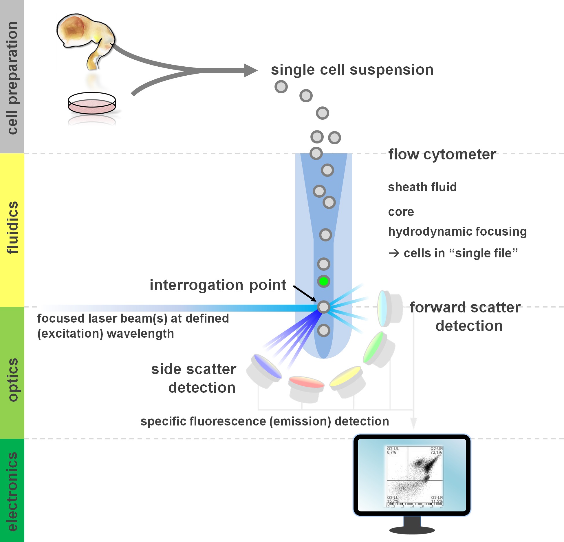
. Figur 1. Principen om flödescytometrianalyser och komponenter i en flödescytometer flödescytometrar omfattar tre huvudsystem: flödeskunskap, optik och elektronik. En strömlinjeformat flöde av celler i suspension (framställd från primär vävnad eller odling in vitro) åstadkommes genom manteln fluid via hydrodynamisk fokusering, begränsa urvalet till dess centrum kärna. De optiska delarna är sammansatta av lasrar som belyser flödet av celler och optiska filter som dirigerar signalen till lämpliga detektorer. Ljussignalerna detekterade omvandlas till elektroniska signaler, därefter behandlas av en dator och visualiseras på en bildskärm för dataanalys och gating. Klicka här för att se en större version av denna siffra.
Användare av flödescytometriska metoder vinst från åtminstone en grundläggande förståelse för de underliggande fundamenta, inklusive en cytometer byggstenar (för översikt se 12,13; se även figur 1). En laserstråle skär med en hydrodynamiskt fokuserad fluidic ström som innehåller celler i suspension, vilket i sin tur passerar genom laserstrålen i "enstaka fil" en efter en. Den interceptipå av en cell (eller någon annan partikel, för den delen) med laserresulterar i spridning av ljus från denna identifieringspunkt. Spritt ljus kan detekteras i fortsättning av laserriktningen (framåtspridning, associerad med storleken av partikeln), såväl som vinkelrätt mot dess riktning (sidospridning, vilket återspeglar granulosity av partikeln / cell). Dessa ovannämnda spridningsegenskaper inte kräver särskild märkning, vilket är anledningen till att en omärkt prov (eller också cellrester, luftbubblor, etc.) kommer att generera en signal (event) på bivariata framåtspridning mot sidopunktdiagram som vanligen används för inledande gating. Genom att använda lämpliga lasrar och filter specifika för motsvarande excitations- och emissionsspektra, kan en cell analyseras för dess positivitet, grad av styrka, eller frånvaro av fluorescerande markörer. Majoriteten av flödesapplikationer cytometrisk har fokuserat på karakterisering via cellytantigener. Till skillnad från det hematopoietiska lineage, har det neurala härstamning förblivit mindre utsträckning definieras enligt ytan epitop uttrycksmönster 5. En fördel med att utnyttja ytantigener är att levande celler kan utsättas för cellsortering paradigm såsom FACS. Däremot kräver intracellulär antigen färgning fixering och permeabilisering steg för att medla epitop-antikroppsinteraktion, utgör hinder tillämpningar efter som kräver levande celler. Notera sådana metoder kan fortfarande för många kvantitativa analyser 14 samt analyser nedströms för RNA och proteinuttryck 15. Hematologi, immunologi och onkologi har ofta använt mer än ett dussin markörer tillsammans för att definiera särskilda subpopulationer 16. Dessutom kan mass cytometri eller CyTOF nu användas för att analysera upp till 30 parametrar samtidigt 17,18.
För neurala stamceller samt primära kulturer 14,19,20 heterogeniteten av celler ivitro är ett vanligt fenomen 21-23. Cellerna inte representerar målgruppen intressanta förkroppsligar ett potentiellt confounding faktor för experimentell avläsning 24,25. Bekvämt, de olika cellulära delmängder som finns inom en heterogen cellsuspension bära distinkta (kända eller ännu Att att dechiffreras) antigen uttryck profiler, som kan användas för att definiera dessa olika populationer. Flödescytometri kan därmed spela en avgörande roll för att lösa cellulär heterogenitet och därigenom underlätta biomedicinska tillämpningar (in vitro-analyser, cellterapi) och optimera kvantitativa avläsning genom att fokusera på de mest relevanta delmängd 24,26. Olika ytantigen kombinationer har identifierats under de senaste åren för att göra det möjligt för kvantifiering och isolering av specifika neurala celltyper. Detta inkluderar CD133 för anrikning av neurala stamceller 27, en kombination av CD15 / CD24 / CD29 ytantigener för isolering av NSC, differentieringted neuron och neurallist celler 28 eller CD15 / CD24 / CD44 / CD184 / CD271 att isolera neurala och gliaceller delmängder 25, bland annat signaturer 29,30. Bortom neuroner, gliaceller markörer inkluderar A2B5 31, CD44 25, NG2 32 och GLAST 33. En färsk publikation har utnyttjat mitthjärnan floorplate gångaren markör CORIN 34,35 att anrika dopaminerga prekursorer vid Parkinsons celltransplantation paradigm 36. CD-molekyler är inte bara markörer, men funktionellt relevanta förmedlare av cell-cell interaktioner och i en cell förmåga att reagera på signaler från extracellulära matrixmolekyler och tillväxtfaktorer 37. En strategi för att ytterligare förbättra den arsenal av kombinatoriska CD antigener att karakterisera neurala härstamning utveckling är att använda kända intracellulära markörer för att screena för och definiera CD antigenkombinationer för en viss celltyp av intresse. Vi har nyligen utnyttjat ett sådant tillvägagångssätt och identifierade CD49f – / CD200 höga kombiuttrycksmönster som en ny metod för att berika neuronala delmängder från neuralt differentierade inducerad pluripotenta stamceller kultursystem 38. Här inkluderar vi och diskuterar det senare protokollet (och valfria variationer därav) där färgning yta och intracellulär färgning kan användas samtidigt för att definiera neurala cellsubpopulationer med flödescytometri.

Figur 2. Flödesdiagram av experimentella protokollalternativ. Figuren visar en schematisk representation av de viktigaste stegen i protokollet. Valfria steg (CFSE dye eller intracellulär antigen märkning) anges med ljusgrå lådor. Efter skörden, är det viktigt att bedöma lönsamheten och cell antal neurala cellsuspensioner före cellytan färgning. Positivt somsåväl som negativa kontroller måste ingå förutom de prover av intresse. Prover kan analyseras genom flödescytometrisk analys och / eller används i cellsortering paradigm. Klicka här för att se en större version av denna siffra.
Medan vi tidigare har använt primära antikroppen i kombination med sekundär antikropp för intracellulär färgning 38, introducerar vi nu icke kovalent märkning av den primära antikroppen via fluorescerande Fab-fragment (Zenon märkning) som en liten variation, vilket minskar stegen cellmanipulation 39. Dessutom som ytterligare ett exempel på protokollet mångsidighet, anställer vi en valfri märkning av en experimentell delmängd av karboxifluorescein succinimidylester (CFSE) före ytan antigen färgning. Sådana CFSE pre-märkning möjliggör omedelbara direkt jämförelse av två cellinjer eller experimentella förhållanden (CFSE-märkta vs. omärkt) inom en enda provrör, minska variansen eller subtila skillnader i inkubationstid och spara antikropp. CFSE är en etablerad fluorescerande färgämne som ofta används för att spåra cell 40, i spridnings 41,42 och streckkoder experiment 43,44. Slutligen medan faktiska sorteringssteg (FACS, immunomagnetisk cellseparations eller immunopanning) är inte en del av detta protokoll, i princip, avverknings- och märkningsförfaranden som beskrivs här gör avkastnings prover som kan utsättas för ytan antigen- eller intracellulära märkning baserade sorterings applikationer 15 25,28.
Med denna artikel vill vi: sammanfatta en livskraftig ytantigen färgningsprotokollet 25,28, sammanfatta ett protokoll för detektion av intracellulära mål samt kombinerad yta och intracellulär antigenanalys 38, presentera en intracellulär CFSE färgämne märkning steg 41,45 som en experimentell alternativ för comjämförande analyser av neurala cellpopulationer, och sammanfatta metoder för att flödescytometrianalyser (lämpliga kontroller 13,46, grind strategi och presentation av data 47).
