Akım sitometri yoğun içsel dağılım özellikleri, hücre yüzey antijeni ifade ve diğer floresan parametreleri 1-3 vasıtasıyla hücre popülasyonlarının tanımlamak için immünoloji, hematoloji ve onkoloji istismar edilmiştir. Kan soy geliştirme ve hastalık halinde bizim anlayışlar ilk uygulama 4,5'ten sonra bu metodolojinin sürekli arıtma önemli ölçüde bir sonucudur. Akış nicel ve genel analitik potansiyeli artan farkındalık sitometri son zamanlarda kök hücre araştırmalarında da daha yaygın kullanımını teşvik etmiştir ve kısa bir zaman dilimi 6 benzer derin ilerleme sağlayabilir. Ancak, özellikle nöral popülasyonları analiz ve izole etmek için akış sitometri uygulaması uzun zorlu olarak algılanmıştır. Doğal olarak süspansiyonda mevcut hematopoietik hücrelerin aksine, sinir hücresi tipleri, tipik olarak glia ve çeşitli o içerebilir aşırı derecede karmaşık kaynaklardan toplanırTher çevreleyen hücreleri ve aynı zamanda işlem taşıyan nöronların karmaşık bir ağdır. Sonuç olarak, nörobiyoloji günlük araştırma rutinleri onun tam potansiyeline flow sitometri çok yönlülüğünü uygulamak için henüz. Ancak nörobiyolojisinde analitik repertuarın değerli unsuru olarak kabul edilebilir olduğu sürece uygun bir tek hücre süspansiyonu elde edilebilir (ve protokoller bu amaçla 7 için tasarlanmış ve optimize edilmiş) halinde, akış sitometrisi ve floresans ile aktive edilen hücre çeşitleme (FACS) 8-11.
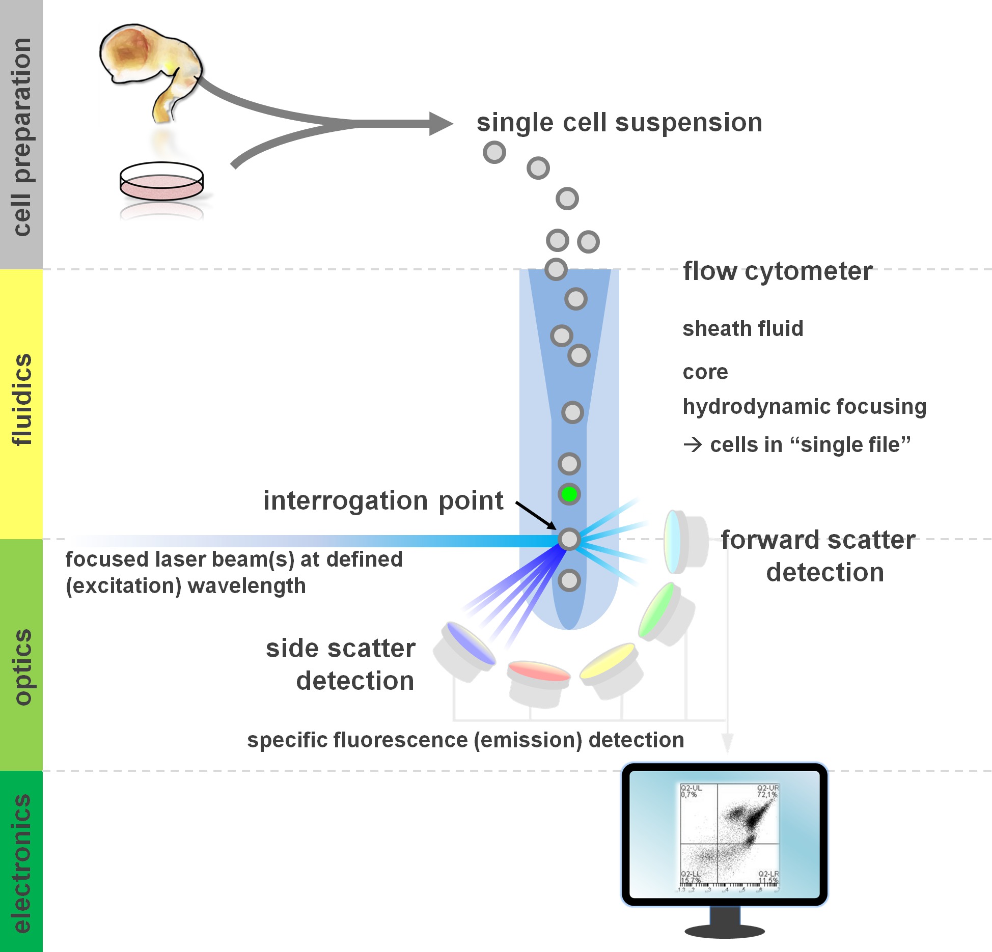
. Fluidik, optik ve elektronik: akım sitometri analizi ve bir akış sitometresinde bileşenlerinin Şekil 1. İlke akış sitometrelerinde üç ana sistemleri içermektedir. Süspansiyon içinde bulunan hücrelerin bir akıcı akış kılıf flui ile gerçekleştirilir (birincil doku ya da in vitro kültürü hazırlanmış)hidrodinamik yoluyla d merkez çekirdek örneği kısıtlayan, odaklama. Optik parçalar uygun dedektör sinyal doğrudan hücrelere ve optik filtrelerin akışı aydınlatmak lazerler oluşmaktadır. elektronik sinyaller, daha sonra bir bilgisayar tarafından işlenir ve veri analizi ve yolluk için bir monitörde görüntülenebilir dönüştürülür tespit ışık sinyalleri. Bu rakamın büyük halini görmek için lütfen buraya tıklayınız.
Bir sitometresinin yapı blokları dahil altta yatan temelleri en azından temel bir anlayış akış sitometri yöntemleri kar Kullanıcılar (inceleme için 12,13 görüyorum; ayrıca bakınız Şekil 1). Bir lazer ışını da 'tek dosya' birbiri ardına lazer ışınının geçmesine süspansiyon hücreleri içeren bir hidrodinamik odaklı akışkan akışı ile kesişir. interceptiBu sorgulama alanına ışık saçılması lazer sonuçları ile hücre (veya bu konuda herhangi bir diğer parçacık, sırasıyla) ile. (Parçacık / hücre granulosity yansıtan tarafında dağılım) Dağınık ışık, hem de dik olan doğrultu (parçacık boyutu ile ilişkili ileri dağılım, hem) lazer yönünde devamında tespit edilebilir. Bu Anılan dağılım özellikleri (aynı zamanda veya hücresel enkaz, hava kabarcıkları, vb) bir etiketsiz örnek yaygın ilk yolluk için kullanılan yan dağılım grafiği iki değişkenli ileri saçılma bir sinyal (olay) üretecektir neden özel etiketleme, gerekmez. Uygun lazerler ve karşılık gelen uyarma ve emisyon spektrumu için özel filtreler kullanılarak, bir hücrenin pozitiflik, yoğunluk seviyesinde veya flüoresan işaretler yokluğu için analiz edilebilir. Akış sitometrik uygulamalarının çoğunluğu hücre yüzey antijenleri üzerinden karakterizasyonu üzerine odaklanmıştır. Hematopoietik lineag aksineE, sinir soy yüzey epitop ifade desenleri 5'e göre daha az kapsamlı tanımlanmış kalmıştır. Yüzey antijenleri istismar bir avantajı, canlı hücreler, FACS gibi sıralama paradigmalar hücre tabi olmasıdır. Buna karşılık, hücre içi antijen boyama canlı hücreler gerektiren mansap uygulamaları engelleyen, epitop-antikor etkileşimi aracılık tespit ve permeabilization adımları gerektirir. Dikkat çekici bir şekilde, bu tür yaklaşımlar yine çok nicel deneyleri 14 yanı sıra, RNA ve protein ekspresyonu 15 alt baş analizler için izin verir. Hematoloji, immünoloji ve onkoloji genellikle belirli alt popülasyonlar 16 tanımlamak için birlikte bir düzineden fazla belirteçleri kullandık. Ayrıca, kitle sitometri veya CyTOF geç 30 parametreleri aynı anda 17,18 kadar analiz etmek için kullanılabilir.
Nöral kök hücre uygulamaları yanı sıra birincil kültürlerin 14,19,20 hücrelerin heterojen olarak içinIn vitro ortak bir olgudur 21-23 olduğunu. ilgi hedef kitleyi temsil etmeyen hücreler deneysel okuma 24,25 için bir potansiyel karıştırıcı faktör somutlaştırmak. Uygun, heterojen bir hücre süspansiyonu içinde bulunan farklı hücresel alt kümeleri bu çeşitli popülasyonları tanımlamak için kullanılabilir farklı (bilinen veya henüz deşifre edilecek) antijen ifade profilleri, ayı. Biyomedikal uygulamaları kolaylaştırmak, dolayısıyla böylece, hücresel heterojenite çözümünde önemli bir rol oynayabilir ve Flow sitometri (in vitro deneyler, hücre tedavisi) ve en alakalı alt kümesi 24,26 odaklanarak kantitatif okuma optimize. Çeşitli yüzey antijen kombinasyonları özel nöral hücre tiplerinin kantitatif ve izole izin vermek için son birkaç yıl içinde tespit edilmiştir. Bu nöral kök hücreler 27 NSC izolasyonu için CD15 / CD24 / CD29 yüzey antijenlerinin kombinasyonu, differentia zenginleştirilmesi için CD133 içerirTed nöron ve nöral krest hücreleri 28 veya CD15 / CD24 / CD44 / CD184 / CD271, diğer imzalar 29,30 arasında nöral ve glial alt kümeleri 25, izole etmek. Nöronlar ötesinde, gliyal belirteçler A2B5 31, CD44, 25 NG2 32 ve GLAST 33 içerir. Son zamanlarda yayın Parkinson hücre nakli dopaminerjik öncüleri zenginleştirmek için orta beyin tabanının habercisi işaretleyici CORIN 34,35 36 paradigmalar istismar etmiştir. CD molekülleri sadece belirteçler değil, hücre-hücre etkileşimleri ve hücrenin yeteneği fonksiyonel ilgili arabulucular dışı matriks molekülleri ipuçlarını cevap ve büyüme faktörleri 37. Ayrıca sinir soy gelişimini karakterize etmek için kombinasyon CD antijenlerinin cephanelik arttırıcı bir strateji, söz konusu özel bir hücre tipi için bir CD antijeni kombinasyonu için taranması ve tanımlamak için bilinen hücre içi belirteçler kullanmaktır. Biz son zamanlarda böyle bir yaklaşım sömürülen ve CD4 belirledik9f – / CD200 nöral ayırt uyarılmış pluripotent kök hücre kültürü sistemleri 38 nöronal alt kümelerini zenginleştirmek için yeni bir yaklaşım olarak yüksek birleştirici ifade desenleri. Burada, içerir ve ikinci protokolü (ve bunların isteğe bağlı varyasyonlar) 'in yüzey boyama ve hücre içi boyama akış sitometrisi ile nöral hücre alt-popülasyonunu tanımlamak için aynı anda kullanılabilir tartışır.

Deneysel protokol seçenekleri Şekil 2. akış diyagramı. Şekil protokolde yer alan önemli adımlardan şematik bir temsilini tasvir etmektedir. İsteğe bağlı adımlar (KAKE boya veya hücre içi antijen etiketleme) açık gri kutular ile gösterilir. Hasattan sonra, bu hücre yüzey boyamadan önce nöral hücre süspansiyonlarının yaşayabilirliği ve hücre sayısını belirlemek için gereklidir. Pozitif olarakde negatif kontroller olarak ilgi örneklere ek olarak dahil edilmesi gerekir. Numuneler akım sitometri ile analiz ve / veya hücre sıralama paradigmalar kullanılabilir. Bu rakamın büyük halini görmek için lütfen buraya tıklayınız.
Daha önce hücre içi boyama 38 için ikincil bir antikor ile kombinasyon halinde birincil antikor kullanmış iken, şimdi, böylece hücre manipülasyonu 39 aşamaları azaltarak küçük bir değişikliği gibi floresan Fab parçaları (Zenon etiketleme) yoluyla birincil antikor kovalent olmayan etiketleme getirmektedir. Ayrıca, protokolün çok yönlü bir başka örnek olarak, antijen lekeleme yüzey önce süksinimidil ester (CFSE) karboksifloresan ile bir deney alt-grubun isteğe bağlı bir etiketleme kullanır. Böyle KAKE ön-etiketleme, iki hücre hatları ya da deneysel koşullar hemen doğrudan karşılaştırma (sağlarVs KAKE-etiketli. Tek bir örnek tüpü, değişiklik veya inkübasyon süresi ince farklılıkların azaltılması ve antikor tasarruf içinde) etiketsiz. CFSE, yaygın üreme 41,42 ve Barkod deneylerde 43,44, hücre takibi 40 için kullanılan kurulu bir floresan boya olan. Gerçek sıralama adımlar (FACS, immünomanyetik hücre ayırma veya immunopanning) Bu protokolün bir parçası değil ise Nihayet, prensip olarak, burada açıklanan hasat ve etiketleme işlemleri antijen ya da hücre içi etiketleme-tabanlı uygulamaları sıralama yüzey tabi olabilir verim örnekleri yapmak 15 25,28.
Bu makale ile, biz hedefliyoruz: bir olarak hücre içi KAKE boya etiketleme adım 41,45 sunmak, bir hücre içi hedeflerin tespiti için protokol yanı sıra kombine yüzeyi ve hücre içi antijen analizi 38 özetlemek, geçerli bir yüzey antijeni boyama protokolü 25,28 özetlemek com için deneysel seçenekkarşılaştırmalı nöral hücre popülasyonlarının analizleri ve sitometrik analizi (strateji ve veri sunumu 47 yolluk uygun kontrolleri 13,46) akış yaklaşımları özetlemek.
