1. Cell Culture and Transfection
- Put a cover glass (22 mm x 22 mm) in a 6-well polystyrene plate. Optionally coat a cover glass with Poly-L-Lysine, 0.1% w/v, in water (see List of Materials/Equipment) to keep mitotic cells on the cover glass following the steps below:
Note: Optimal conditions must be determined for each cell line and application.- Aseptically coat culture surface with Poly-L-Lysine, 0.1% w/v, in water (0.4 ml/well of a 6-well polystyrene plate). Rock gently to ensure even coating of the culture surface.
- After 5 min, remove solution by aspiration and thoroughly rinse surface with sterile tissue culture grade water.
- Dry at least 2 hr before introducing cells and medium.
- Seed HeLa cells17 or HeLa Tet-Off cells17,22 on a cover glass (22 mm x 22 mm) put in a 6-well polystyrene plate. Check that cell density is 5.4 x 10⁵ per well. Culture cells in high-glucose DMEM with 10% FBS and 1% penicillin-streptomycin.
Note: For optimal results, empirically determine the cell density to use in seeding. - Incubate the cells at 37 °C in an atmosphere of 5% CO2 for 18 hr.
Note: In the case of HeLa Tet-Off cells, transcription of the exogenous gene is active in the absence of the inducer (i.e., tetracycline/doxycycline) therefore culture cells without tetracycline/doxycycline and transfect transiently with the pTRM4 overexpression vector (Table 2), whose transcription is regulated by the TRE promoter (see below). - Eighteen hours after seeding, transfect cells as follows:
- Make solution A by mixing 1.5 µl siRNA oligo (20 µM annealed stock; Table 1) and/or 2.0 µg plasmid (Table 2) in 50 µl reduced serum medium (see List of Materials/Equipment), and incubate at RT for 5 min.
Note: In this analysis, CA-UTR siRNAs (a mixture of 5' and 3' UTR siRNA; Table 1) were co-transfected for use in Protocols 3 and 4 (see Discussion). - Make solution B by mixing 0.75 µl transfection reagent I (see List of Materials/Equipment) in 50 µl reduced serum medium, and incubate at RT for 5 min.
Note: An optional step is the addition of 1.0 µl transfection reagent II (see List of Materials/Equipment). - Mix solutions A and B together, and incubate at RT for 15 min.
- Wash the cultured cells once with PBS, and then add 500 µl reduced serum medium to each well of the 6-well polystyrene plate. Add the mixture of solutions A and B (i.e., RNA and /or DNA-lipid complex) directly to each of the individual well.
Note: Final concentration is 3.3 µg/ml (plasmid); 50 nM (siRNA). - Incubate the cells at 37 °C in an atmosphere of 5% CO2 for 4.5 hr. Change the medium to high-glucose DMEM with 10% FBS and 1% penicillin-streptomycin.
- Incubate the cells at 37 °C in an atmosphere of 5% CO2 for 48-72 hr after transfection.
Note: For optimal results, the incubation period for cell growth before fixation must be determined empirically, and protein depletion and/or expression must be confirmed by Western blot analysis (see Protocol 6). - If mitotic cell analysis is of interest, add paclitaxel (10 nM) to cultured cells 24 hr before fixation, or add TN16 (0.5 µM) to the cultured cells 2.5 hr before fixation.
- Make solution A by mixing 1.5 µl siRNA oligo (20 µM annealed stock; Table 1) and/or 2.0 µg plasmid (Table 2) in 50 µl reduced serum medium (see List of Materials/Equipment), and incubate at RT for 5 min.
2. Cell Fixation and Immunofluorescent Staining to Detect Endogenous Centromere-kinetochore Proteins (Paraformaldehyde Fixation)
- Preparation of fixative, buffers, and reagents.
- Freshly prepare 50 ml of 4% paraformaldehyde solution in PBS, pH 7.4.
- Add 40 ml of 1× PBS to a glass beaker on a stir plate in a ventilated hood. Heat while stirring to approximately 60 °C. Add 2 g of paraformaldehyde powder to the heated PBS.
- Raise slowly the pH by adding 1 ml of 1 N NaOH in total, because the powder will not immediately dissolve.
Note: The solution clears after the addition of NaOH. - Once paraformaldehyde has dissolved, cool and filter the solution.
- Adjust the pH with 1 N HCl to pH 7.4 and the volume of the solution with 1× PBS to 50 ml.
Note: Aliquots of the solution can be frozen or stored at 2-8 °C for as long as 1 week. The solution should be ice-cold or stored at 4 °C until it is to be used.
- Prepare 50 ml of buffer KB1, which consists of 10 mM Tris-HCl, pH 7.5; 150 mM NaCl; 0.5% BSA; and 0.5% Triton X-100.
Note: BSA should be freshly added.
Note: The buffer KB series are based on buffer KB described in previous reports.1,2,4 - Prepare 50 ml of buffer KB2, which consists of 10 mM Tris-HCl, pH 7.5; 150 mM NaCl; and 0.5% BSA.
Note: BSA should be freshly added. - Prepare 1 ml of buffer KB3 containing DAPI (50 ng/ml).
- Prepare 100 ml of mounting medium, which consists of 1 mg/ml p-phenylenediamine; 10% PBS, and 90% glycerol.
- Adjust the pH of 1× PBS to 9.8 with 1 N NaOH, dissolve p-phenylenediamine in the solution, and then add glycerol.
- Store 1 ml aliquots at -80 °C. Protect from light.
- Freshly prepare 50 ml of 4% paraformaldehyde solution in PBS, pH 7.4.
- Remove the culture medium by aspiration at 48-72 hr post transfection (see Protocol 1) for cell fixation. Rinse cells once with PBS. Apply PBS to the side of the culture wells to avoid disturbing the surface of the cells.
Note: The optimal time point for cell fixation must be determined empirically. - Fix the cells in 4% paraformaldehyde in PBS for 30 min at 4 °C. Rinse the cells twice with buffer KB2 to sufficiently remove residual 4% paraformaldehyde.
- Permeabilize the samples in buffer KB1 for 30 min at RT. Rinse the cells once with buffer KB2, and add buffer KB2 for 5 min at RT to additional block sites of nonspecific binding.
Note: Buffer KB1 also contributes significantly to blocking. - Remove a cover glass (22 mm x 22 mm) from a 6-well polystyrene plate using forceps. Using a hydrophobic barrier pen (see List of Materials/Equipment), draw a pale green square or circle to form a hydrophobic barrier around each cover glass sample. Do not touch or get too close to the cells with the hydrophobic barrier pen. Put the cover glass in new 6-well polystyrene plate.
- Dilute a primary antibody to centromere or kinetochore protein (dilution ratio of 1:100 to 1:200; see also List of Materials/Equipment) and either an anti-CENP-B antibody (dilution ratio of 1:400) or an anti-centromere antibody (ACA) (dilution ratio of 1:2,000) as a centromere location marker in buffer KB2.
Note: For optimal results, the final concentration of the primary antibody in this solution must be determined empirically. - Apply a sufficient volume (ca. 30 µl) of the diluted primary antibody to immerse the cell sample. Incubate the cell sample for 1 hr at 37 °C. Rinse the cells 3 times with buffer KB2.
- Using buffer KB2, dilute a fluorophore-conjugated secondary antibody (dilution ratio of 1:100 to 1:200) directed against each primary antibody.
Note: For optimal results, the final concentration of the secondary antibody in this solution must be determined empirically. - Apply a sufficient volume (a drop of ca. 30 µl on the cover glass) of the diluted secondary antibody to immerse the cell sample. Incubate the cell sample for 1 hr at 37 °C. Rinse the cells 5 times with buffer KB2 during a period of 30 min (five 6 min washes).
Note: For optimal results (i.e., for minimal loss of cells), the optimal washing condition must be determined empirically. - Apply a sufficient volume of buffer KB3 containing DAPI (50 ng/ml) to immerse the cell sample. Incubate the cell sample for 5 min at RT. Rinse the cells 1-2 times with buffer KB2.
- Mount the cover glass that contains the cell sample onto the micro slide.
- Place a drop of mounting medium in the center of the micro slide.
- Remove liquid from the cell sample and, using hands or forceps, position the sample in the center of the micro slide. Avoid air bubbles.
- Remove excess mounting medium with a paper towel.
3. Cell Fixation and Immunofluorescent Staining of C-terminal Flag-tagged CENP-A Proteins (Acetone Fixation)
- Preparation
- Prepare ice-cold 75% acetone.
- Freshly prepare 50 ml of PBS (pH 7.4) containing 0.5% skim milk and 0.5% BSA.
- Freshly prepare 50 ml of PBS (pH 7.4) containing 0.1% skim milk and 0.1% BSA.
- Prepare 1 ml of PBS (pH 7.4) containing DAPI (50-100 ng/ml).
- Prepare 100 ml of mounting medium as described in 2.1.5.
- Remove the culture medium by aspiration at 48-72 hr post transfection (see Protocol 1) for cell fixation. Rinse cells once with PBS. Apply PBS to the side of the culture well to avoid disturbing the surface of the cells.
Note: The optimal time point for cell fixation must be determined empirically. - Fix the cells in ice-cold 75% acetone, and incubate the cells for 10 min at -20 °C. Dry cells on the cover glass in a fume hood for 30-60 min at RT.
Note: For optimal results, the length of time needed for fixation and cell drying must be determined empirically. - Using the hydrophobic barrier pen (see List of Materials/Equipment), draw a pale green square or circle to form a hydrophobic barrier around each cover glass sample. Do not touch or get too close to the cells with the hydrophobic barrier pen.
- Block nonspecific binding sites on the cells by adding PBS containing 0.5% skim milk and 0.5% BSA for 5 min at RT.
- Using PBS containing 0.1% skim milk and 0.1% BSA, dilute an anti-Flag antibody (1:1,000 dilution ratio) and either an anti-CENP-B antibody (dilution ratio of 1:200) or ACA (1:2,000 dilution ratio) as a centromere location marker.
Note: For optimal results, the final concentration of the primary antibody in this solution must be determined empirically. - Apply a sufficient volume (ca. 30 µl) of the diluted primary antibody to immerse the cell sample. Incubate the cell sample for 1 hr at 37 °C. Rinse the cells 5 times with the blocking buffer during a period of 30 min.
Note: Cells can be rinsed with PBS. For optimal results (i.e., to avoid loss of cells), the optimal washing conditions must be determined empirically. - Dilute a fluorophore-conjugated secondary antibody (dilution ratio of 1:100 to 1:200) directed against the each primary antibody in PBS containing 0.1% skim milk and 0.1% BSA.
Note: For optimal results, the final concentration of the secondary antibody in this solution must be determined empirically. - Apply a sufficient volume (ca. 30 µl) of the diluted secondary antibody to immerse the cell sample. Incubate the cell sample for 1 hr at 37 °C. Rinse the cells 2 times with PBS containing 0.1% skim milk and 0.1% BSA.
Note: PBS alone can also be used to wash the cells. - Apply a sufficient volume of PBS containing DAPI (50-100 ng/ml) to immerse the cell sample. Incubate the cell sample for 5 min at RT. Rinse the cells 1-2 times with PBS.
- Mount the cover glass containing the cell sample onto the micro slide as described in 2.11.
4. Cell Fixation and Immunofluorescent Staining of N-terminal Flag-tagged CENP-A Proteins (Methanol Fixation)
- Preparation
- Prepare ice-cold methanol.
- Prepare TBS (pH 7.4) containing 4% goat serum.
- Prepare 1 ml of TBS (pH 7.4) containing DAPI (50-100 ng/ml).
- Prepare 100 ml of mounting medium as described in 2.1.5.
- Remove the culture medium by aspiration at 48-72 hr post transfection (see Protocol 1) for cell fixation. Rinse cells once with TBS. Apply TBS to the side of the culture wells to avoid disturbing the surface of cells.
Note: The optimal time point for cell fixation must be determined empirically. - Fix the cells in ice-cold methanol, and incubate the cells for 6 min at -20 °C. Rinse the cells twice with TBS to sufficiently remove residual methanol.
- Using a hydrophobic barrier pen (see List of Materials/Equipment), draw a pale green square or circle to create a hydrophobic barrier around each cover glass sample. Do not touch or get too close to the cells with the hydrophobic barrier pen.
- Block nonspecific binding sites on the cells by adding TBS containing 4% goat serum. Incubate for 10 min at RT.
- Dilute an anti-Flag antibody (1:1,000 dilution) and either an anti-CENP-B antibody (dilution ratio of 1:200) or ACA (1:2,000 dilution ratio) as a centromere location marker in TBS containing 4% goat serum.
Note: For optimal results, the final concentration of the primary antibody in this solution must be determined empirically. - Apply a sufficient volume (ca. 30 µl) of the diluted primary antibody to immerse the cell sample. Incubate the cell sample for 1 hr at 37 °C. Rinse the cells 5 times with the blocking buffer during a period of 30 min. For optimal results (i.e., the minimal loss of cells), the optimal washing condition must be determined empirically.
- Dilute a fluorophore-conjugated secondary antibody (dilution ratio of 1:100 to 1:200) directed against each primary antibody in TBS containing 4% goat serum.
Note: For optimal results, the final concentration of the secondary antibody in this solution must be determined empirically. - Apply a sufficient volume (ca. 30 µl) of the diluted secondary antibody to immerse the cell sample. Incubate the cell sample for 1 hr at 37 °C. Rinse the cells 3 times with the blocking buffer.
- Apply a sufficient volume of TBS containing DAPI (50-100 ng/ml) to immerse the cell sample. Incubate the cell sample for 5 min at RT. Rinse the cells 1-2 times with TBS.
- Mount the cover glass that contains the cell sample onto the micro slide as described in 2.11.
5. Immunofluorescence Image Observation, Acquisition, Quantitation, and Analysis
- Observe the cell sample through a motorized fluorescence microscope equipped with an 63X and 100X oil immersion lens, an external compact light source, and a digital CCD camera.
- Perform image acquisition and processing, including deconvolution, by using Software A, or Softwares B1 and B2 (see List of Materials/Equipment). Please see Supplemental Code Files (5.2.1) for all commands used in Software A. For all commands used in Softwares B1 and B2, please see (5.2.2) in Supplemental Code Files.
- Use a previously described method23-25 to quantify signals of centromere-kinetochore proteins (e.g., remaining signals of CENP-A at the centromere) with the following minor modifications:
- Select area of the centromere-kinetochore proteins and that of the background as follows:
- Mitotic cell: (centromere-kinetochore region) Select area overlapping with chromosomes stained with DAPI; (background region) and area outside chromosomes but inside the identical single cell (i.e., cytosolic region).
- Interphase cell: (centromere-kinetochore region) Select area overlapping with chromatin stained with DAPI; (background region) and area outside chromatin but inside the identical single cell (i.e., cytosolic region).
Note: See also Supplemental Code Files (5.2.1.4.3) or (5.2.2.6.4) for area selection with Software A or Software B2, respectively.
- Quantify the percentage of remaining signals at the centromeres by using Software A or B. For this task, use the following formula:
Remaining signals of centromere-kinetochore protein at the centromere-kinetochore

where s is the signal brightness of the selected area, which is confirmed by ACA or CENP-B staining; b is the background signal brightness; rsample is the reference ACA or CENP-B signals for siRNA(s)-treated cells; and rctrl is the reference ACA or CENP-B signals for Luc siRNA-transfected cells.
Note: In this analysis, CENP-B signals were used as reference signals for CUL4A and RBX1 as described in previous reports.17- Use an Excel file for this calculation after copying and pasting the raw data from the signal quantitation software described above. See also Supplemental Code Files: (5.2.1.4) and (5.2.2.6) for details. One example of calculation is shown in Table 3.
- Analyze at least 20 cells to eliminate variation in staining and image acquisition for each measurement level. Optionally use "averaged value" of remaining centromere-kinetochore signals for each analyzed cell to compare these values among different centromere-kinetochore proteins.
- Select area of the centromere-kinetochore proteins and that of the background as follows:
6. Western Blot Analysis of Total Protein
- Resuspend the cells in denaturing buffer A (20 mM Tris-HCl, pH 7.4; 50 mM NaCl; 0.5% Nonidet P-40; 0.5% deoxycholate; 0.5% SDS; 1 mM EDTA; and complete EDTA-free protease inhibitor cocktail),26 subject the suspension to a sonication and freeze-thaw process, and measure protein concentrations as follows:
- For the sonication process, add 50 µl of buffer A to the cells collected from two wells of a 6-well polystyrene plate at 48-72 hr post transfection (see Protocol 1). Operate a sonicator equipped with disruptor horn and microtip (see List of Materials/Equipment) for total 15 sec intermittent-pulse duration (duty cycle 50%) per one sample.
- For freeze-thaw process, freeze cells with liquid nitrogen and thaw cells at RT.
- Measure protein concentrations using a commercial protein assay reagent I or II (see List of Materials/Equipment).
Note: Lysates are diluted with ratio of 1:10 in the measurement of protein concentrations with either reagent I or II. At this dilution, the SDS present in buffer A shows little or no interference in this measurement.
- Mix the lysate containing 20-30 µg of total protein with 2× or 4× SDS-PAGE loading buffer.27 Boil the samples for 5 min and then load them on a 12.0%-15.0% denaturing SDS-polyacrylamide gel for electrophoresis.
- Transfer the proteins separated by SDS-PAGE onto a PVDF membrane by using a Western blotting method described previously.17,24,27-31
- Block the nonspecific binding sites on the membrane with 5% non-fat milk in 1× PBS, and then incubate the membrane with solutions of diluted primary antibodies for 1 hr at RT. See List of Materials/Equipment for detailed information (e.g., dilution ratio) of each primary antibody.
- After washing the membrane 3-4 times (each 3- to 5-min incubation with shaking) with PBS-T buffer (1× PBS and 0.1% Tween-20), incubate the membrane in a mixture of near-infrared (IR) fluorescent dye-conjugated secondary antibodies (dilution ratio of 1:20,000), DyLight-conjugated secondary antibodies (dilution ratio of 1:20,000), and/or horseradish peroxidase (HRP)-conjugated secondary antibodies (diluted in PBS-T; dilution ratio of 1:10,000) for 1 hr at RT. See List of Materials/Equipment for detailed information (e.g., dilution ratio) of each secondary antibody.
- Wash the membrane 3 times, and then scan the membrane to analyze the proteins with the infrared imaging system and/or the chemiluminescence imager for immunoblot detection (see List of Materials/Equipment).
- For using the infrared imaging system, see (6.4.1) in Supplemental Code Files.
- For using the chemiluminescence imager, see (6.4.2) in Supplemental Code Files. Use an ultra-sensitive enhanced chemiluminescent (ECL) substrate (see List of Materials/Equipment) for this system.
Immunofluorescence analysis of endogenous CENP-A supports the hypothesis that CUL4A-E3 ligase is required for localization of CENP-A to centromeres
Our recent studies showed that CUL4A-RBX1-COPS8 E3 ligase activity is required for ubiquitylation of lysine 124 (K124) on CENP-A and localization of CENP-A to centromeres.17 Initially, the interphase-centromere complex (ICEN) was isolated by anti-CENP-A:native chromatin immunoprecipitation,32-34 and it has been hypothesized that some of the ICEN proteins may play a role in localizing CENP-A to centromeres. Therefore, siRNA knockdown experiments were performed to screen for proteins whose absence of expression induced the delocalization of CENP-A at centromeres17 (data not shown): asynchronously growing HeLa cells were transfected with Cullin 4A (CUL4A) siRNA or RBX1 siRNA for the times required for optimal protein depletion (48 hr for CUL4A and 72 hr for RBX1). Cells transfected with CUL4A siRNA showed a significant delocalization of CENP-A at centromeres (Figure 1A-C). As seen in Figure 2G, the protein levels of CENP-A in total cell lysates of CUL4A siRNA-transfected cells were similar to CENP-A levels in lysates from luciferase (Luc) siRNA-transfected cells under the same culture conditions. The possibility of off-target effects of CUL4A siRNA was excluded, because ectopic expression of CUL4A-Flag rescued the reduction of CENP-A at centromeres when CUL4A siRNA targeted the 3' UTR (Figure 2A-C). The protein levels of CENP-A in total cell lysates were confirmed to be similar to CENP-A levels in lysates from Luc siRNA-transfected cells under the same culture conditions regardless of the cell cycle stage (Figures 1B, 2G); thus, the possibility that CUL4A depletion caused CENP-A protein degradation was eliminated.
Cullin-RING-E3 ubiquitin ligases (CRLs) are the most prominent class of ubiquitin ligases35 and contain 3 major elements: a cullin scaffold, a RING finger protein (RBX1 or RBX2), and a ubiquitin-charged E2 enzyme that is recruited by RBX1 or RBX2,36 A RING finger protein also recruits a substrate adaptor that places substrates in proximity to the E2 enzyme to facilitate ubiquitin transfer.36 RBX1 siRNAs induced a significant reduction of CENP-A at centromeres (Figure 1D-F) under the conditions in which the protein levels of CENP-A in total cell lysates of RBX1 siRNA-transfected cells were similar to CENP-A levels in lysates from Luc siRNA-transfected cells (Figure 2G). Degradation of CENP-A protein either by RBX1 depletion or by the combination of CUL4A and RBX1 depletion under the same culture conditions was not observed (Figure 2G). The possibility of off-target effects of RBX1 siRNA was excluded, because ectopic expression of Flag-RBX1 rescued the reduction of CENP-A at centromeres when RBX1 siRNA targeted the 3' UTR (Figure 2D-F). These findings suggested that CUL4A-RBX1-E3 ligase is specifically required for the localization of CENP-A to centromeres.
Immunofluorescence analysis of exogenous CENP-A–Flag proteins indicates that CENP-A K124 ubiquitylation is essential for localization of CENP-A to centromeres
Previously, our immunoprecipitation-mass spectrometry analysis showed that lysine 124 (K124) of CENP-A-Flag is ubiquitylated in HeLa cells.17 In the crystal structure of the CENP-A nucleosome, K124 resides in the α3 helix, although the site is not within the CATD region.17 In addition, K124 is conserved among mammals, birds, lizards, plants, and a group of fungi (e.g., budding yeast).17 CENP-A lysine mutants (Figure 3A) were constructed and tested their ability to localize to the centromere. Substantial abrogation of the centromeric localization of exogenous CENP-A K124R mutant with diffuse signals in both mitotic and interphase HeLa cells was observed (Figure 3B, C). Centromere localization was not significantly affected neither by K9A mutations (K9 corresponds to histone H3 K9 methylation) nor K77R mutations (K77 is a unique lysine site in CATD) (Figure 3B, C).
Based on these results, two hypotheses are proposed regarding the in vivo function of CENP-A ubiquitylation at K124. First, the role of K124 ubiquitylation is for ubiquitin-mediated proteolysis to eliminate overexpressed and/or mislocalized CENP-A to euchromatin. Second, the role of CENP-A ubiquitylation on K124 is for loading CENP-A onto centromeres. To test the first possibility, the stabilities of CENP-A-Flag wild type and the K124R mutant as well as endogenous CENP-A after cycloheximide (CHX) treatment of CUL4A- or RBX1-depleted cells were addressed. These data suggested that ubiquitylation of K124 is probably not involved in ubiquitin-mediated proteolysis to eliminate overexpressed and/or mislocalized CENP-A (data not shown).17. Furthermore, it was confirmed that the K124R mutation abrogates putative monoubiquitylation and diubiquitylation bands, both in in vivo and in vitro ubiquitylation assays (data not shown).17 Collectively, these data suggest that the CUL4A-RBX1 complex contributes to "signaling" ubiquitylation, which is required for CENP-A localization at centromeres.
Immunofluorescence analysis of exogenous Flag–CENP-A proteins indicates that monoubiquitin fusion is sufficient to load CENP-A K124R at centromeres
Based on the results above, it was hypothesized that covalently linked monoubiquitin serves as a signal for CENP-A loading onto centromeres. To test this hypothesis, an N-terminal Flag-tagged and C-terminal ubiquitin-fused wild-type CENP-A and a K124R mutant CENP-A was constructed (Figure 4A). The monoubiquitin mutant Ub (K48R), which lacks a major site for polyubiquitylation37-39 was also used to prevent ubiquitin-fused CENP-A protein from potential polyubiquitylation. By capturing anti-Flag immunofluorescent signals, the centromeric localization of proteins encoded by these constructs was tested. Whereas Flag-CENP-A (K124) substantially abrogated centromere localization of CENP-A (Figure 4B and 4C, column [6]), both Flag-CENP-A (WT) and Flag-CENP-A (WT)-Ub (WT) maintained their centromere localization (Figure 4B and 4C, columns [1] and [2]). The Flag-CENP-A (K124R)-Ub (K48R) protein presumably mimicked monoubiquitylated CENP-A, as this protein substantially restored localization to centromeres (Figure 4C, compare columns [5] and [6]) more efficiently than did CENP-A (K124R)-Ub (WT) (Figure 4C, compare columns [4]-[6]). These data demonstrated that monoubiquitylation is sufficient for recruitment of CENP-A to centromeres.

Figure 1. Immunofluorescence analysis of endogenous CENP-A supports the hypothesis that CUL4A-E3 ligase is required for localization of CENP-A to centromeres (Figures were adapted from Niikura et al.17). (A) CUL4A siRNA induced delocalization of CENP-A at centromeres. HeLa cells were transfected with CUL4A or luciferase (Luc) siRNA and incubated for 48 hr (Table 1). Scale bar represents 10 μm. (B) Western blot analysis of HeLa whole cell lysates using the same culture condition as in (A). Cells were harvested 48 hr after transfection with CUL4A siRNA or Luc siRNA (Table 1). GAPDH served as a loading control. (C) Quantified endogenous CENP-A signals at centromeres shown in (A). Normalization of signals was performed by using Luc siRNA-transfected cells, and the mean percentages (±SD) are shown. ****P < 0.0001 vs Luc siRNA-transfected cells (Student's t-test). (D) RBX1 siRNA induced delocalization of CENP-A from centromeres. HeLa cells were transfected with RBX1 or Luc siRNA(s) and incubated for 72 hr (Table 1). Scale bar represents 10 μm. (E) Western blot analysis of HeLa whole cell lysates using the same culture condition as in (D). Cells were harvested 72 hr after transfection with RBX1 or Luc siRNA(s) (Table 1). GAPDH served as a loading control. (F) Quantified endogenous CENP-A signals at centromeres shown in (D). Normalization of signals was performed by using Luc siRNA-transfected cells, and the mean percentages (±SD) are shown. ****P < 0.0001 vs Luc siRNA-transfected cells (Student's t-test). Please click here to view a larger version of this figure.

Figure 2. Supplemental information related to Figure 1 centromeres (Figures were adapted from Niikura et al.17). (A) Exogenous CUL4A-Flag rescued the delocalization of CENP-A when CUL4A siRNA targeted the 3' UTR. HeLa Tet-Off cells were fixed at 48 hr after cotransfection with siRNA (CUL4A #2: 3' UTR target or Luc; Table 1) plus the plasmid construct (pTRM4-CUL4A-Flag or vector; Table 2). 4-color immunostaining: staining for DAPI (blue); Flag; endogenous CENP-B (red); and endogenous CENP-A (green), was performed and Flag-positive cells were sorted, but Flag images are omitted for simplicity. Note: CUL4A #2 is the identical target as that shown in Figure 1A-C. Scale bar represents 10 μm. (B) Western blot analysis of HeLa Tet-Off whole cell lysates following the same culture condition as shown in (A). GAPDH served as a loading control. (C) CENP-A signals at centromeres shown in (A) were quantified by microscopy after the sorting of cells to isolate those bound by an anti-Flag antibody. Normalization of signals was performed with cells transfected with Luc siRNA plus vector, and the mean percentages (±SD) are shown. ****P < 0.0001 vs cells transfected with Luc siRNA plus vector (left column, Student's t-test). (D) Exogenous Flag-RBX1 rescued the delocalization of CENP-A at the centromere when RBX1 siRNA targeted the 3' UTR. HeLa cells were fixed at 72 hr after cotransfection with siRNA (RBX1 #2: 3' UTR target or Luc; Table 1) plus the plasmid construct (pcDNA3-Flag-RBX1 or vector; Table 2). 4-color immunostaining: staining for DAPI (blue); Flag; endogenous CENP-B (red); and endogenous CENP-A (green), was performed and Flag-positive cells were sorted, but Flag images are omitted for simplicity. Scale bar represents 10 μm. (E) Western blot analysis of HeLa whole cell lysates following the same culture condition as in (D). GAPDH served as a loading control. (F) CENP-A signals at centromeres shown in (D) were quantified by microscopy after the sorting of cells to isolate those bound by an anti-Flag antibody. Normalization of signals was performed with cells transfected with Luc siRNA plus vector, and the mean percentages (±SD) are shown. ****P < 0.0001 vs cells transfected with Luc siRNA plus vector (left column, Student's t-test). (G) Depletion of CUL4A and RBX1 did not affect levels of endogenous CENP-A protein. Levels of endogenous CENP-A protein were measured in HeLa whole cell lysates harvested 72 hr after transfection with CUL4A, RBX1, CUL4A plus RBX1, or Luc siRNA. Forty-eight hours after transfection, cells were either untreated (Asyn) or treated with paclitaxel (+Tax) to induce arrest in mitosis, and they were cultured for 24 hr. GAPDH served as a loading control. Please click here to view a larger version of this figure.

Figure 3. Immunofluorescence analysis of exogenous CENP-A-Flag proteins indicated that CENP-A K124 ubiquitylation is required for CENP-A localization at centromeres (Figures were adapted from Niikura et al.17). (A) Overexpression of CENP-A-Flag constructs (WT and KR mutants) was confirmed by Western blot analysis of HeLa Tet-Off whole cell lysates. Cells were harvested 48 hr after transfection with pTRM4-CENP-A-Flag WT, KR mutants, or pTRM4 vector (Table 2). Overexpression of CENP-A-Flag was detected by an anti-Flag antibody. GAPDH served as a loading control. Putative CENP-A-Flag dimer (##) and CENP-A-Flag monomer (#) are shown. Note: SDS-resistant CENP-A dimers have been reported previously.40,41 (B) The CENP-A K124R mutant showed diffused delocalization from centromeres. Signals from DAPI (blue), Flag (green), and endogenous CENP-B (red; a centromere location control) are shown. Note: Unlike endogenous CENP-A in cells transfected with CUL4A or RBX1 siRNA, diffuse signals appeared in cells that overexpressed exogenous CENP-A K124R-Flag as a phenotype of the inability to be targeted to centromeres, presumably because its expression level is approximately 10- to 25-fold higher than that of endogenous CENP-A (data not shown).17 Scale bar represents 10 μm. (C) Histograms of the localization patterns shown in (B). More than 50 pro/prometaphase and metaphase cells and more than 200 interphase cells per experiment were counted (n ≥ 3 experiments). The mean percentages (±SD) are shown. "Others (Non-centromere)" indicates mostly damaged cells, dead cells, or cells with nucleolar localization (only in interphase), presumably because of transfection or other treatments. ***P < 0.001 vs CENP-A WT-Flag (Student's t-test). Please click here to view a larger version of this figure.
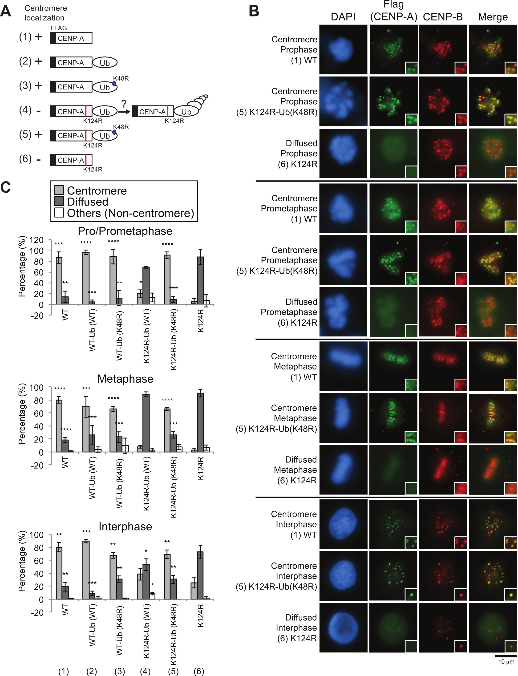
Figure 4. Immunofluorescence analysis of exogenous Flag-CENP-A proteins indicated that CENP-A monoubiquitin fusion rescued delocalization of the CENP-A K124R mutant from centromeres (Figures were adapted from Niikura et al.17). (A) Schematic cartoons of each construct used in this study (see also Table 2). The K124R mutation site on CENP-A (red) and the K48R mutation site on monoubiquitin (blue) are shown. (1) WT: Flag-CENP-A WT, (2) WT-Ub (WT): Flag-CENP-A WT-Ub (WT), (3) WT-Ub (K48R): Flag-CENP-A WT-Ub (K48R), (4) K124R-Ub (WT): Flag-CENP-A K124R-Ub (WT), (5) K124R-Ub (K48R): Flag-CENP-A K124R-Ub (K48R), (6) K124R: Flag-CENP-A K124R. (B) Exogenous CENP-A monoubiquitin fusion rescued delocalization of the CENP-A K124R mutant from centromeres. DAPI (blue), Flag (green), and endogenous CENP-B (red; a centromere location control) are shown. Scale bar represents 10 µm. (C) Histograms of the localization patterns shown in (C). More than 50 pro/prometaphase and metaphase cells and more than 200 interphase cells per experiment were counted (n ≥ 3 experiments). The mean percentages (±SD) are shown. ****P < 0.0001 and ***P < 0.001 vs non-fused Flag-CENP-A K124R (column [6]) (Student's t-test). Please click here to view a larger version of this figure.
| siRNA target | Indication in present study | Target type | siRNA database number | Forward sequence(s) | Source / Reference | |
| Luciferase (GL3) | - | 1 target | RKK9 | CUUACGCUGAGUACUUCGAdTdT | Elbashir et al., (2001) | |
| CENP-A | CA-UTR | siRNA pool (2 targets mixture) | RKK375 / 5' UTR t1 | CGAGCGGCGCGGACUUCUGCCdTdT | Niikura et al., (2015) | |
| RKK383 / 3' UTR t1 | UCCUGCACCCAGUGUUUCUGUdGdT | Niikura et al., (2015) | ||||
| CUL4A | #1 | 1 target | CRKK178 / RKK309 / t1 | AGCGAUCGUAAUCAAUCCUGA | Niikura et al., (2015) | |
| #2 (3'UTR) | 1 target | CRKK181 / RKK331 / t4 | AUGCGGGUUUGAAAUAUGACA | Niikura et al., (2015) | ||
| RBX1/ROC1 | #1 | siRNA pool (4 targets mixture) | CRKK198 / RKK411 / t1 | GAAGCGCUUUGAAGUGAAA | Niikura et al., (2015) | |
| CRKK198 / RKK413 / t2 | GCAUAGAAUGUCAAGCUAA | Niikura et al., (2015) | ||||
| CRKK198 / RKK415 / t3 | GCAAGAAGCGCUUUGAAGU | Niikura et al., (2015) | ||||
| CRKK198 / RKK417 / t4 | CAACAGAGAGUGGGAAUUC | Niikura et al., (2015) | ||||
| #2 (3'UTR) | 1 target | CRKK206 / RKK425 / t8 | UUCCCUGCUGUUACCUAAUUA | Niikura et al., (2015) | ||
Table 1. siRNA sequences used in this study.
| B number | Relevant characteristic(s) | Source/Reference |
| B288 | pTRM4 | Niikura et al., (2006) |
| B2067 | pTRM4-human CENP-A-Flag | Niikura et al., (2015) |
| B2281 | pTRM4-human CENP-A K9A-Flag | Niikura et al., (2015) |
| B2387 | pTRM4-human CENP-A K77R-Flag | Niikura et al., (2015) |
| B2388 | pTRM4-human CENP-A K124R-Flag | Niikura et al., (2015) |
| B2512 | pTRM4-Flag-human CENP-A | Niikura et al., (2015) |
| B2579 | pTRM4-Flag-human CENP-A K124R | Niikura et al., (2015) |
| B2513 | pTRM4-Flag-human CENP-A-Ub (WT) | Niikura et al., (2015) |
| B2559 | pTRM4-Flag-human CENP-A K124R-Ub (WT) | Niikura et al., (2015) |
| B2515 | pTRM4-Flag-human CENP-A-Ub (K48R) | Niikura et al., (2015) |
| B2560 | pTRM4-Flag-human CENP-A K124R-Ub (K48R) | Niikura et al., (2015) |
| B2759 | pTRM4-human CUL4A-Flag | Niikura et al., (2015) |
| B2624 | pcDNA3-Flag-human RBX1 | Niikura et al., (2015) |
| B1491 | pcDNA3-Flag | Niikura et al., (2015) |
Table 2. Plasmid vectors used in this study.
| R (anti-CENP-B) sample | b (anti-CENP-B) | R sample (subtracted) | Ratio (S sample : R sample) | Corrected Ratio (S sample : R sample) |
| 520 | 514 | 6 | -4.167 | 0.000 |
| 535 | 445 | 90 | -1.033 | 0.000 |
| 515 | 431 | 84 | 0.274 | 0.274 |
| 562 | 483 | 79 | 0.506 | 0.506 |
| 902 | 562 | 340 | 0.509 | 0.509 |
| 1203 | 703 | 500 | -0.560 | 0.000 |
| 1014 | 589 | 425 | -0.819 | 0.000 |
| 768 | 510 | 258 | -1.345 | 0.000 |
| 555 | 458 | 97 | -1.845 | 0.000 |
| 781 | 576 | 205 | -1.146 | 0.000 |
| 556 | 436 | 120 | 0.758 | 0.758 |
| 534 | 493 | 41 | -1.634 | 0.000 |
| 702 | 543 | 159 | 0.667 | 0.667 |
| 482 | 429 | 53 | -1.000 | 0.000 |
| 654 | 531 | 123 | 0.740 | 0.740 |
| 1190 | 607 | 583 | 0.384 | 0.384 |
| 552 | 454 | 98 | 0.969 | 0.969 |
| 489 | 413 | 76 | 0.539 | 0.539 |
| 511 | 452 | 59 | -3.186 | 0.000 |
| 485 | 475 | 10 | -2.700 | 0.000 |
| 481 | 441 | 40 | -1.475 | 0.000 |
| Sum | 5.347 | |||
| Average | 0.255 | |||
| R (anti-CENP-B) ctrl | b (anti-CENP-B) | R ctrl (subtracted) | Ratio (S ctrl : R ctrl) | Corrected Ratio (S ctrl : R ctrl) |
| 543 | 483 | 60 | 3.017 | 3.017 |
| 526 | 466 | 60 | 2.667 | 2.667 |
| 532 | 460 | 72 | 1.792 | 1.792 |
| 507 | 457 | 50 | 3.520 | 3.520 |
| 491 | 458 | 33 | 4.273 | 4.273 |
| 545 | 464 | 81 | 2.160 | 2.160 |
| 518 | 484 | 34 | 3.559 | 3.559 |
| 461 | 404 | 57 | 2.404 | 2.404 |
| 487 | 447 | 40 | 3.550 | 3.550 |
| 534 | 477 | 57 | 3.965 | 3.965 |
| 528 | 456 | 72 | 3.097 | 3.097 |
| 707 | 601 | 106 | 1.868 | 1.868 |
| 615 | 528 | 87 | 2.828 | 2.828 |
| 660 | 481 | 179 | 1.620 | 1.620 |
| 565 | 476 | 89 | 1.607 | 1.607 |
| 527 | 455 | 72 | 3.583 | 3.583 |
| 560 | 474 | 86 | 2.930 | 2.930 |
| 510 | 451 | 59 | 4.017 | 4.017 |
| 729 | 586 | 143 | 2.678 | 2.678 |
| 634 | 576 | 58 | 3.655 | 3.655 |
| 507 | 476 | 31 | 3.935 | 3.935 |
| Sum | 62.725 | |||
| Average | 2.987 | |||
| Remaining CENP-A signals at centromere (%) | 8.5 |
Table 3. One example of calculation of remaining CENP-A signals at the centromere (%). An example of Pro/Prometaphase in Figure 2F is shown: sample is RBX1 #2 (3'UTR) + Vec (center column in Figure 2F) and control is Luc + Vec (left column in Figure 2F). As a result, remaining CENP-A signals at centromere (%) = (0.255 / 2.987) x 100 = 8.5% is obtained (Or [5.347 / 62.725] x 100 = 8.5% with the same total cell number [i.e., 21 cells] analyzed between two samples). Note that if b value is more than S value (i.e., if subtracted value is negative), corrected ratio value is set to 0.00.
