Here, we present a protocol for the ex vivo qualitative detection of antigen-specific CD8+ T cells. Analysis is possible with single cell suspensions from organs or from small amounts of blood. A broad range of studies require the analysis of cytotoxic T cell responses (vaccination and cancer immunotherapy studies).
Method Article
Simultaneous Quantification of Anti-vector and Anti-transgene-Specific CD8+ T Cells Via MHC I Tetramer Staining After Vaccination with a Viral Vector
In This Article
Summary
Abstract
Upon viral infection, antigen-specific CD8+ cytotoxic T cells (CTLs) arise and contribute to the elimination of infected cells to prevent the spread of pathogens. Therefore, the frequency of antigen-specific CTLs is indicative of the strength of the T cell response against a specific antigen. Such analysis is important in basic immunology, vaccine development, cancer immunobiology and the adaptive immunology. In the vaccine field, the CTL response directed against components of a viral vector co-determines how effective the generation of antigen-specific cells against the antigen of interest (i.e., transgene) is. Antigen-specific CTLs can either be detected by stimulation with specific peptides followed by intracellular cytokine staining or by the direct staining of antigen-specific T cell receptors (TCRs) and analysis by flow cytometry. The first method is rather time-consuming since it requires sacrificing of animals to isolate cells from organs. Also, it requires isolation of blood from small animals which is difficult to perform. The latter method is rather fast, can be easily done with small amounts of blood and is not dependent on specific effector functions, such as cytolytic activity. MHC tetramers are an ideal tool to detect antigen-specific TCRs.
Here, we describe a protocol to simultaneously detect antigen-specific CTLs for the immunodominant peptides of the viral vector VSV-GP (LCMV-GP, VSV-NP) and transgenes (OVA, HPV 16 E7, eGFP) by MHC I tetramer staining and flow cytometry. Staining is possible either directly from blood or from single cell suspensions of organs, such as spleen. Blood or single cell suspensions of organs are incubated with tetramers. After staining with antibodies against CD3 and CD8, antigen-specific CTLs are quantified by flow cytometry. Optionally, antibodies against CD43, CD44, CD62L or others can be included to determine the activation status of antigen-specific CD8+T cells and to discriminate between naïve and effector cells.
Introduction
The aim of this method is to assess the frequency of cytotoxic T lymphocyte (CTL) responses to (multiple) antigens in the mouse by flow cytometric analysis without the need for time-consuming peptide stimulation. This method analyses the phenotype and antigen specificity of CTL subsets, in a single staining. We optimized the Major Histocompatibility Complex I (MHC I) tetramer staining protocol to analyze efficacy of new vaccine approaches, such as VSV-GP, a new variant of the vesicular stomatitis virus (VSV), where the glycoprotein G of VSV has been replaced by the glycoprotein GP of the lymphocytic choriomeningitis virus (LCMV)1,2. In addition to the humoral response, an important read-out of the effectivity of a vaccine is the induction of a CTL response against one or several antigens. As the consistency and durability of the cellular response are important in this context, it is favorable to monitor kinetics of CTL responses from the same animal. This will also lead to a reduction of animal numbers, an important aspect regarding the principles of the "3Rs"3. Hence, analysis from as little as 20 µL of blood is optimal for this purpose.
Tetramers were developed in the late 90s4 and became an important tool in the field of T cell immunology. Tetramers are fluorescently-labeled complexes of four MHC I/peptide molecules, which bind to TCRs, specific for a single peptide. Nowadays, they can be either bought ready-made5, custom-ordered free of charge at the NIH Tetramer Core Facility at Emory University6 or produced in the lab7. MHC I and II tetramers are available, i.e. for CD8+ and CD4+ T cells, respectively. The potency of tetramer staining lies in time-savings, rather simple and easy to standardize8 protocols, and sensitivity9. Also, if working with blood, animals do not need to be sacrificed and minimal amounts of sample are required. One measurement is not limited to a single antigen, but several antigens can be analyzed in one staining when combining tetramers conjugated with different fluorophores. Newly discovered antigens, e.g. from peptide screens, can easily be incorporated in tetramers and used for quantification of the T cell subset.
Tetramer staining will not give information about CTL functionality (i.e., cytokine production, effector functions), but only specificity. To gain information about T cell functionality, intracellular cytokine staining (ICS) or Enzyme Linked Immuno Spot (ELISpot) Assay needs to be performed8,10. Tetramer staining and ICS/ELISpot, however, are not redundant but rather complement each other. In vitro stimulation to induce cytokine production for ICS/ELISpot will alter the original T cell phenotype. Tetramer staining, in contrast, leaves the T cell untouched; the original phenotype is preserved and can be analyzed. Also, another big advantage of tetramers is that staining can be combined with magnetic sorting and enrichment of antigen-specific cells11. This allows for the analysis of rare populations, as well as culturing of sorted cells with defined antigen-specificities — a feature that is not be possible with other methods.
Using the protocol described here, tetramer staining, as well as ICS/ELISpot can be performed from one organ, because only very little material (blood: 20 µL; spleen: 1 x 10⁶ cells) is required for tetramer staining. However, depending on the frequency of the antigen-specific cells of interest, the strength of the respective TCR and the experimental context, the amount of cells required might need to be scaled up or magnetic enrichment might need to be applied.
Tetramers are widely used, for example to assess effectivity of (antitumor) vaccines12,13,14,15 or immunotherapy16,17, phenotypic analysis and spatial localization of antigen-specific T cell subsets18,19,20,21,22,23. The method described here is suited for studies, which aim to include the quantification and phenotypic analysis of murine antigen-specific CD8+ T cells in their analysis in a fast and convenient way.
Protocol
All methods described were performed in compliance with the Austrian National Animal Experimentation Law (“Tierversuchsgesetz”), and animal trial permission was granted by Austrian National Authorities.
1. Buffer Preparation and Sample Collection
NOTE: The mouse strain used depends on the epitope analyzed. Choose an appropriate tetramer that binds to an MHC type expressed in the mice, e.g., H-2Kb for C57BL/6 mice. The gender and age of the animals will depend on the scientific question. For most of the experiments described here, use female mice at 6–8 weeks of age at the start of the experiment, i.e., first immunization.
- Prepare FACS Buffer (Phosphate-buffered saline (PBS) + 1% fetal calf serum (FCS) + 0.1% sodium azide + 2 mM Ethylenediaminetetraacetic acid (EDTA) and FACS fixing buffer (1.5% (v/v) formaldehyde in PBS).
NOTE: It is recommended that both buffers are prepared in advance. Stored at 4 °C until use. - Blood: Collect 20 µL of blood per mouse from the tail vein of the mouse in EDTA-coated tubes, as described previously24.
NOTE: Blood may also be collected by other routes, e.g., vena facialis or retro-orbital sinus. However, the method of blood collection has to be in compliance with the national animal experimentation law and animal trial applications. Collection of blood from the tail vein is ideal for studies where repeatedly small amounts of blood are needed. Additional material is required for compensation controls and the non-stained control. - Spleen: Isolate the organ and, with the help of the plunger of a syringe, press through a 70 nm and 40 µm cell strainer. Perform lysis of erythrocytes, as described in step 6 and count. Adjust the concentration to 1 x 10⁷ cells/mL in PBS. Per sample, 1 x 10⁶ cells are required.
NOTE: Always include some mock immunized or control vector immunized animals as negative control. For ovalbumin (OVA)-tetramer, a sample from OT-1 mice might be used as positive control. Do not forget unstained and compensation controls in the calculation. If necessary, samples from different animals in the experiment might be pooled for this. - After sample collection, directly proceed with the staining.
2. Staining Set-up
- Prepare one FACS tube for each sample. Label tubes properly and transfer 100 μL of organ suspension (1 x 10⁶ cells) or 20 μL of blood into each tube.
- Spleen: Centrifuge for 5 min at 600 x g at 4–8 °C and discard the supernatant. Vortex to resuspend the cell pellet.
NOTE: This will result in a remaining volume of around 20 µL, similar as the volume for the blood samples. - For each channel to be used also prepare one FACS tube for a compensation sample. Prepare one additional sample as an unstained control.
3. Tetramer Staining
- For each sample, use 50 μL of tetramer dilution. For suggested tetramers and optimized dilutions, refer to Table 1.
NOTE: When working with tetramers or antibodies, turn off the light of the safety cabinet and protect samples from light.- Prepare a tube with FACS buffer (volume = 50 μL × number of samples plus additional 10% of the total volume to compensate for pipetting errors).
- Add tetramer(s) at optimal dilution, as listed in Table 1. Vortex the solution.
NOTE: When using the whole antibody panel (CD3, CD8, CD43, CD44, CD62L) with listed fluorophores, two tetramers (in PE and APC) can be included. Both tetramers can be combined in a single staining, i.e., staining of cells with both tetramers can be performed simultaneously.
| Tetramer | Peptide sequence | Allele | Fluorophore | Dilution |
| MHC I/OVA | SIINFEKL | H-2Kb | APC | 1:25 |
| MHC I/VSV-NP | RGYVYQGL | H-2Kb | PE | 1:25 |
| MHC I/EGFP | HYLSTQSAL | H-2Kd | PE | 1:25 |
| MHC I/LCMV-GP | KAVYNFATC | H-2Db | APC | 1:25 |
| MHC I/HPV 16 E7 | RAHYNIVTF | H-2Db | APC | 1:10 |
Table 1: Suggested tetramers and optimal dilutions. Recommended tetramers for some immunodominant peptides of model antigens (Ovalbumin (OVA) and enhanced green fluorescent protein (eGFP)) or pathogen components (Vesicular stomatitis virus (VSV) nucleoprotein (NP), Lymphocytic Choriomeningitis Virus (LCMV) Glycoprotein (GP) and human papillomavirus (HPV) E7 oncoprotein (E7)). For each, the peptide sequence and corresponding allele, as well as recommended fluorophore and optimized dilution is listed.
- Add 50 μL of tetramer dilution to each sample and vortex gently. Add FACS buffer only (without tetramer) to compensation controls and to the unstained sample.
- Incubate samples for 20 min at 37 °C, protected from light. To ensure a seamless transition from tetramer to antibody staining, prepare the antibody mix as described in step 4 during the incubation time.
NOTE: For each individual tetramer, optimal conditions (dilution, incubation time and temperature) need to be adjusted.
4. Preparation of Antibodies
- For each sample, prepare 50 µL of antibody mix.
- Prepare a tube with FACS buffer (volume = 50 μL × number of samples plus additional 10% of the total volume to compensate pipetting errors).
- Add antibodies in the dilutions as listed in Table 2.
NOTE: Depending on the scientific question, other marker combinations apart from the one described here might be used. Make sure to always include antibodies against CD3 and CD8 in the panel. - Vortex the solution.
| Antibody | Fluorophore | Dilution | µg/sample | Marker |
| CD3 | PE-Cy7 | 1:200 | 0.05 | CTLs (CD3+CD8+) |
| CD8 | Pacific Blue | 1:750 | 0.013 | |
| V450 | 1:100 | 0.1 | ||
| CD43 | FITC | 1:100 | 0.25 | Activation (CD43+) |
| CD44 | PE-Cy5 | 1:250 | 0.04 | Naive (CD44-CD62L+) & effector (CD44+CD62L-) |
| CD62L | APC-Cy7 | 1:500 | 0.02 |
Table 2: Antibodies used in this protocol and optimal dilutions. Recommended surface markers (CD3, CD8, CD43, CD44 and CD62L) are listed in the first column. For each, the recommended fluorophore, optimized dilution and amount of antibody/sample are listed. In the last column, the cell type identified with each marker is specified.
- Prepare antibodies for compensation controls. For each compensation control, use an antibody against CD8 in the respective color.
- For each channel, prepare a tube with 200 μL of FACS buffer and add 1 μL of a 1:200 dilution antibody against CD8 in the respective color.
- Vortex the tubes.
- Immediately proceed with staining.
5. Staining of Samples
- Wash samples once by adding ~1 mL of FACS buffer and centrifuge for 5 min at 600 x g at 4–8 °C. After centrifugation, discard the supernatant and drain off remaining liquid on a stack of paper towels.
NOTE: When working with blood, be cautious when draining off remaining liquid. Prior to the lysis of erythrocytes, blood will not stick to the bottom of the FACS tube. Alternatively, aspirate the supernatant. - Add 50 μL of the antibody mix to each cell pellet and vortex gently.
- Add 50 μL of each compensation mix to the cell pellet of the corresponding compensation control and vortex gently.
- Add 50 μL of FACS buffer to the cell pellet of the unstained control and vortex gently.
- Incubate all samples for 30 min at 4 °C, protected from light.
- When working with organs: skip step 6. Wash once by adding ~1–2 mL of FACS buffer and centrifuge for 5 min at 600 x g at 4–8 °C. After centrifugation, discard the supernatant and drain off remaining liquid on a stack of paper towels.
- When working with blood: proceed to step 6 (lysis of erythrocytes).
6. Lysis of Erythrocytes
- Add 500 μL of ACK (Ammonium-Chloride-Potassium) buffer25 to each sample and gently vortex.
NOTE: ACK buffer will lead to osmotic swelling and lysis, specifically of erythrocytes. - Incubate for 5 min at room temperature in the dark.
- Add 1 mL of FACS buffer and centrifuge for 5 min at 600 x g at 4–8 °C. After centrifugation, discard the supernatant and drain off the remaining liquid on a stack of paper towels.
Note: When the pellet is rather red, repeat the lysis of erythrocytes. - Wash once by adding ~1–2 mL FACS buffer and centrifuge for 5 min at 600 x g at 4–8 °C. After centrifugation, discard the supernatant and drain off remaining liquid on a stack of paper towels.
7. Flow Cytometric Measurement and Analysis
- Add 150–300 μL of FACS fixing buffer to each tube and mix by vortexing. For 20 µL of blood, 150 µL of buffer is sufficient.
NOTE: Before fixation, make sure that cells are well re-suspended in order to prevent formation of clumps. Proceed with flow cytometric measurement as quickly as possible. - Measure the compensation controls and correct any spectral overlaps.
- Set up sequential gates, as depicted in Figure 1 to select for CD3+/CD8+ cells.
- Gate on lymphocytes using forward and sideward scatter (Area) (non-logarithmic scale).
- Within the lymphocyte population, gate on single cells using forward scatter width vs area (non-logarithmic scale).
- Plot single cell lymphocytes using CD3 and CD8 channels (logarithmic scale). Identify CD8+ T cells by gating on CD3+/CD8+ cells.
- Plot CD8+ vs Tetramer+ cells and gate on CD8+ Tetramer+ cells, as depicted in Figure 2.
- If possible, record 20,000 cells (at least 5,000 cells from blood) in the CD3+/CD8+ gate for each sample and save as a FCS file.
NOTE: The amount of cells to record might need to be adjusted according to the frequency of the antigen-specific cells of interest. - Analyze FCS files with appropriate analysis software. Use the gating strategy, as described above (section 7.3) and quantify CD8+ Tetramer+ cells.
Results
Figure 1 demonstrates how to gate correctly on the target cells of this protocol, namely CD3+/CD8+ cells. It is to note that activated cells often downregulate the T cell receptor26,27 and, therefore, CD3low cells should also be included in the gating. After gating the CD3+/CD8+ cells, tetramer positive cells can be identified (Figure 2). Representative blots for a negative control (naïve) mouse, as well as animals either vaccinated with either OVA-secreting Adenovirus 5 (Adeno-OVA) or OVA-expressing VSV-GP (VSV-GP-OVA) are shown. As seen in the lower blots two different tetramers can be combined in the same tube for staining. This allows simultaneous quantification of two different CTL specificities: virus-specific (VSV N) and transgene-specific (OVA) CTLs. We confirmed that single and double tetramer stainings give similar percentages of positive cells for each tetramer. Using this protocol, other virus-specific (e.g., LCMV GP, HPV 16 E7) or transgene-specific (e.g., GFP) T cell populations can be analyzed (Supplemental Figure 1). In Supplemental Table 1, results for five animals after immunization with VSV-GP-OVA are shown — indicating robustness of tetramer staining.
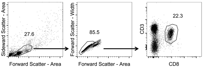
Figure 1: Representative gating strategy to analyze CD8+ T cells in blood. Schematic representation of the gating strategy used for flow cytometric analysis. After tetramer staining and flow cytometric measurement, data was analyzed. Lymphocytes were identified with forward and sideward scatter (Area) (non-logarithmic scale). From those, single cells were identified by applying forward scatter width vs area (non-logarithmic scale). CD8+ T cells were identified by gating on CD3+/CD8+ cells (logarithmic scale). Please click here to view a larger version of this figure.
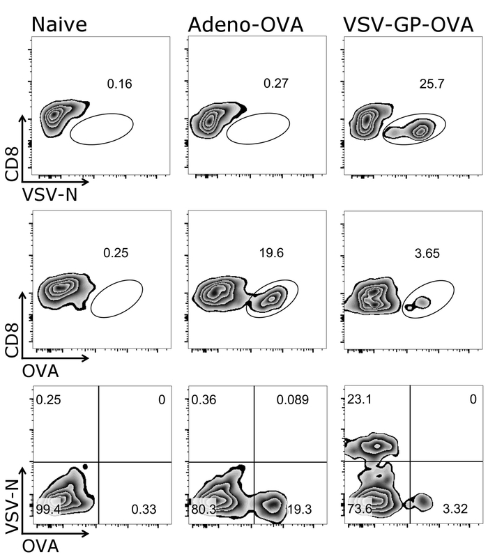
Figure 2: Representative gating strategy to quantify OVA- and N-specific CD8+ T cells in blood. Schematic representation of the gating strategy used for flow cytometric quantification of tetramer+ cells. CD3+/CD8+ cells were used for tetramer analysis. Upper and middle panel: the CD8 marker was plotted against respective tetramer (logarithmic scale). Lower panel: both tetramers were plotted against each other (logarithmic scale). Left: control (naïve) mouse; middle: mouse was immunized with OVA-secreting Adenovirus 5 (Adeno-OVA); right: mouse was immunized with ovalbumin (OVA)-expressing VSV-GP (VSV-GP-OVA). Blood was collected from tail vein at day 7 after immunization. Please click here to view a larger version of this figure.

Supplemental Figure 1: Representative result of CD3+/CD8+ tetramer+ cells after vaccination. Schematic representation of flow cytometric quantification of tetramer+ cells. CD3+/CD8+ cells were used for tetramer analysis and the CD8 marker was plotted against respective tetramer (logarithmic scale). Left: mice were naïve, right: mice were immunized with VSV-GP (upper panel), enhanced Green Fluorescent Protein (eGFP)-expressing VSV-GP (middle panel) or VSV-GP expressing human papillomavirus (HPV) E7 oncoprotein (E7) (lower panel). Blood was collected from tail vein at day 7 after immunization and stained with tetramers (LCMV-GP, eGFP and HPV E7). Please click here to view a larger version of this figure.
One big advantage of the protocol described here is that T cell responses from the same mouse can be followed over time as only small amounts of blood are needed for each measurement. Figure 3 shows exemplary results for T cell responses over time. In addition to quantities of antigen-specific CTLs, their phenotype can also be analyzed using this protocol (Figure 4).
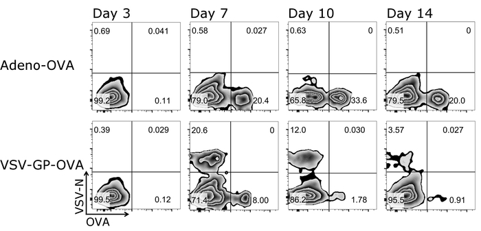
Figure 3: Representative result of CD8+ T cell kinetic in blood after vaccination. Schematic representation of the gating strategy used for flow cytometric quantification of tetramer+ cells. CD3+/CD8+ cells were used for tetramer analysis and both tetramers were plotted against each other (logarithmic scale). Upper panels: mouse was immunized with OVA-secreting Adenovirus 5 (Adeno-OVA); lower panels: mouse was immunized with ovalbumin (OVA)-expressing VSV-GP (VSV-GP-OVA). Blood was collected from tail vein at day 3, 7, 10 and 14 after immunization. Please click here to view a larger version of this figure.

Figure 4: Representative result of CD8+ T cell activation and differentiation into naïve and effector cells after vaccination. Schematic representation of the gating strategy used for flow cytometric quantification of activated (CD43+), naïve (CD44-/CD62L+) and effector (CD44+/CD62L-) CD3+/CD8+ cells (logarithmic scale). Left: control (naïve) mouse; middle: mouse was immunized with OVA-secreting Adenovirus 5 (Adeno-OVA); right: mouse was immunized with ovalbumin (OVA)-expressing VSV-GP (VSV-GP-OVA). Blood was collected from tail vein at day 7 after immunization. Please click here to view a larger version of this figure.
| Mouse # | % CD43+ | % VSV-N Tetramer+ | % OVA Tetramer+ | |
| mock | 1 | 1.7 | 0.45 | 0.41 |
| 2 | 0.77 | 0.69 | 0.15 | |
| 3 | 1.34 | 0.35 | 0.31 | |
| 4 | 1.68 | 0.83 | 0.26 | |
| 5 | 0.84 | 0.47 | 0.23 | |
| VSV-GP-OVA | 1 | 23 | 13.2 | 3.69 |
| 2 | 32.6 | 15.9 | 3.54 | |
| 3 | 22.1 | 11.7 | 2.43 | |
| 4 | 15.1 | 8.89 | 1.79 | |
| 5 | 29.1 | 14.7 | 5.64 |
Supplemental Table 1: Percentages of activated and antigen-specific CD3+/CD8+ cells after vaccination. Mice were either naïve or immunized with ovalbumin (OVA)-expressing VSV-GP (VSV-GP-OVA) (n = 5). Blood was collected from tail vein at day 7 after immunization and stained with tetramers (VSV-N and OVA). Activated (CD43+) and antigen-specific (tetramer+) CD3+/CD8+ cells were quantified by flow cytometry.
Discussion
Tetramer staining is a rather straightforward and uncomplicated protocol to analyze phenotype and peptide specificity of a T lymphocyte. The usage of blood for analysis, as described here, is minimally invasive and allows continuous monitoring, for example in vaccination studies. In the field of vaccination, the quantification of vector- and antigen-specific responses is of interest, as vector-specific responses might hinder an effective immune response against the vaccine antigen28. Of note is that with the protocol described here, both populations can be quantified simultaneously in a single tetramer staining, thereby reducing staining variability and sample amounts. However, a few steps need to be done carefully to ensure proper measurement and reliable data. If using blood from the tail vein for analysis, one should make sure to pre-warm the animals to induce vasodilation24. Thereby, sufficient blood can be collected in a short time, stress on the animals is reduced and the analysis is much better, as compared to if the blood is collected slowly. Also, after sample collection (either blood or organs), direct staining is recommended to avoid false negative results due to TCR downregulation. The same applies for all subsequent steps: the procedure should not be interrupted and all washing steps reduced to the minimal number (as stated in the protocol). To ensure proper staining, care should be taken to vortex all solutions and samples prior to and after incubation. This is especially important before fixing the samples to avoid clumping of cells.
In terms of modifying the protocol, other surface markers and tetramers can be used, depending on the aim of the analysis. However, all reagents then need to be titrated, optimally in combination with the whole staining panel. For some of the tetramers specified here, optimization revealed that we can increase the dilution recommended by the manufacturer (1:10 recommended, optimized 1:25) (Table 1). To compensate spectral overlap, compensation beads can be used instead of stained cells. Regarding the choice of the tetramer-coupled fluorochrome, one should envisage to use bright fluorochromes, as this facilitates detection - especially when the signal is low. As Dolton and colleagues29, we prefer to use PE- or APC-coupled tetramers, which can be perfectly combined in a single staining and CD8+ T cells with a single antigen specificity can be nicely detected (Figure 2). Regarding temperature and incubation times, a variety of tetramer staining conditions exist. In our optimization process, we addressed this issue and performed tetramer staining at different conditions (e.g. 4 °C, room temperature or 37 °C). From the results obtained, we recommend to stain for 20 min at 37 °C, which is in concordance with literature30,31. Prolonged incubation should be avoided, as this can lead to internalization of the tetramer30 and false negative results.
The choice of the right antibody for detection of CD8+ cells is another important issue, which has to carefully considered (and potentially adapted). This arises from the fact that certain anti-CD8 antibody clones block binding of tetramers to the TCR, in human32 as well as mouse33 samples. For our tetramer staining protocol, we selected clone 53–6.7 to stain for murine CD8+ cells — a clone which does not block, but rather enhances tetramer staining.
Tetramer staining is rather uncomplicated when analyzing prominent immune responses at the peak of the T cell response, for example. However, there might be populations which are a bit more 'problematic'. Such examples include cells specific for low affinity antigens (tumor, self), recently activated cells which subsequently down-regulated their receptors or rare cell subsets (e.g. naïve precursor or memory cell populations). In these cases, the classical tetramer staining protocol might need to be improved or combined with other methods. For example, the protein kinase inhibitor (PKI) dasatinib inhibits TCR internalization and might be included prior to tetramer staining. Tetramers can also be stabilized by including anti-fluorochrome unconjugated primary Abs after tetramer staining. Additionally, fluorescence intensity can be increased by addition of a second anti-Ab fluorochrome-conjugated Ab29,34,35,36. We optimized the conditions selectively for the tetramers specified in this protocol and did not include PKI or additional Abs. However, for any other tetramer, the optimal conditions have to be adjusted individually. With regard to rare populations, tetramer staining might need to be combined with magnetic enrichment11.
To facilitate and validate FACS analysis of tetramer staining, negative and positive controls should be included. As a negative control, we always stain cells of a naïve mouse of the same strain with our tetramer of interest. Alternatively, samples can be stained with tetramers with irrelevant peptides, but with the same fluorochrome as the tetramer of interest. Such controls are essential to exclude false positive signals, e.g., originating from dying cells. In addition to this, it is recommended to include a live/dead stain, such as propidium iodide (PI). This is of special importance if cells are not stained directly after isolation. Another strategy to remove autofluorescence background might be to include several non-T cell markers in one channel. By excluding cells positive in this channel, non-T cell populations can be excluded. As a positive control, a sample from an OT-1 mouse can be used for OVA tetramer, for example. For other tetramers, this has to be chosen individually (e.g., sample from a mouse, which was immunized several times). Alike others37, we also observe a down-regulation of the CD8 receptor during CTL activation at day 7 of the effector T cell response. Therefore, to avoid loss of activated tetramer+ effector T cells, we recommend to include the CD8low cells in the analysis (at least if measuring in the effector phase).
The quality and amount of information one can retrieve from this protocol is dependent on the knowledge about the antigen to be studied, the availability and specificity of tetramer and the quality of the FACS machine (number of lasers and available detectors). If working with animal samples, variation in the immune response is natural and inevitable. Therefore, to gain meaningful results from tetramer staining, at least 3–5 animals should be analyzed. If done so, the protocol described here will give reliable and reproducible results (exemplary result from one experiment can be found in Supplemental Table 1). As mentioned before, this method is perfectly suited to quantify the phenotype and antigen-specificity of CD8+ T cells (Figure 3, 4; Supplemental Figure 1), not only in the mouse but also in humans. However, to analyze CD8+ T cell effector functions, such as granzyme-induced cell death, ICS and/or ELISpot need to be performed. However, one should keep in mind that T cell functions, as measured by in vitro stimulation might not represent the actual situation in vivo. In vivo, a suppressive environment might prevent T cell functions which are measured in vitro.
On its own, tetramer staining does not provide all information, but it evolved to become an essential method to characterize T cell responses and quantify T cell subsets in vitro in a very sensitive manner38. Tetramers cannot only be used to quantify certain subsets, but also to isolate those39, localize them by in situ hybridization19,20 and study low-affinity antigens, such as tumor-associated40,41. Since the discovery of tetramer technology4, tetramer staining has become an essential tool in T-cell analysis and the range of applications.
Disclosures
Dorothee von Laer is an inventor of VSV-GP and holds minority shares in the biotech company ViraTherapeutics GmbH, which holds the intellectual property rights for VSV-GP. For the other authors, no competing financial interests exist.
Acknowledgements
This project was funded by the FWF Austrian Science Fund (project number P 25499-B13) and the European Union's Horizon 2020 research and innovation program under grant agreement No. 681032. The following reagent was obtained through the NIH Tetramer Core Facility: Class I MHC Tetramer for vesicular stomatitis virus nucleoprotein (RGYVYQGL).
Materials
| Name | Company | Catalog Number | Comments |
|---|---|---|---|
| Safety cabinet class 2 | VWR | LBCP302411030 | |
| Flow cytometer (e.g. FACSCanto II) | BD | 338962 | |
| Analysis platform for flow cytometry analysis (e.g. FlowJo) | Fisher Scientific Co. L.L.C. | NC0887833 | |
| Binocular microscopes, VisiScope 100 | VWR | 630-1553 | |
| Vortex mixer | Phoenix Instrument | RS-VA 10 | |
| Centrifuge suitable for FACS tubes (e.g. Rotanta 460R) | Hettich | 5660 | |
| Sterile Scalpel Blades Nr. 10 | Braun | BB510 | |
| Cell strainer 40 µm | Sigma | CLS431750 | |
| Cell strainer 70 µm | Sigma | CLS431751 | |
| Neubauer counting chamber | VWR | 630-1506 | |
| Pipettes (20 μL, 200 µL and 1000 μL) | Eppendorf | 4924000037, 4924000061, 4924000088 | |
| Pipette tips, sterile (20 µL, 200 µL, 1,000 µL) | Biozym | 770050, 770200, 770400 | |
| Pipet Boy | Integra | 155 000 | |
| Sterile pipettes (5 mL, 10 mL, 25 mL) | Sarstedt | 86.1253.001, 86.1254.001, 86.1685.001 | |
| Multistep Pipette, HandyStep S | BRAND | 705110 | |
| 12.5 mL Combitips for Multistep Pipette | BrandTech Scientific | 702378 | |
| Microvette CB 300 K2E | Sarstedt | 16.444 | |
| Sterile reaction tubes (1.5 mL, 50 mL) | Sarstedt | 72.692.005, 62.547.254 | |
| FACS tubes (non-sterile) | Szabo Scandic | BDL352008 | |
| PBS | Lonza | LONBE17-516F | |
| Heat-inactivated FCS | ThermoFisher Scientific | 10500064 | |
| Formaldehyde | Roth | 4979.1 | |
| Sodium azide | Roth | K305.1 | |
| PE-Cy7 Rat Anti-Mouse CD3 Molecular Complex | BD | 560591 | Clone 17A2; Lot # 7235504 |
| Pacific Blu Rat Anti-Mouse CD8a | BD | 558106 | Clone 53-6.7; Lot # 5058904 |
| V450 Rat anti-Mouse CD8a | BD | 560469 | Clone 53-6.7; Lot # 5205945 |
| FITC anti-mouse CD43 | BioLegend | 121206 | Clone 1B11; Lot # B233778 |
| PE-Cy5 Rat Anti-Mouse CD44 | BD | 553135 | Clone IM7; Lot # 85660 |
| APC-Cy7 Rat Anti-Mouse CD62L | BD | 560514 | Clone MEL-14; Lot # 7215801 |
| OVA-tetramer/APC | MBL | TB-5001-2 | SIINFEKL, H-2Kb; Lot # T1702008 |
| VSV NP-tetramer/PE | MBL | TS-M529-1 | RGYVYQGL, H-2Kb; Lot # 007 |
| EGFP-tetramer/PE | MBL | TS-M525-1 | HYLSTQSAL, H-2Kd; Lot # 004 |
| LCMV-GP-tetramer/APC | MBL | TB-5002-2 | KAVYNFATC, H-2Db; Lot # T1412006 |
| HPV 16 E7-tetramer/APC | MBL | TB-5008-2 | RAHYNIVTF, H-2Db; Lot # T1804003 |
References
- Tober, R., et al. VSV-GP: a potent viral vaccine vector that boosts the immune response upon repeated applications. Journal of virology. 88 (9), 4897-4907 (2014).
- Muik, A., et al. Re-engineering vesicular stomatitis virus to abrogate neurotoxicity, circumvent humoral immunity, and enhance oncolytic potency. Cancer Reseaerch. 74 (13), 3567-3578 (2014).
- Eales, L. J., Farrant, J., Helbert, M., Pinching, A. J. Peripheral blood dendritic cells in persons with AIDS and AIDS related complex: loss of high intensity class II antigen expression and function. Clinical and Experimental Immunology. 71, 423-427 (1988).
- Altman, J. D., et al. Phenotypic analysis of antigen-specific T lymphocytes. Science. 274 (5284), 94-96 (1996).
- Tetramers and Monomers. , Available from: https://www.mblintl.com/products/research/monomer-tetramers/filter/product_type/monomer,peptide,tetramer (2016).
- NIH Tetramer Core Facility. , Available from: http://tetramer.yerkes.emory.edu/ (2010).
- Class I MHC Tetramer Preparation: Overview. , Available from: http://tetramer.yerkes.emory.edu/support/protocols (2010).
- Wolfl, M., et al. Quantitation of MHC tetramer-positive cells from whole blood: evaluation of a single-platform, six-parameter flow cytometric method. Cytometry A. 57 (2), 120-130 (2004).
- Burrows, S. R., et al. Peptide-MHC class I tetrameric complexes display exquisite ligand specificity. The Journal of Immunology. 165 (11), 6229-6234 (2000).
- Sims, S., Willberg, C., Klenerman, P. MHC-peptide tetramers for the analysis of antigen-specific T cells. Expert Review of Vaccines. 9 (7), 765-774 (2010).
- Legoux, F. P., Moon, J. J. Peptide:MHC tetramer-based enrichment of epitope-specific T cells. The Journal of Visualized Experiments. (68), (2012).
- Xie, Y., et al. A novel T cell-based vaccine capable of stimulating long-term functional CTL memory against B16 melanoma via CD40L signaling. Cellular & Molecular Immunology. 10 (1), 72-77 (2013).
- Nanjundappa, R. H., Wang, R., Xie, Y., Umeshappa, C. S., Xiang, J. Novel CD8+ T cell-based vaccine stimulates Gp120-specific CTL responses leading to therapeutic and long-term immunity in transgenic HLA-A2 mice. Vaccine. 30 (24), 3519-3525 (2012).
- Bowers, E. V., Horvath, J. J., Bond, J. E., Cianciolo, G. J., Pizzo, S. V. Antigen delivery by alpha(2)-macroglobulin enhances the cytotoxic T lymphocyte response. Journal of Leukocyte Biology. 86 (2), 1259-1268 (2009).
- Guo, H., Baker, S. F., Martinez-Sobrido, L., Topham, D. J. Induction of CD8 T cell heterologous protection by a single dose of single-cycle infectious influenza virus. The Journal of Immunology. 88 (20), 12006-12016 (2014).
- Sakai, K., et al. Dendritic cell-based immunotherapy targeting Wilms' tumor 1 in patients with recurrent malignant glioma. Journal of Neurosurgery. 123 (4), 989-997 (2015).
- Rosaely, C. G., et al. Immune responses to WT1 in patients with AML or MDS after chemotherapy and allogeneic stem cell transplantation. International Journal of Cancer. 138 (7), 1792-1801 (2016).
- Shane, H. L., Reagin, K. L., Klonowski, K. D. The Respiratory Environment Diverts the Development of Antiviral Memory CD8 T Cells. The Journal of Immunology. , (2018).
- Li, S., Mwakalundwa, G., Skinner, P. J. In Situ MHC-tetramer Staining and Quantitative Analysis to Determine the Location, Abundance, and Phenotype of Antigen-specific CD8 T Cells in Tissues. The Journal of Visualized Experiments. (127), e56130(2017).
- De Vries, I. J. M., et al. In situ detection of antigen-specific T cells in cryo-sections using MHC class I tetramers after dendritic cell vaccination of melanoma patients. Cancer Immunology Immunotherapy. 56 (10), 1667-1676 (2007).
- Tan, H. X., et al. Induction of vaginal-resident HIV-specific CD8 T cells with mucosal prime-boost immunization. Mucosal Immunology. , (2017).
- Huang, H., et al. CD8(+) T Cell Immune Response in Immunocompetent Mice during Zika Virus Infection. Journal of Virology. 91 (22), (2017).
- Hensel, M. T., et al. Selective Expression of CCR10 and CXCR3 by Circulating Human Herpes Simplex Virus-Specific CD8 T Cells. Journal of Virology. 91 (19), (2017).
- Diehl, K. H., et al. A good practice guide to the administration of substances and removal of blood, including routes and volumes. Journal of Applied Toxicology. 21 (1), 15-23 (2001).
- ACK Lysis Buffer. , Available from: http://www.cshprotocols.cshlp.org/content/2014/11/pdb.rec083295.short (2014).
- San Jose, E., Borroto, A., Niedergang, F., Alcover, A., Alarcon, B. Triggering the TCR complex causes the downregulation of nonengaged receptors by a signal transduction-dependent mechanism. Immunity. 12 (2), 161-170 (2000).
- Dietrich, J., Hou, X., Wegener, A. M., Geisler, C. CD3 gamma contains a phosphoserine-dependent di-leucine motif involved in down-regulation of the T cell receptor. EMBO Journal. 13 (9), 2156-2166 (1994).
- Schone, D., et al. Immunodominance of Adenovirus-Derived CD8(+) T Cell Epitopes Interferes with the Induction of Transgene-Specific Immunity in Adenovirus-Based Immunization. Journal of Virology. 91 (20), (2017).
- Dolton, G., et al. More tricks with tetramers: a practical guide to staining T cells with peptide–MHC multimers. Immunology. 146 (1), 11-22 (2015).
- Whelan, J. A., et al. Specificity of CTL Interactions with Peptide-MHC Class I Tetrameric Complexes Is Temperature Dependent. The Journal of Immunology. 163 (8), 4342-4348 (1999).
- Wooldridge, L., et al. Tricks with tetramers: how to get the most from multimeric peptide–MHC. Immunology. 126 (2), 147-164 (2009).
- Denkberg, G., Cohen, C. J., Reiter, Y. Critical role for CD8 in binding of MHC tetramers to TCR: CD8 antibodies block specific binding of human tumor-specific MHC-peptide tetramers to TCR. The Journal of Immunology. 167 (1), 270-276 (2001).
- Daniels, M. A., Jameson, S. C. Critical role for CD8 in T cell receptor binding and activation by peptide/major histocompatibility complex multimers. The Journal of Experimental Medicine. 191 (2), 335-346 (2000).
- Tungatt, K., et al. Antibody stabilization of peptide-MHC multimers reveals functional T cells bearing extremely low-affinity TCRs. The Journal of Immunology. 194 (1), 463-474 (2015).
- Lissina, A., et al. Protein kinase inhibitors substantially improve the physical detection of T-cells with peptide-MHC tetramers. Journal of Immunological Methods. 340 (1), 11-24 (2009).
- Rius, C., et al. Peptide-MHC Class I Tetramers Can Fail To Detect Relevant Functional T Cell Clonotypes and Underestimate Antigen-Reactive T Cell Populations. The Journal of Immunology. 200 (7), 2263-2279 (2018).
- Xiao, Z., Mescher, M. F., Jameson, S. C. Detuning CD8 T cells: down-regulation of CD8 expression, tetramer binding, and response during CTL activation. The Journal of Experimental Medicine. 204 (11), 2667-2677 (2007).
- McMichael, A. J., O'Callaghan, C. A. A New Look at T Cells. The Journal of Experimental Medicine. 187 (9), 1367-1371 (1998).
- Hunsucker, S. A., et al. Peptide/MHC tetramer-based sorting of CD8(+) T cells to a leukemia antigen yields clonotypes drawn nonspecifically from an underlying restricted repertoire. Cancer Immunology Research. 3 (3), 228-235 (2015).
- Pittet, M. J., et al. Ex vivo analysis of tumor antigen specific CD8+ T cell responses using MHC/peptide tetramers in cancer patients. International Immunopharmacology. 1 (7), 1235-1247 (2001).
- Cohen, C. J., et al. Isolation of neoantigen-specific T cells from tumor and peripheral lymphocytes. The Journal of Clinical Investigation. 125 (10), 3981-3991 (2015).
Reprints and Permissions
