

ERRATUM NOTICE
Important: There has been an erratum issued for this article. Read more …
Summary
These protocols will help users probe mitochondrial energy metabolism in 3D cancer cell-line-derived spheroids using Seahorse extracellular flux analysis.
Abstract
Three-dimensional (3D) cellular aggregates, termed spheroids, have become the forefront of in vitro cell culture in recent years. In contrast to culturing cells as two-dimensional, single-cell monolayers (2D culture), spheroid cell culture promotes, regulates, and supports physiological cellular architecture and characteristics that exist in vivo, including the expression of extracellular matrix proteins, cell signaling, gene expression, protein production, differentiation, and proliferation. The importance of 3D culture has been recognized in many research fields, including oncology, diabetes, stem cell biology, and tissue engineering. Over the last decade, improved methods have been developed to produce spheroids and assess their metabolic function and fate.
Extracellular flux (XF) analyzers have been used to explore mitochondrial function in 3D microtissues such as spheroids using either an XF24 islet capture plate or an XFe96 spheroid microplate. However, distinct protocols and the optimization of probing mitochondrial energy metabolism in spheroids using XF technology have not been described in detail. This paper provides detailed protocols for probing mitochondrial energy metabolism in single 3D spheroids using spheroid microplates with the XFe96 XF analyzer. Using different cancer cell lines, XF technology is demonstrated to be capable of distinguishing between cellular respiration in 3D spheroids of not only different sizes but also different volumes, cell numbers, DNA content and type.
The optimal mitochondrial effector compound concentrations of oligomycin, BAM15, rotenone, and antimycin A are used to probe specific parameters of mitochondrial energy metabolism in 3D spheroids. This paper also discusses methods to normalize data obtained from spheroids and addresses many considerations that should be considered when exploring spheroid metabolism using XF technology. This protocol will help drive research in advanced in vitro spheroid models.
Introduction
Advances in in vitro models in biological research have rapidly progressed over the last 20 years. Such models now include organ-on-a-chip modalities, organoids, and 3D microtissue spheroids, all of which have become a common focus to improve the translation between in vitro and in vivo studies. The use of advanced in vitro models, particularly spheroids, spans several research fields, including tissue engineering, stem cell research, cancer, and disease biology1,2,3,4,5,6,7, and safety testing, including genetic toxicology8,9,10, nanomaterials toxicology11,12,13,14, and drug safety and efficacy testing8,15,16,17,18,19.
Normal cell morphology is critical to biological phenotype and activity. Culturing cells into 3D microtissue spheroids allows cells to adopt a morphology, phenotypic function, and architecture, more akin to that observed in vivo but difficult to capture with classical monolayer cell culture techniques. Both in vivo and in vitro, cellular function is directly impacted by the cellular microenvironment, which is not limited to cellular communication and programming (e.g., cell-cell junction formations, opportunities to form cell niches); cell exposure to hormones and growth factors in the immediate environments (e.g., cellular cytokine exposure as part of an inflammatory response); composition of physical and chemical matrices (e.g., whether cells are grown in stiff tissue culture plastic or an elastic tissue environment); and most importantly, how cellular metabolism is impacted by nutrition and access to oxygen as well as the processing of metabolic waste products such as lactic acid.
Metabolic flux analysis is a powerful way to examine cellular metabolism within defined in vitro systems. Specifically, XF technology allows for the analysis of live, real-time changes in cellular bioenergetics of intact cells and tissues. Given that many intracellular metabolic events occur within the order of seconds to minutes, real-time functional approaches are paramount for understanding real-time changes in cellular metabolic flux in intact cells and tissues in vitro.
This paper provides protocols for cultivating cancer-derived cell lines A549 (lung adenocarcinoma), HepG2/C3A (hepatocellular carcinoma), MCF-7 (breast adenocarcinoma), and SK-OV-3 (ovarian adenocarcinoma) as in vitro 3D spheroid models using forced-aggregation approaches (Figure 1). It also (i) describes in detail how to probe mitochondrial energy metabolism of single 3D spheroids using the Agilent XFe96 XF analyzer, (ii) highlights ways to optimize XF assays using single 3D spheroids, and (iii) discusses important considerations and limitations of probing 3D spheroid metabolism using this approach. Most importantly, this paper describes how datasets are collected that allow the calculation of oxygen consumption rate (OCR) to determine oxidative phosphorylation and thus mitochondrial function in cellular spheroids. Though not analyzed for this protocol, extracellular acidification rate (ECAR) is another parameter that is measured alongside OCR data in XF experiments. However, ECAR is often poorly or incorrectly interpreted from XF datasets. We provide a commentary as to the limitations of calculating ECAR following basic approaches from the technology manufacturer.
Subscription Required. Please recommend JoVE to your librarian.
Protocol

Figure 1: Graphical workflow for the generation of cellular spheroids, extracellular flux analysis and downstream assays. Four cancer cell lines were selectively cultured as monolayers (A), detached from tissue culture flasks, and seeded into ultralow attachment 96-well microplates to form spheroids (B). A549 lung carcinoma, HepG2/C3A liver carcinoma, SK-OV-3 ovarian adenocarcinoma, and MCF-7 breast carcinoma cells were seeded at 1 × 10³-8 × 10³ cells/well and grown up to 7 days to form single spheroids and optimize spheroid seeding density and cultivation time by continuous observation and planimetric measurements. Once formed, single spheroids were washed into a serum-free XF medium and carefully seeded into spheroid assay microplates, precoated with poly-D-lysine (C). Spheroids were subjected to extracellular flux analysis using the XFe96 analyzer using several protocols to address: (1) optimal spheroid size for basal mitochondrial respiration response; (2) optimized titration of mitochondrial respiratory inhibitors; (3) optimization of spheroid placement within microplate wells. (D) Post XF analyses, phase contrast microscopy, and spheroid DNA quantification were used for data normalization and other downstream in vitro assays. Please click here to view a larger version of this figure.
1. Cultivation of cancer cell lines as 3D in vitro spheroids
| Cell line | Description | Culture medium | Source |
| A549 | Lung carcinoma cell line | RPMI 1640 | European Collection of Authenticated Cell Cultures (ECACC) |
| Sodium pyruvate (1 mM) | |||
| Penicillin- Streptomycin - (100 U/mL – 100 mg/mL) | |||
| 10 % (v/v) FBS | |||
| HepG2/C3A | Hepatic carcinoma cell line, a clonal derivative of the parent HepG2 cell line | DMEM | American Tissue Culture Collection (ATCC) |
| Penicillin- Streptomycin - (100 U/mL – 100 mg/mL) | |||
| 10 % (v/v) FBS | |||
| MCF7 | Breast adenocarcinoma cell line | RPMI 1640 | European Collection of Authenticated Cell Cultures (ECACC) |
| Sodium pyruvate (1 mM) | |||
| Penicillin- Streptomycin - (100 U/mL – 100 mg/mL) | |||
| 10 % (v/v) FBS | |||
| SK-OV-3 | Ovarian adenocarcinoma cell line | RPMI 1640 | European Collection of Authenticated Cell Cultures (ECACC) |
| Sodium pyruvate (1 mM) | |||
| Penicillin- Streptomycin - (100 U/mL – 100 mg/mL) | |||
| 10 % (v/v) FBS | |||
| Component | RPMI assay medium (50 mL final volume) | ||
| Base Medium | Agilent Seahorse XF RPMI, pH 7.4 | ||
| Glucose (1 M sterile stock) | 11 mM (0.55 mL stock solution) | ||
| L-glutamine (200 mM sterile stock) | 2 mM (0.5 mL of stock solution) | ||
| Sodium pyruvate (100 mM sterile stock) | 1 mM (0.5 mL of stock solution) |
Table 1: Cancer cell line media and XF media compositions.
- Culture all cell lines using standard aseptic tissue culture technique and confirm that they are free of mycoplasma using a suitable assay kit.
- Culture the cell lines in T75 tissue culture flasks or equivalent, using the recommended medium (Table 1). Culture the cell lines to 65-80% confluency and passage them regularly up to a maximum of 25 passages.
- Rinse the cell culture flasks twice in Dulbecco's modified phosphate-buffered saline (DBPS).
- Detach the cells from the flasks with 3 mL of the cell dissociation reagent (see the Table of Materials) for 5 min at 37 °C and confirm the detachment by microscopy.
- Aspirate the detached cell suspension gently to ensure a single-cell suspension and deactivate the cell dissociation reagent with 7 mL of complete tissue culture medium.
- Collect the cells by centrifugation at 300 × g for 5 min, discard the supernatant, and resuspend the cells in complete medium.
- Count the cells using a hemocytometer or an automated cell counter and titrate to the desired cell density required for seeding.
NOTE: To seed an entire 96-well plate at 100 µL/well at 4 × 10³ cells/well, cells should be titrated to 4 × 10⁴ cells/mL in a recommended volume of 12 mL. - Decant the cell suspension into a sterile reservoir and dispense 100 µL of the cell suspension into each well of a cell-repellent microplate using a multichannel pipettor.
NOTE: Only the inner 60 wells of a microplate should be seeded and the remainder filled with DPBS. This will form an evaporation barrier, ensure spheroid homogeneity across the plate, and minimize plate edge effects. - Centrifuge spheroid microplates at 300 × g for 15 min to force the cells into loose aggregates.
- Incubate the plates at 37 °C, 5% CO2 for a minimum of 3 days to ensure spheroid formation.
- Perform phase-contrast microscopy using standardized laboratory practices to monitor the growth of spheroids. Replenish the cell culture medium every 3 days or twice weekly by performing a half-volume medium exchange.
2. Probing mitochondrial energy metabolism of single spheroids using Extracellular Flux (XF) Technology
- Assay preparation (one day prior)
- Check spheroid viability using an inverted light microscope with phase contrast at 4x magnification to ensure intact spheroid structure, morphology, and overall uniformity between samples.
- Hydrate the sensor cartridge.
- Aliquot ~20 mL of the calibrant into a conical tube.
- Place the conical tube containing the calibrant in a non-CO2 37 °C incubator overnight.
- Remove the contents from the assay kit.
- Remove the sensor cartridge from the utility plate and place it upside down on the worktop next to the utility plate.
- Pipette 200 µL of sterile ddH2O into each well of the sensor cartridge utility plate using a multichannel P300 pipette.
- Place the sensor cartridge on top of the utility plate.
- Check that the water level in each well is high enough to submerge the sensor probes.
- Transfer the assembled sensor cartridge to a non-CO2 37 °C incubator and leave it overnight.
NOTE: This step can be performed 12-72 h prior to assay commencement.
- Coat spheroid assay microplate
- Using aseptic techniques, add 30 µL/well of sterile Poly-D-Lysine (0.1 mg/mL) solution to the spheroid microplate and incubate it for 30 min at room temperature.
- Aspirate the solution from each well of the spheroid microplate, invert the plate, and tap it firmly onto tissue paper to remove any residual solution.
- Wash the plate twice with 200 µL/well of sterile ddH2O.
- After the final wash, invert the microplate and tap it firmly onto tissue paper to remove any residual water.
- Allow the plate to air-dry for 30 min before using or storing it at 4 °C for future use.
NOTE: The spheroid assay microplate should be coated with a molecular adhesive to ensure that the spheroids are fixed at the bottom of the microplate. Without a molecular adhesive, spheroids can become dislodged and interfere with assay results. Other molecular adhesives can also be used as an alternative to Poly-D-Lysine for precoating plates. Precoated plates can be stored at 4 °C but should be left to equilibrate to room temperature before assay commencement.
- Prepare XF Assay medium
- Prepare XF RPMI medium, as detailed in Table 1, and sterile-filter with a 0.22 μm syringe filter
- Assay preparation (1 h prior to assay)
- Prewarm the supplemented XF RPMI assay medium to 37 °C.
- Prewarm the coated spheroid assay microplate in a non-CO2 37 °C incubator or dry bath.
- Prepare the sensor cartridge.
- Take out the conical tube containing the calibrant and the sensor cartridge from the air incubator.
- Remove the sensor cartridge from the utility plate and place it upside down on the work surface.
- Using a P300 multichannel pipette, aspirate the water from the utility plate and discard it.
- Pour the calibrant solution into a sterile reagent reservoir and add 200 µL/well of the prewarmed calibrant to the utility plate using a P300 multichannel pipette.
- Pick up the sensor cartridge and place it back on top of the utility plate, ensuring the sensors are well submerged in the calibrant.
- Transfer the assembled sensor cartridge back into the non-CO2 37 °C incubator until ready to load the port injection solutions.
- Wash the spheroids with the assay medium.
- Remove the spheroid culture plate from the 37 °C, 5% CO2 incubator and observe the spheroids under the microscope to ensure their integrity prior to the spheroid transfer steps.
- Load all wells of the spheroid plate with 180 µL/well of prewarmed assay medium, including any background correction wells.
- Partially fill a 7 cm Petri dish with 3 mL of the assay medium.
- Using a multichannel pipette loaded with wide orifice pipette tips, transfer the spheroids from the 96-well culture plate into 7 cm Petri dishes by setting the pipettor at an aspiration volume of 10-50 µL.
- Seed spheroids into the pre-coated spheroid assay microplate.
- Using a dissection microscope and a lightbox apparatus, transfer the spheroids from the Petri dish to the spheroid assay microplate as detailed below.
- Set the volume of a single-channel pipettor fitted with a wide orifice pipette tip to 20 µL and carefully aspirate a single spheroid. Place the tip directly in the center of each well of the spheroid assay microplate and allow gravity to elute a single spheroid into the center of each well, i.e., do not expel any medium from the pipette tip and allow capillary action to withdraw the spheroid from the pipette tip. To confirm elution, the contents of the pipettor can be pipetted back into the 7 cm Petri dish under the microscope.
NOTE: Gravity elution of a single spheroid typically takes 15-30 s depending on spheroid size/density. During this time, the pipettor should not be removed. Any background correction wells should be free of spheroids and only contain assay medium. Under the microscope, confirm the position of each spheroid. Each spheroid should ideally be positioned within the center of each well. - Once all the spheroids have been transferred to the spheroid assay microplate, transfer the plate to a non-CO2 incubator at 37 °C for a minimum of 1 h prior to the assay.
- Set the volume of a single-channel pipettor fitted with a wide orifice pipette tip to 20 µL and carefully aspirate a single spheroid. Place the tip directly in the center of each well of the spheroid assay microplate and allow gravity to elute a single spheroid into the center of each well, i.e., do not expel any medium from the pipette tip and allow capillary action to withdraw the spheroid from the pipette tip. To confirm elution, the contents of the pipettor can be pipetted back into the 7 cm Petri dish under the microscope.
- Using a dissection microscope and a lightbox apparatus, transfer the spheroids from the Petri dish to the spheroid assay microplate as detailed below.
3. Preparation and loading of compounds into the sensor cartridge for XF assays
| Injection Strategy | Compound (Port) | XFe96 microwell starting volume (µL) | Desired final well concentration | Port Volume (µL) | Final XFe96 microwell volume post injection (µL) | Working stock concentration |
| 1 | Oligomycin (A) | 180 | 3 ug/mL | 20 | 200 | 30 µg/mL |
| Rotenone (B) | 200 | 2 µM | 20 | 220 | 22 µM | |
| Antimycin A (B) | 200 | 2 µM | 20 | 220 | 22 µM | |
| 2 | BAM15 (A) | 180 | 5 µM | 20 | 200 | 50 µM |
| Rotenone (B) | 200 | 2 µM | 20 | 220 | 22 µM | |
| Antimycin A (B) | 200 | 2 µM | 20 | 220 | 22 µM |
Table 2: Mitochondrial compound concentrations for probing mitochondrial energy metabolism of single 3D spheroids using the XFe96 Analyzer.
- Prepare working stock concentrations of each compound as noted in Table 2 using fully supplemented, prewarmed XF RPMI assay medium.
- Orient the cartridge plate (coupled to the utility plate) column-wise, 1-12 from left to right.
- If using a loading guide, place it atop the cartridge plate according to the well-loading procedure, e.g., if port A is be loaded first, ensure that A is visible in the upper-left corner of the guide.
- Transfer the working solution of each compound into a suitable reservoir and, using a calibrated P100 multichannel pipette, dispense 20 µL into all corresponding ports. Repeat for each compound into the remaining ports.
NOTE: If any ports are not used on the sensor cartridge plate, these can be left empty or filled with assay medium. If only a selection of a specific port letter is being used, ensure that the other ports corresponding to that letter are loaded with assay medium; otherwise, air will be injected into the well, compromising the results in those wells. - After port loading, remove the plate-loading guides (if used) and prepare the analyzer for loading the sensor cartridge.
NOTE: If the assay is not being run immediately after loading the ports, place the lid back on the sensor cartridge and put the plate back in the 37 °C air incubator until ready to load into the machine.
4. Assay design, injection strategies, and data acquisition
- Running the assay
- Power on the analyzer and connect to controller (computer).
NOTE: This can be verified by the instrument connection status in the widget panel of the Wave Controller software. - Navigate to the templates page in the WAVE software, find the assay template file for the experiment and double-click to open it.
NOTE: If the assay template does not appear on the Templates view, import the template file into the template folder from a shared network drive or USB flash drive. - To start the assay, click the Run Assay tab.
NOTE: If the group definitions have been correctly allocated within the plate map, the assay will be ready to run as indicated by the green tick on the right-hand side of the page. At this stage, any additional information can be input on the assay summary page or the page left blank; proceed to the next step. Due to the delayed penetration of mitochondrial modulators in 3D microtissue spheroids (Figure 2), use the measurement protocol information described in Table 3.
- Power on the analyzer and connect to controller (computer).
| Measurement Period | Injection Number and Port | Measurement Details | Period Duration (h:min:s) |
| Calibration | Not applicable | XF analysers always perform this calibration to make sure measurements are accurate | 00:20:00 (this is an average and can vary between machines) |
| Equillibration | Not applicable | Equilibration occurs after Calibration and it is recommended. | 00:10:00 |
| Basal | Not applicable | Cycles = 5 | 00:30:00 |
| Mix = 3:00 | |||
| Wait = 0:00 | |||
| Measure = 3:00 | |||
| Oligomycin / BAM15 | Injection 1 (Port A) | Cycles = 10 | 01:00:00 |
| Mix = 3:00 | |||
| Wait = 0:00 | |||
| Measure = 3:00 | |||
| Rotenone + antimycin A | Injection 2 (Port B) | Cycles = 10 | 01:00:00 |
| Mix = 3:00 | |||
| Wait = 0:00 | |||
| Measure = 3:00 | |||
| Total Time: | 03:00:00 | ||
Table 3: Protocol setup for probing mitochondrial energy metabolism of single 3D spheroids using the XFe96 Analyzer.
- Click start run to bring up the save location dialog box.
- Enter the save location for the result file, and place the assembled sensor cartridge onto the thermal tray that appears from the door on the side of the analyzer. Wait for the thermal tray to open automatically and the screen to display the Load Calibrant Utility Plate message. Before following the on-screen prompts, ensure i) proper fit of the sensor cartridge on the Utility plate, ii) the lid is removed from the sensor cartridge, and iii) correct orientation of the sensor cartridge on the utility plate.
- Follow the on-screen commands to initiate sensor cartridge calibration.
NOTE: The time taken to complete calibration is approximately 10-20 min (for assays at 37 °C). - After sensor cartridge calibration, load the spheroid microplate into the analyzer by following the on-screen instructions on the Wave Controller to initiate the 12 min equilibration step.
NOTE: Green boxes with white ticks indicate a 'good' calibration for that well. If any wells fail to provide a 'good' calibration, they will be indicated with a red box and white cross. Such wells should be noted and excluded from any analysis after the assay is completed using the modification assay tab. - Wait for the analyzer to automatically begin acquiring baseline measurements after the machine has completed the equilibration step (as outlined in the instrument protocol).
- To complete the experiment, follow the on-screen commands on the WAVE controller.
NOTE: Once the spheroid microplate has been removed from the analyzer, discard the sensor cartridge and set aside the spheroid plate for further analysis if necessary (e.g., double-stranded (ds) DNA quantification). If the microplate is not required for further analysis, it can be discarded along with the sensor cartridge. - Wait for the assay dialog to appear and view the results or return to the templates view.
5. Data normalization and analysis strategies - post assay normalization and downstream assays (optional steps)
- Data normalization
- To normalize spheroid data, refer to the series of protocols pertinent to data normalization strategies for calculating spheroid size and volume and quantifying dsDNA in spheroid assays. These have been included as supplemental files; see Supplemental file 1 and Supplemental file 2.
- Data analysis
- To export data into one of the automated analysis generators, follow the data export commands on the WAVE controller and select the export generator that matches the assay type. Alternatively, export the data file and upload it into Seahorse analytics.
NOTE: The downside of report generators and Seahorse analytics is that data analysis is limited to how the XF assay is designed and does not allow for averages to be taken across measurement cycles. Manual export of datasets from the instrument software allows for user preference in this regard. Given that the injection strategy for assessing mitochondrial respiration of 3D spheroids will likely differ from that of a typical 'MitoStress' test, a series of spreadsheet templates have been developed to help analyze these datasets, specific to 3D cell cultures and will be provided upon request. These data template files will provide data on the key mitochondrial respiratory parameters detailed and explained in Figure 2. - To analyze the data, export the data as a spreadsheet report from the WAVE controller software and use an independent spreadsheet template for analysis.
- To export data into one of the automated analysis generators, follow the data export commands on the WAVE controller and select the export generator that matches the assay type. Alternatively, export the data file and upload it into Seahorse analytics.

Figure 2: Schematic descriptors for parameters derived from extracellular flux data analyses. Abbreviation: OCR = oxygen consumption rate. Please click here to view a larger version of this figure.
Subscription Required. Please recommend JoVE to your librarian.
Representative Results
To obtain well-formed, compact spheroids, each cell line was optimized individually for seeding density and duration of cultivation (Figure 3). A549, HepG2/C3A, and SK-OV-3 cell lines initially formed loose aggregates that did not progress to round spheroids with clearly defined perimeters until after 7 days in culture. Conversely, MCF-7 cells could form spheroids within 3 days. There was a clear correlation between the initial cell seeding density and spheroid volume after the culture period for all spheroid models. Spheroid size and morphology were optimized to seeding density. Morphology and circularity began to decline with increased spheroid size in all models. Seeding strategies for cell lines were optimized at 4 × 10³ cells/well for A549 and SK-OV-3 cells; HepG2/C3A cells have been previously optimized elsewhere to 1 × 10³ cells/well, and MCF-7 cells were used at 4 × 10³ cells/well in all assays. At optimized seeding strategies, spheroid volume was between 5.46 × 107 µm3 (SK-OV-3) and 1.45 × 108 µm3 (A549) (Figure 3B). All spheroid types had a linear correlation between the initial seeding density and spheroid volume where A549 and HepG2/C3A had R2 values of 0.957 and 0.947, respectively. MCF-7 and SK-OV-3 spheroid volumes were both found to have a greater correlation with the initial seeding density, R2=0.977 (Figure 3A).
Spheroid circularity was calculated using image planimetry within FIJI analysis software using the long and short spheroid diameters. Perfect spheroid symmetry had circularity = 1.0; deviation from 1.0 indicated a loss of circularity (Figure 3C). Circularity was greater in MCF-7 spheroids than other models where circularity was maintained between 0.83 and 0.9 at all seeding densities. By comparison, the outer perimeter of SK-OV-3 spheroids was not as clearly defined, and spheroid volume was significantly smaller even after 7 days in culture, yielding spheroids with a maximum circularity of 0.61 at a seeding density of 4 × 10³/well. HepG2/C3A cells were also found to form tight, well-formed spheroids with an even morphology across the surface area of all spheroids, with circularity maintained at 0.79 for cells seeded at 1 × 10³ cells/well. A549 cells appeared to follow a trend wherein spheroid circularity and morphology were enhanced with seeding density; however, circularity was not greater than 0.63 at the density used in these experiments.
Basal mitochondrial respiration was calculated as OCR measured from spheroids seeded at 1 × 10³, 2 × 10³, 4 × 10³, or 8 × 10³ cells/well in ultralow attachment spheroid culture microplates (Figure 3D). For all spheroid types, OCR increased with spheroid size and was linearly correlated to spheroid volume with R2 highest in MCF-7 spheroids at 0.988 and lowest in SK-OV-3 spheroids at 0.744 (Figure 3E). The measured OCR was statistically different between all experimental groups. A549 had the lowest OCR, achieving only 18 pmol/min/well at the largest spheroid size (Figure 3D). Conversely, MCF-7 spheroids yielded a similar OCR at the smallest spheroid size after only 3 days in culture, reaching a maximum baseline OCR of 53 pmol/min/well for the largest spheroid size (Figure 3D). HepG2/C3A yielded OCR data highly consistent with spheroid size and morphology. In HepG2/C3A spheroids seeded from 1 × 10³ cells/well, baseline OCR reached an average of 15 pmol/min/well, increasing to a maximum of 52 pmol/min/well in the largest spheroids (Figure 3D). OCR in SK-OV-3 spheroids was only significant between spheroids grown from 4 × 10³ cells/well and 8 × 10³ cells/well, with little difference seen in spheroids grown between 1 × 10³, 2 × 10³, or 4 × 10³ cells. Despite size differences, OCR data were highly similar between HepG2/C3A and MCF-7 spheroids at all size points. Relative to spheroid size (µm3), baseline OCR by MCF-7 spheroids was comparable to that of HepG2/C3A spheroids grown over 7 days from 1,000 cells per well.

Figure 3: Spheroid growth parameters determine baseline mitochondrial respiration. (A) A549, HepG2/C3A, MCF-7, and SK-OV-3 spheroids were optimized for seeding density and their growth monitored at 1 × 10³, 2 × 10³, 4 × 10³, and 8 × 10³ cells/well in each of the photomicrographs, from upper left to lower right, respectively; scale bars = 500 μm. (B) Spheroid size was calculated using planimetric data from collected photomicrographs and compared using Pearson's correlation statistic; dotted lines represent the distribution of 95% confidence interval. (C) Spheroid morphology was compared by calculation of circularity. (D) OCR was measured 5x, after which rotenone-antimycin A was added to account for nonmitochondrial respiratory rate using the Agilent Seahorse XFe96 analyzer. OCRbasal measured as OCR - OCRr/a was compared between seeding density (D) and spheroid volume (E). Data are averages ± SEM from 5-8 well replicates per spheroid type and cell seeding density. Abbreviations: OCR = oxygen consumption rate; OCRbasal = Basal mitochondrial respiration; OCRr/a = OCR after addition of rotenone-antimycin A. Please click here to view a larger version of this figure.
The concentration and time course for exposure to respiratory modulators in XF analysis is a critical step in assay optimization. Respiratory modulator compounds-oligomycin, BAM15, a mixture of rotenone-antimycin A-or a DMSO vehicle control were sequentially injected through the sensor cartridge injection ports into microplate wells containing the MCF-7 spheroids (Figure 4A). Four measurement cycles were completed to determine the average basal OCR of 30-40 pmol/min/well for all sample groups. For the remainder of the assay duration, respiratory modulators were sequentially added every 5 measurement cycles to achieve final well concentrations of 0.5 µM (0.5 µg/mL oligomycin) after injection 1; 2.0 µM (2 µg/mL oligomycin) after injection 2; 5 µM (5 µg/mL oligomycin) after injection 3; and finally, a maximum well concentration of 11 µM (11 µg/mL oligomycin) after the fourth sequential addition. MCF-7 spheroids did not respond to the vehicle control throughout the experiment (Figure 4A). Basal OCR immediately changed after the first injection of each respective compound at the lowest concentration of 0.5 µM or 0.5 µg/mL oligomycin (Figure 4B). OCR in MCF-7 spheroids was lowered with oligomycin from 41 pmol/min/well to 23 pmol/min/well after 5 measurement cycles following the first injection of 0.5 µg/mL (Figure 4B).
In response to 0.5 µm BAM15, OCR was increased from 33 to 41 pmol/min/well before the second injection (Figure 4C). Comparatively, the combination of rotenone plus antimycin A lowered OCR from 37 to 13 pmol/min/well before the second injection (Figure 4D). Kinetic traces further revealed a steady linear decrease (oligomycin and rotenone-antimycin A) or increase (BAM15) in OCR. For all compound dosing regimens, a steady-state OCR was achieved within 10-12 complete measurement cycles (60-72 min) at a total well concentration of 2 µM BAM15, 2 µM rotenone, 2 µM antimycin A, and 2 µg/mL oligomycin (Figure 4A). Oxygen consumption rate reached a steady-state plateau at ~19 pmol/min/well (oligomycin), 52 pmol/min/well (BAM15), and 10 pmol/min/well (rotenone-antimycin A) (Figure 4A). Increasing the compound concentration of oligomycin, BAM15, or rotenone plus antimycin A further had no obvious effect on OCR, which remained constant throughout the remainder of the assay. These data demonstrate that both compound concentration and time course of exposure to respiratory modulator compounds should be considered for assay optimization when using 3D spheroids.

Figure 4: Titration of respiratory modulator compounds as an important step for optimizing extracellular flux analysis. (A) MCF-7 spheroids were seeded at 4 × 10³ cells/well and cultured over 3 days before being placed in wells of a spheroid assay microplate containing XF RPMI and probed for OCR ± mitochondrial modulators using the XFe96 analyzer. OCR was measured 5x, after which titrations of either vehicle control, oligomycin (B), BAM15 (C), or rotenone-antimycin A (D) were added to inhibit mitochondrial ATP synthase, determine maximal respiratory capacity, or establish nonmitochondrial respiratory rate, respectively. The concentration of each mitochondrial modulator was increased over 4 individual titration injection strategies (0.5 µM, 1.5 µM, 3 µM, and 6 µM; units for oligomycin are µg/mL) to determine maximal steady-state OCR in response to optimal compound concentration. OCR was measured for 5 measurement cycles between each injection. Data are averages ± SEM from 5-8 individual well replicates. Abbreviation: OCR = oxygen consumption rate. Please click here to view a larger version of this figure.
One of the main benefits of XF technology is the ability to probe mitochondrial function in intact cells and tissues. To examine specific aspects of mitochondrial function in cells and tissues, mitochondrial modulators are added sequentially to wells of the sample microplate through the 4 available injection ports on the sensor cartridge. The typical sequence of modulators used to probe mitochondrial parameters in XF assays are oligomycin, a protonophore (e.g., FCCP or BAM15), and a combination of rotenone plus Antimycin A, which are added sequentially to inhibit the mitochondrial ATP synthase, determine maximal respiratory capacity, and correct for nonmitochondrial respiratory rate, respectively. This typical sequence of modulator additions is termed the MitoStress test by the assay technology manufacturer. Given that oligomycin can inhibit uncoupler-stimulated respiration in some cell monolayers20, we examined this with cancer-derived 3D spheroids by measuring uncoupled-stimulated OCR (OCRmax) before (single) and after (sequential) oligomycin injection (Figure 5A-D). OCRmax was not significantly limited by the addition of oligomycin in spheroids formed from HEPG2/C3A or SK-OV-3 (Figure 5E and Figure 5G). However, OCRmax was significantly lowered in A549 and MCF-7 spheroids following a sequential injection of BAM15 after oligomycin compared to OCRmax achieved from a single injection of BAM15 (Figure 5F and Figure 5H). Unless otherwise known, it is therefore recommended to use separate wells to treat with oligomycin and uncoupler, with a final addition of rotenone and antimycin A when exploring mitochondrial energy metabolism of 3D spheroids. This approach still allows for the calculation of all mitochondrial parameters as with a typical MitoStress test where compounds are added sequentially.

Figure 5: Single or sequential injection of mitochondrial respiratory compounds. Cancer-cell-derived spheroids of MCF-7, HEPG2/C3A, SK-OV-3, and A549 were placed into wells of an XFe96 spheroid microplate in XF RPMI and probed for OCR using the Agilent Seahorse XFe96 analyzer. OCR was measured 5x, after which 2 µg/mL oligomycin (injection Port A: green trace) or 5 µM BAM15 (injection Port A: blue trace or injection port B: green trace) to inhibit the mitochondrial ATP synthase and determine maximal respiratory capacity, respectively. Kinetic OCR data are expressed as % basal (A-D). Maximal respiratory capacity (OCRmax) was calculated as a factor of basal OCR by the equation: OCRmax = OCRBAM15 / OCRbasal. OCRmax was obtained from OCR averages across measurement cycles 8-10 post BAM15 injection with (green bars) and without (blue bars) oligomycin. Data are averages ± SEM from 3-8 individual well replicates across the spheroid assay microplate. Abbreviations: OCR = oxygen consumption rate. Please click here to view a larger version of this figure.
Using optimal cell seeding densities, compound concentrations, injection strategy, and measurement cycle period determined in these optimization experiments (Table 3), we have developed a detailed protocol for accurately probing basal mitochondrial respiration: OCRbasal (Figure 6A), ADP phosphorylation respiration: OCRADP (Figure 6B), leak respiration: OCRomy (Figure 6C), coupling efficiency (Figure 6D), maximal respiratory capacity: OCRmax (Figure 6E), and spare respiratory capacity: OCRspare (Figure 6F) using cancer-derived 3D spheroids.

Figure 6: Probing OCR with XF technology to establish mitochondrial energy metabolism of cancer-derived spheroids. Cancer-cell-derived spheroids of MCF-7, HEPG2/C3A, SK-OV-3, and A549 were placed in wells of a spheroid assay microplate in XF RPMI and probed for OCR using the Agilent Seahorse XFe96 analyzer. OCR was measured 5x, after which 2 µg/mL oligomycin, or 5 µM BAM15, and RA was added to inhibit the mitochondrial ATP synthase, determine maximal respiratory capacity, and calculate the nonmitochondrial respiratory rate, respectively. (A) Basal mitochondrial respiration (OCRbasal) was calculated as the average of OCR from the 3 measurement cycles before port A injection. (B) Coupling efficiency of oxidative phosphorylation was approximated by expressing OCRADP (OCRbasal- OCRleak) as a percentage of OCRbasal. (C) ADP phosphorylation respiration (OCRADP) was measured as oligomycin-sensitive OCR, calculated from the averaged OCR across measurement cycles 11-13 prior to BAM15 injection. (D) Leakomy respiration (OCRleak) was measured as OCR insensitive to oligomycin, calculated from the mean averaged OCR across measurement cycles 11-13. (E) Maximal respiratory capacity (OCRmax) was measured as the average maximum OCR measured following BAM15 injection. (F) Spare respiratory capacity was calculated by expressing OCRspare (OCRmax - OCRbasal)as a percentage of OCRbasal. OCR after rotenone-antimycin A injection (OCRr/a) was subtracted from all rates to correct for nonmitochondrial OCR. Data are averages ± SEM from 3-8 individual well replicates across the XFe96 spheroid plate. Abbreviations: OCR = oxygen consumption rate; RA = 2 µM rotenone-2 µM antimycin A. Please click here to view a larger version of this figure.
MCF-7 spheroids grown from 4 × 10³ cells/well over 3 days were used as a model to determine optimum transfer, placement, and analysis within spheroid assay microplates. Using dimensions provided for the spheroid microplate from the manufacturer, the well surface was split into three zone-areas for optimum spheroid placement (Figure 7A), where zone 1 was highlighted as the optimal zone at the center of the well. With careful pipetting using wide-orifice pipette tips, spheroids were transferred into the spheroid plates and randomly distributed across the well-surfaces by gravity elution (Figure 7B). Where spheroids were carefully transferred using gravity elution, most spheroids could typically be found in zones 1-2 of the microplate, using the recommended transfer techniques from the manufacturer. Where spheroids were forced out of the pipette tip by aspiration, spheroids were often placed beyond these zones and could not be seen using microscopy.
To compare spheroid placement positions, MCF-7 spheroids were transferred into the spheroid assay microplates in designated zones 1-3 or out of zone (Figure 7A). These 4 wells were tracked through a kinetic experiment OCR at baseline and after the addition of oligomycin, BAM15, or rotenone-antimycin A (Figure 7C). OCR was calculated from the mean of three cycle readings before each injection (Figure 7B). OCR was measured kinetically over 200 min in the 4 selected wells (Figure 7C) and baseline-corrected (Figure 7D). Where spheroids were placed in zone 3 or out of zone, baseline OCRs were significantly lower than spheroids placed in zones 1 and 2 (Figure 7C). The effects of respiratory compounds oligomycin, BAM15, and rotenone-antimycin A also differed dramatically between spheroids placed in zones 1 and 2 compared with zone 3 and out-of-zone regions. An increase in OCR was seen with oligomycin in spheroids placed in zone 3 or out of zone (Figure 7E). Moreover, spheroids placed in zone 3 or out of zone experienced an excessively high response to BAM15 with OCR higher than baseline following rotenone-antimycin A injection (Figure 7E). Despite an almost two-fold increase in basal OCR (Figure 7C) with spheroids placed in zone 2 versus zone 1, the fold-changes in response to all respiratory compounds were very similar (Figure 7E), suggesting that differences in basal OCR between spheroids placed in zones 1 or 2 are unlikely to be the result of placement within the well.

Figure 7: Placement of spheroids within the spheroid assay microplate dictates basal OCR and mitochondrial modulator effects using XF technology. MCF-7 spheroids were seeded at 4 × 10³ cells/well and cultured over 3 days before being placed into the wells of the spheroid microplate containing XF RPMI and probed for OCR ± mitochondrial modulators using the Agilent Seahorse XFe96 analyzer. (A) Photomicrographs of spheroid zone positions in spheroid assay microplates after assay duration; scale bar = 500 µm and OCR captured from corresponding wells over time expressed as either pmol/min-1/well-1 (B) or % basal (C). (D) Mitochondrial modulator effects of MCF-7 spheroids placed in different zones within the spheroid assay microplate; data expressed as fold change from basal. (E) Example kinetic trace highlighting which OCR data measurements (red circles) are used to calculate the response of each mitochondrial modulator for data presented in E. Data shown are from individual well responses. Abbreviation: OCR = oxygen consumption rate. Please click here to view a larger version of this figure.
The selection criteria for the background are of high importance; the use of outermost wells for background correction is not representative of all microplate wells, which may lead to incorrect data assumptions being drawn and erroneous data conclusions due to edge effects across the spheroid microplate. To assess this observation, MCF-7 spheroids were used to compare the assay correction procedures to derive OCR values in response to the addition of a vehicle control, oligomycin, BAM15, or rotenone-antimycin A (Figure 8). All respiratory compounds yielded the expected kinetic OCR profiles for the selected compounds, revealing an average steady basal respiration rate of 20-30 pmol/min/well (Figure 8A). However, where the assay data were analyzed using the outermost wells for background temperature correction (A1, A12, H1, and H12), values revealed for OCR after the addition of respiratory compounds were especially low; OCR yielded negative values for rotenone-antimycin A. In response to these observations, alternative analysis was performed using a series of empty wells, randomly distributed across the spheroid microplate, as background temperature correction wells (Figure 8B). Where alternative background correction was applied, all relative compound effects on OCR were the same in both analysis sets; however, absolute OCR values increased by approximately 10 pmol/min/well (Figure 8). These data highlight the power and importance of background temperature correction on spheroid assay microplates and emphasize the importance of user optimization for XF analysis.

Figure 8: Random selection of wells for background correction to improve the control for temperature gradients across the spheroid assay microplate. OCR data extrapolated from Figure 2A using recommended wells for background correction (A) versus randomly assigned wells for background correction (B). Abbreviation: OCR = oxygen consumption rate. Please click here to view a larger version of this figure.
Unlike cell monolayers, spheroids represent a heterogeneous aggregation of cells in a 3D space and therefore require thorough consideration with respect to analysis, particularly when normalizing these data. This paper presents three approaches to normalize XF data acquired from MCF-7 spheroids (Figure 9). When unnormalized, OCR positively correlates (R2 = 0.98) with spheroid size (determined by initial cell seeding density) significantly when compared statistically with Pearson correlation coefficient, P = 0.0057 (Figure 9A). This linear relationship is lowered when OCR is normalized to the initial cell seeding density (R2 = 0.78) and no longer significantly correlates with spheroid size (P = 0.117, Figure 9B). This is also the case when normalized to spheroid volume (R2 = 0.77; Pearson correlation coefficient P = 0.120, Figure 9C) and nuclear dsDNA content (R2 = 0.58; Pearson correlation coefficient P= 0.233, Figure 9D). These data highlight the importance of normalizing XF data when probing the mitochondrial metabolism of spheroids, especially if they are of different sizes.

Figure 9: Normalization of extracellular flux data acquired from cellular spheroids. (A) Raw OCR data were obtained from MCF-7 cultured over 3 days and plotted using Pearson's model to obtain a correlation coefficient between spheroid seeding density and OCR; P value set at 0.05. (B) Raw OCR data were normalized against initial spheroid seeding density; (C) MCF-7 spheroid volume obtained from microscopy planimetry; and (D) nuclear ds DNA content compared using Pearson's correlation coefficient. Abbreviations: OCR = oxygen consumption rate; ds DNA = double-stranded DNA. Please click here to view a larger version of this figure.
| Cell line | Seeding density (well) | Spheroid growth (days) | Final spheroid volume (µM3) | Basal OCR (pmolO2/min/well) | Sensitivity for basal OCR met (YES/NO) |
| SKOV | 1000 | 5 | 9.52E+06 | 28 ± 3.5 | YES |
| SKOV | 2000 | 5 | 2.38E+07 | 26 ± 1.4 | YES |
| SKOV | 4000 | 5 | 4.92E+07 | 36 ± 3.1 | YES |
| SKOV | 8000 | 5 | 1.11E+08 | 50 ± 7.9 | YES |
| HepG2 | 1000 | 5 | 1.11E+07 | 15 ± 0.7 | NO |
| HepG2 | 2000 | 5 | 2.88E+07 | 23 ± 1.8 | YES |
| HepG2 | 4000 | 5 | 5.46E+07 | 31 ± 1.7 | YES |
| HepG2 | 8000 | 5 | 1.21E+08 | 52 ± 2.8 | YES |
| A549 | 1000 | 5 | 2.11E+07 | 30 ± 2.5 | YES |
| A549 | 2000 | 5 | 3.57E+07 | 41 ± 1.6 | YES |
| A549 | 4000 | 5 | 6.93E+07 | 53 ± 7.2 | YES |
| A549 | 8000 | 5 | 1.44E+08 | 65 ± 8.4 | YES |
| MCF-7 | 1000 | 3 | 1.60E+07 | 29 ± 0.8 | YES |
| MCF-7 | 2000 | 3 | 2.52E+07 | 37 ± 1.7 | YES |
| MCF-7 | 4000 | 3 | 6.00E+07 | 46 ± 1.7 | YES |
| MCF-7 | 8000 | 3 | 1.06E+08 | 66 ± 2.9 | YES |
Table 4: Optimized parameters for determining basal OCR measurements in single 3D spheroids. Abbreviation: OCR = oxygen consumption rate.
Supplemental File 1: Analysis of spheroid size and volume. Please click here to download this File.
Supplemental File 2: Quantification of double-stranded DNA from spheroids in the spheroid microplate. Please click here to download this File.
Supplemental File 3: Recommendations for the number of replicates required to obtain reliable XF assay datasets. Please click here to download this File.
Subscription Required. Please recommend JoVE to your librarian.
Discussion
Main findings and outputs
This paper provides a detailed protocol to probe mitochondrial energy metabolism of single 3D spheroids using a series of cancer-derived cell lines with the XFe96 XF Analyzer. A method is developed and described for the rapid cultivation of A549, HepG2/C3A, MCF7, and SK-OV-3 cellular spheroids using cell-repellent technologies for forced aggregation. This protocol addresses many considerations of probing spheroid metabolism with XF technology, including (1) optimization of spheroid culture protocols and the handling and transfer of spheroids into specific spheroid assay microplates from the technology manufacturer from their original culturing vessels; (2) the concentration of respiratory compounds to be used and time-dependency of compound penetration; (3) injection strategies to be used; and (4) ways to normalize data between experimental groups. All these considerations have been examined in the current paper and are discussed in further detail below. These methods are presented as simplified approaches to generating consistent metabolic oxygen flux data using single 3D spheroids with the XFe96 Flux analyzer. This experimental approach can be used as a starting point and rubric for use in other spheroid models that are easily implemented within a basic laboratory setting.
Considerations
Spheroid growth, size, and sensitivity of XF technology
To establish reproducible data with XF technology, it is essential to characterize and optimize the assay for the specific model. This approach is relatively simple in a basic monolayer of cells; however, this presents additional challenges when cultivating cells as 3D spheroids. During the experiments presented here, RPMI medium from the manufacturer was supplemented upon use. While it is noted that some cell lines, namely HepG2/C3A, were cultured in DMEM growth medium, during these relatively short assays (~3-5 h), substitution with RPMI-DMEM formulations had limited impact on XF analysis. The formulation of the two media are very similar, and users could 'tune' Seahorse RPMI media to match the matrix of their cell culture mediums through supplementation, e.g., increased glucose, further addition of carbohydrate sources. Critical to the final formulation of all XF buffers and mediums is the absence of phenol-red, which is likely to interfere with the fluorescent probes within the XF probe cartridge plate, and sodium bicarbonate, which will lead to alkalinity due to the lack of CO2 buffering present in cell culture incubators. Other media and buffers can be purchased and/or made in-house. For example, Krebs Ringer HEPES buffer is a simple buffer that can be used to assess respiration in many different cells, including spheroid models. However, users of XF assays should note that a change in medium/buffer and its supplementation may change its overall buffering capacity. This is of particular concern when users may be interested in measuring ECAR, in which the buffer factor of the medium needs to be assessed to allow ECAR transformation to proton efflux rate (PER).
As cellular OCR measured by XF technology is proportional to cell density when the cell number in the well is within the sensitivity of the system, it was important to investigate this relationship using single 3D spheroids. By probing OCR of single 3D spheroids cultured from 4 different cancer cell lines seeded at densities of 1,000, 2,000, 4,000, or 8,000 cells per well, we show that the XFe96 analyzer is sensitive enough to pick up changes in the rate of mitochondrial respiration between 3D spheroids grown from different cell seeding densities (Figure 3). We show that the optimal range of cell seeding density, and thus spheroid volume for forming 3D spheroids for probing OCR, differ depending on cell type. This is shown by the linear relationship between OCR and seeding density or spheroid volume (Figure 3). For A549 and HepG2/C3A cells, the optimal seeding density for OCR sensitivity was between 1,000 and 8,000 cells/well; it was 2,000-8,000 cells/well for MCF-7 and 4,000-8,000 cells/well for SK-OV-3 cells. These data demonstrate that optimization of spheroid size is of particular importance when assessing OCR using XF technology.
Considerations on minimal and maximal spheroid volumes and basal OCR
In general, there will always be minimum and maximum thresholds for measurable OCR parameters recommended by the manufacture for these experiments. For the XFe96 analyzer, basal OCR between 20 pmol O2/min/well and 200 pmol O2/min/well are the lower and upper limits, respectively. This is the case with monolayer cells and spheroids, and where the experimental model sits within this dynamic OCR range will depend on the amount of biological material available, e.g., the number of cells as a monolayer or the size of spheroids. See Table 4 for an example of how OCR thresholds were achieved by the spheroid models used here. It may be prudent to check the oxygen level within the well for which these data are also available from these measurements as the level data. This should be viewed routinely from each experiment for quality control purposes. If there is oxygen depletion in the well, this will be made evident within the data. Should this be the case, adjusting the measurement cycles within the experiment may be necessary; for example, increasing the mixing step such that the oxygen level in the well is recovered before the next measurement period within the measurement cycle. Though possible, we have found this to be very unlikely for single-spheroid experiments using the cell lines described.
Choice of mitochondrial uncouplers for extracellular flux assays
Proton ionophores, such as carbonyl cyanide 4-(trifluoromethoxy) phenylhydrazone (FCCP)21, carbonyl cyanide m-chlorophenyl hydrazone (CCCP)22 or BAM1523, are potent small-molecule chemicals capable of disrupting the electrochemical proton gradient across mitochondrial membranes, inhibiting the production of ATP, and ultimately uncoupling mitochondrial respiration24. New small molecules continue to be developed for these purposes, particularly in the treatment of metabolic disease25,26,27; refer to two excellent reviews28,29. Conversely, uncoupling of oxidative respiration has been linked with undesirable off-target toxicity30. However, within in vitro cellular assays, the molecule FCCP depolarizes mitochondrial membrane potential and exerts off-target effects such as plasma membrane depolarization, disrupting NA+ ion flux31; interference with cellular protein processing32, and even inducing cellular senescence33. BAM15 was originally introduced in 2013 as a mitochondrial uncoupler with minimal influence on plasma membranes23, with protonophoric activity in the micromolar range in whole cells and nanomolar range in isolated mitochondria23,34.
Given the potency of FCCP on plasma membrane depolarization, BAM15 is a more reliable protonophore for uncoupling respiration in intact whole cells in extracellular flux assays. Although FCCP and its counterpart, CCCP, have been used for over 50 years to assay maximal respiratory capacities and continue to be used widely in XF studies, the use of these small molecules often underestimates mitochondrial and cellular metabolic capacity. This is partly linked to why so many publications using XF technology fall into the trap of reporting negative spare respiratory capacities or underestimate true mitochondrial respiratory capacities when FCCP is used. The added potency of FCCP in intact cells and tissues often leads to compromised mitochondrial function, and cells can struggle to operate appropriately to sustain a maximum respiratory capacity across multiple measurement cycles following their addition, even at very low concentrations35. Therefore, the response of cells to FCCP can be found in many studies to drop off following the initial measurement cycle period. While FCCP has been routinely used for XF analysis, BAM15 is used preferentially in cases involving whole cells or spheroid models, given that it can maintain a maximum respiratory capacity in fully depolarized mitochondria at concentrations as high as 10 μM 3. Moreover, BAM15 induces effects on extracellular acidification, which coincides with that of nutrient oxidation through the hydration of CO2 to form HCO3- and H+ to a greater extent than FCCP3. Nevertheless, in the case of isolated mitochondria and permeabilized cells, any of these uncouplers should perform as well as BAM15 for mitochondrial uncoupling if titrated at the correct concentration.
Kinetics of compound penetration and assay cycling
The concentrations, penetration, and kinetic profiles of chemical compounds used to conduct a typical MitoStress test with 3D spheroids using the XF analyzer are more complex to address. Given that spheroids present 3D structure, the penetration of molecules across the diameter of the spheroid is an infinitely more complex process than across cell monolayers. For example, the kinetic penetration and, therefore, sensitivity to the chemotherapeutic sorafenib was determined by spheroid age and, therefore, size in a HepG2 spheroid model36. The ability of small-molecule chemicals (e.g., drugs, nanoparticles) to reach a biological target depends on several underlying factors, including the complexity of the system to be dynamically penetrated and diffused through37,38. This is particularly true for drugs targeting tumor tissue39. Similar to tumor targeting in the context of a 3D spheroid, size, compactness, and other phenotypic responses such as the expression of drug transporter proteins can govern the penetration time and concentration of a compound required to elicit a biological response.
In this protocol, we addressed the issue around penetration time and small-molecule concentration in response to the ATP synthase inhibitor oligomycin, the protonophore and mitochondrial uncoupler BAM15, and the combination of the Complex I and Complex III inhibitors rotenone and antimycin A. By probing the OCR of single MCF-7 spheroids exposed to multiple titrations of these common respiratory compounds, we demonstrate that the optimal concentration of each compound required to induce a steady-state respiratory rate falls within a similar range to that of monolayer cells (Figure 4). Importantly, and differing from their monolayer counterparts, it is shown that increasing the number of measurement cycles between injections is key to achieving a steady-state OCR in single 3D spheroids. These data highlight the importance of compound penetration and their respective kinetic profiles when exploring mitochondrial respiratory parameters of 3D spheroids using these approaches. Using spheroid optimization properties, concentrations of compounds, and measurement cycle times informed by the data presented in Figure 3, Figure 4, and Figure 5, a validated MitoStress test was established for probing specific parameters of mitochondrial oxidative metabolism in a range of cancer-derived 3D spheroids (Figure 6). Of importance, and like some monolayer cancer cell lines40, the maximal respiratory capacity (rate of uncoupled-stimulated respiration) of certain cancer-derived 3D spheroids was inhibited by oligomycin (Figure 5). Specifically, 3D spheroids grown from either A549 or MCF-7 cells showed a significantly lower maximal rate of respiration when uncoupled with BAM15 following oligomycin injection compared to being uncoupled by BAM15 without oligomycin (Figure 5F and Figure 5H). Given that this effect may be present within other 3D spheroid cultures, we suggest that unless a previously validated protocol is employed, maximal respiratory capacity in 3D spheroids should be estimated without oligomycin.
Simultaneous collection of ECAR data as a measure of glycolytic flux in cellular spheroids
As typically seen in the literature or information from the technology manufacturer, the glycolytic rate of spheroids, measured as ECAR, is a secondary parameter that can be captured alongside OCR. Calculating ECAR alone is not a useful or meaningful parameter in any XF experiment as it is not corrected for the buffering capacity of the XF assay buffer or the addition of mitochondrial acidification, which arises from the hydration of CO2 to HCO3- and H+. ECAR is only insightful once these data corrections are applied, after which it becomes possible to provide more accurate conclusions about glycolytic flux. To correct for the buffering capacity to generate more meaningful PER data, one must know the volume of the microchamber for the spheroid microplate. The manufacturer has been unable to provide a true volume for this with the spheroid microplate, and therefore, PER data cannot be determined easily. Indeed, although these measurements could be achieved empirically, this was beyond the scope of this manuscript. However, with the appropriate corrections and knowing the volume of the microchamber for a given spheroid size present (e.g., obtaining a measure of spheroid density) in the well, ECAR data would become meaningful, and calculations of glycolytic PER could be made. Hence, XF data could then be more informative for investigating glycolytic and oxidative metabolism in spheroids, but only if these parameters were considered in depth.
Spheroid formation, handling, transfer, and movement
Some cell lines are better suited to the formation of spheroids than others and may not form spheroids at all, e.g., MCF-7 ovarian cancer cells41,42 form highly circular spheroids compared to other cell lines (Figure 3). As another example, Capan-1 pancreatic cancer cells have been shown to form better spheroids than Panc-1 or BxPC343. Similarly, hepatic carcinoma cell lines are known to have variable abilities to form compact spheroids5,44, with an observed change in phenotype such as enhanced drug metabolism or the production of albumin, as is the case for HepG2 versus HepG2/C3A9,45,46 or HepaRG spheroids17,47,48. Therefore, users should optimize spheroid culturing techniques accordingly and perform titration experiments to determine optimal seeding density and cultivation time course. In addition, the formulation and composition of assay media have been shown to impact spheroid formulation, including the addition of methylcellulose, often added to media to increase matrix viscosity43,49,50. Hence, the optimal cell medium composition should be determined empirically for all cell lines used.
The number of medium exchanges throughout spheroid culture is determined by the cell line used. However, typically, half-volume medium exchanges every 2-3 days is applicable in most cases to replenish nutrients. We used the forced-aggregate approach to generate 3D spheroids using cell-repellent microplates from commercially available sources for rapid development and deployment of spheroid models in XF analyses studies. However, alternative platforms may be better suited to generate spheroids from other cell types, e.g., hanging-drop or matrix-embedded approaches. In resource-limited laboratories, users may wish to look toward the agarose-liquid overlay technique for the formation of cell-repellent microplate surfaces51,52 to significantly reduce the economic costs of initial spheroid method development steps. The movement of spheroids between culture vessels is necessary to perform XF analysis and other downstream assays. Ease of transfer is typically dictated by spheroid size and overall density. We recommend using a P200 or P1000 wide-orifice pipette tip to maintain spheroid integrity; smaller-bore pipette tips risk mechanical disruption of the spheroid, which can be bought sourced commercially or, with care, made by simply trimming the end of the pipette tip to increase the orifice. However, this approach may be liable to introduce furring to the plastic around the end of the tip, which could cause mechanical disruption during handling. The use of a backlight or lightbox is also useful for spheroid handling and observation under a dissection microscope as an essential step to ensure the successful transfer of spheroids into the spheroid assay microplate. Moreover, the spheroid position within the well of a spheroid assay microplate is of particular importance and directly impacts OCR and compound effect during a typical MitoStress test (Figure 7), most likely due to the relationship of the spheroid position and the sensor probe fluorophores.
Background correction and temperature control wells
The use of microplate-based assays is a widely used approach in several research areas; however, their use presents several practical challenges. As is true in other experimental approaches, particularly those that use the 96 (or greater) array format, microplate geometry and positioning can influence temperature and gas-exchange gradients across the plate over time, often referred to as 'edge effects'53,54. We found the same to be true of the spheroid assay microplate. Under the manufacturer's guidelines and protocols, the outermost corner wells: A1, A12, H1, and H12 are always designated as background correction and temperature control wells for the XFe96 analyzer. Conversely, with the 24-well array format, A1 and D6 are designated as control wells, alongside two other wells evenly spread across the middle of the plate at B4 and C3. On performing XF spheroid analysis, we found significant deviation in data initially collected using the manufacturer's guidance. This was despite the inclusion of the necessary steps to ensure assay preequilibration to temperature and CO2 content prior to commencing the acquisition of data, often yielding negative values for OCR following the injection of certain respiratory inhibitors (Figure 8).
We found these observations likely to be due to edge effects across the spheroid assay microplate. In Figure 8, we found that redistributing background control wells across the microplate, XF data were adjusted approximately 2-fold. Two most likely reasons are (1) due to evaporation effects at the edge wells resulting in a smaller total volume chamber for the XFe96 probe to sample from, and (2) from inadequate temperature equilibrations between those wells designated for background correction and sample wells, resulting in datasets that either mask or over-inflate OCR. To avoid such outcomes, it is therefore recommended, especially within the context of spheroid analysis, that users redistribute wells designated for background correction across the entirety of the spheroid assay microplate and take necessary steps to pre-equilibrate their assay prior to acquiring XF data.
Normalization of data
In addition to providing a detailed protocol for probing mitochondrial energy metabolism of single 3D spheroids with XF technology, this paper also presents possible ways to normalize mitochondrial respiratory rate data obtained with 3D spheroids. Using respiratory rate data obtained with MCF-7 spheroids cultured at different cell seeding densities (Figure 3), we present basal mitochondrial respiratory rates from MCF-7 spheroids of increasing size and diameter when normalized to initial cell seeding density, spheroid volume, and dsDNA content (Figure 9). The appropriate normalization method is paramount for the accurate interpretation of XF datasets, particularly when comparing in vitro 3D spheroid models and different cell types. Poor normalization can lead to erroneous results that simply cannot be compared between datasets. Protein content is not preferred for normalization of spheroid XF data, as pretreatments may impact rates of protein synthesis without significant effect on respiratory rate. Moreover, significant, inconsistent amounts of protein can bind to spheroid microplates upon cell lysis, introducing variation in protein content between wells. This may be further complicated in XF analyses using spheroids or nonadherent cells that require biomolecular glues to bind to, which may contain protein.
Contrary to intracellular protein content, nuclear DNA content is independent of cell type and is proportional to cell number (Figure 9D)-a more accurate and less time-consuming approach than the disaggregation of spheroids for cell number quantification. Conversely, Yepéz et al.55, conducting XF analyses in monolayers of fibroblasts cells, found that normalizing XF data to cell number introduced greater dispersion of data than before normalization. Nuclear DNA content is independent of differentiated state or phenotype and hence is more accurate for the normalization of spheroid data in XF assays than protein content. DNA content has also been a proven strategy for the analysis of other metabolism-linked datasets56. However, it is important to note that nuclear DNA content is quantified from all cells present within the spheroid; therefore, normalization to DNA content is not recommended for XF datasets wherein spheroids undergo treatments that may result in significant loss of cell viability. For such datasets, if feasible, normalization to cell viability is preferred, or the data can be baseline-corrected to basal respiration.
Using spare respiratory capacity as an exemplar for the importance of data normalization
Spare respiratory capacity is a measure of the rate of maximal mitochondrial respiratory capacity minus basal mitochondrial respiratory rate (Figure 6). However, the issue with reporting data of this type as a rate, i.e., pmolO2/min/well within certain experiments, is that the data are void of normalization. Even if one normalizes spheroid data to cell density/DNA content, this often excludes the key parameter that needs to be normalized for-mitochondrial density within the cells. Given that a change in mitochondrial density will lead to a proportional change in basal and maximal respiration, spare capacity will also increase. For example, if spheroid OCRbasal is 200 and OCRmax is 400, spare capacity is reported as 200; if OCRbasal is 100 and OCRmax, then spare capacity is also 100; however, as a percentage, they are both 50% of maximal (or 100% of basal). Therefore, the spare capacity is not changed between these two examples, despite differences in rates of 200 and 100 when calculated as pmols O2/min/well. Internally normalized values are more reliable and insightful to make XF data more comparable across studies and projects. To do this for spare respiratory capacity, we have chosen to present this as a percentage of maximal respiration instead of an absolute rate. This could also be presented as a percentage of basal respiration. This would be the case if working with cells or spheroids. However, given that the location of the spheroid in the microwell plate may alter the absolute OCR but not the relative changes with inhibitors or uncouplers, it is more important to look at internally normalized responses in spheroids as fold change or percentages.
The spheroid models generated here present a range of cell types and architecture that cannot be captured in classical 2D models. These include heterogeneous, spatial arrangement of cells in three dimensions, enhanced cell-cell contacts (e.g., formation of gap junctions and extracellular matrices), and biochemical gradients across the spheroid diameter (e.g., pH gradients, oxygen diffusion access to nutrients). Using extracellular flux to study in vitro spheroid biology could allow optimal targets for drug therapies to be identified through metabolic perturbation observations. These could be extrapolated from in vitro spheroids to in vivo tumors and identify pathways that may target spheroid-tumor metabolism, e.g., carbohydrate utilization during spheroid growth. Therapeutic modalities may be effective in targeting spheroids in early growth phases but prove less effective in the later phases of spheroid growth as metabolic network complexity matures. To conclude, the development of 3D cell culture models and sophisticated analysis technologies in biological research will continue to be a dynamic and rapidly changing field with unsurpassed potential. Extracellular flux analysis of in vitro cell culture spheroids could be employed as a cutting-edge research method to advance research outcomes that could be extrapolated to understand human-relevant biology better, reduce the use of animal models in research, and enhance patient-centric research.
Subscription Required. Please recommend JoVE to your librarian.
Disclosures
The authors have no conflicts of interest to declare.
Acknowledgments
N.J.C was supported by a BBSRC MIBTP CASE Award with Sygnature Discovery Ltd (BB/M01116X/1, 1940003)
Materials
| Name | Company | Catalog Number | Comments |
| A549 | ECACC | #86012804 | Lung carcinoma cell line |
| Agilent Seahorse XF RPMI Medium, pH 7.4 | Agilent Technologies Inc. | 103576-100 | XF assay medium with 1 mM HEPES, without phenol red, sodium bicarbonate, glucose, L-glutamine, and sodium pyruvate |
| Agilent Seahorse XFe96 Extracellular Flux Analyzer | Agilent Technologies Inc. | - | Instrument for measuring rates of spheroid oxygen uptake in single spheroids |
| Antimycin A | Merck Life Science | A8674 | Mitochondrial respiratory complex III inhibitor |
| BAM15 | TOCRIS bio-techne | 5737 | Mitochondrial protnophore uncoupler |
| Black-walled microplate | Greiner Bio-One | 655076 | For fluorescence-based assays |
| CELLSTAR cell-repellent surface 96 U well microplates | Greiner Bio-One | 650970 | Microplates for generating spheroids |
| CellTiter-Glo 3D Cell Viability Assay | Promega | G9681 | Assay for the determination of cell viability in 3D microtissue spheroids |
| Cultrex Poly-D-Lysine | R&D Systems a biotechne brand | 3439-100-01 | Molecular cell adhesive for coating XFe96 spheroid microplates to facillitate attachment of spheroids |
| D-(+)-Glucose | Merck Life Sciences | G8270 | Supplement for cell culture growth and XF assay medium |
| Dulbecco’s Modified Eagle Medium (DMEM) | Gibco | 11885084 | Culture medium for HepG2/C3A spheroids |
| EVOS XL Core Imaging System | Thermo Fisher Scientific | AMEX1000 | Phase-contrast imaging microscope |
| EZ-PCR Mycoplasma test kit | Biological Industries | 20-700-20 | Mycoplasma screening in cell cultures |
| FIJI Is Just Image J | Analysis of collated images | ||
| Foetal bovine serum | Merck Life Science | F7524 | Supplement for cell culture medium |
| HepG2/C3A | ATCC | #CRL-10741 | Hepatic carcinoma cell line, a clonal derivative of the parent HepG2 cell line |
| Lactate-Glo | Promega | J5021 | Assay for measurement of lactate within spheorid culture medium |
| L-glutamine (200 mM solution) | Merk Life Sciences | G7513 | Supplement for cell culture growth and XF assay medium |
| M50 Stereo microscope | Leica Microsytems | LEICAM50 | Stereo dissection micrscope; used for spheorid handling |
| MCF-7 | ECACC | #86012803 | Breast adenocarcinoma cell line |
| Oligomycin from Streptomyces diastatochromogenes | Merck Life Science | O4876 | ATP Synthase Inhibitor |
| Penicilin-Streptomycin | Gibco | 15140122 | Antibiotics added to cell culture medium |
| Quant-iT PicoGreen dsDNA Assay Kit | Initrogen | P7589 | Analysis of dsDNA in spehroids |
| Rotenone | Merck Life Science | R8875 | Mitochondrial Respiratory Complex I Inhibitor |
| RPMI 1640 | Gibco | 21875091 | Culture medium for A549, MCF7, and SK-OV-3 spheroids |
| Seahorse Analytics | Agilent Technologies Inc. | Build 421 | https://seahorseanalytics.agilent.com |
| Seahorse XFe96 Spheroid FluxPak | Agilent Technologies Inc. | 102905-100 | Each Seahorse XFe96 Spheroid FluxPak contains: 6 Seahorse XFe96 Spheroid Microplates (102978-100), 6 XFe96 sensor cartridges, and 1 bottle of Seahorse XF Calibrant Solution 500 mL (100840-000) |
| Serological pipette: 5, 10, and 25 mL | Greiner Bio-One | 606107; 607107; 760107 | Consumables for cell culture |
| SK-OV-3 | ECACC | #HTB-77 | Ovarian adenocarcinoma cell line |
| Sodium pyruvate (100 mM solution) | Merck Life Science | S8636 | Supplement for cell culture growth and XF assay medium |
| T75 cm2 cell culture flask | Greiner Bio-One | 658175 | Tissue culture treated flasks for maintaining cell cultures |
| TrypLExpress | Gibco | 12604-021 | Cell dissociation reagent |
| Wave controller software | Agilent Technologies Inc. | - | |
| Wide orifice tip | STARLAB International GmbH | E1011-8400 | Pipette tips with wide opening for spheroid handling |
References
- Correa de Sampaio, P., et al. A heterogeneous in vitro three dimensional model of tumour-stroma interactions regulating sprouting angiogenesis. PLoS One. 7 (2), 30753 (2012).
- Amann, A., et al. Development of an innovative 3D cell culture system to study tumour-stroma interactions in non-small cell lung cancer cells. PLoS One. 9 (3), 92511 (2014).
- Russell, S., Wojtkowiak, J., Neilson, A., Gillies, R. J. Metabolic profiling of healthy and cancerous tissues in 2D and 3D. Scientific Reports. 7 (1), 15285 (2017).
- Zanoni, M., et al. 3D tumor spheroid models for in vitro therapeutic screening: a systematic approach to enhance the biological relevance of data obtained. Scientific Reports. 6, 19103 (2016).
- Song, Y., et al. Patient-derived multicellular tumor spheroids towards optimized treatment for patients with hepatocellular carcinoma. Journal of Experimental and Clinica Cancer Research. 37 (1), 109 (2018).
- Courau, T., et al. Cocultures of human colorectal tumor spheroids with immune cells reveal the therapeutic potential of MICA/B and NKG2A targeting for cancer treatment. Journal for ImmunoTherapy of Cancer. 7 (1), 74 (2019).
- Ivanova, E., et al. Use of ex vivo patient-derived tumor organotypic spheroids to identify combination therapies for HER2 mutant non-small cell lung cancer. Clinical Cancer Research. 26 (10), 2393-2403 (2020).
- Mandon, M., Huet, S., Dubreil, E., Fessard, V., Le Hegarat, L. Three-dimensional HepaRG spheroids as a liver model to study human genotoxicity in vitro with the single cell gel electrophoresis assay. Scientific Reports. 9 (1), 10548 (2019).
- Stampar, M., et al. Hepatocellular carcinoma (HepG2/C3A) cell-based 3D model for genotoxicity testing of chemicals. Science of the Total Environment. 755, 143255 (2020).
- Coltman, N. J., et al. Application of HepG2/C3A liver spheroids as a model system for genotoxicity studies. Toxicology Letters. 345, 34-45 (2021).
- Tchoryk, A., et al. Penetration and uptake of nanoparticles in 3D tumor spheroids. Bioconjugate Chemistry. 30 (5), 1371-1384 (2019).
- Leite, P. E. C., et al. Suitability of 3D human brain spheroid models to distinguish toxic effects of gold and poly-lactic acid nanoparticles to assess biocompatibility for brain drug delivery. Partical Fibre Toxicology. 16 (1), 22 (2019).
- Elje, E., et al. Hepato(Geno)toxicity assessment of nanoparticles in a HepG2 liver spheroid model. Nanomaterials. 10 (3), 545 (2020).
- Conway, G. E., et al. Adaptation of the in vitro micronucleus assay for genotoxicity testing using 3D liver models supporting longer-term exposure durations. Mutagenesis. 35 (4), 319-330 (2020).
- Wang, Z., et al. HepaRG culture in tethered spheroids as an in vitro three-dimensional model for drug safety screening. Journal of Applied Toxicology. 35 (8), 909-917 (2015).
- Proctor, W. R., et al. Utility of spherical human liver microtissues for prediction of clinical drug-induced liver injury. Archives of Toxicology. 91 (8), 2849-2863 (2017).
- Basharat, A., Rollison, H. E., Williams, D. P., Ivanov, D. P. HepG2 (C3A) spheroids show higher sensitivity compared to HepaRG spheroids for drug-induced liver injury (DILI). Toxicology and Applied Pharmacology. 408, 115279 (2020).
- Benning, L., Peintner, A., Finkenzeller, G., Peintner, L. Automated spheroid generation, drug application and efficacy screening using a deep learning classification: a feasibility study. Scientific Reports. 10 (1), 11071 (2020).
- Mittler, F., et al. High-content monitoring of drug effects in a 3D spheroid model. Frontiers in Oncology. 7, 293 (2017).
- Brand, M. D., Nicholls, D. G.
- Benz, R., McLaughlin, S. The molecular mechanism of action of the proton ionophore FCCP (carbonylcyanide p-trifluoromethoxyphenylhydrazone). Biophysical Journal. 41 (3), 381-398 (1983).
- Kasianowicz, J., Benz, R., McLaughlin, S. The kinetic mechanism by which CCCP (carbonyl cyanide m-chlorophenylhydrazone) transports protons across membranes. The Journal of Membrane Biology. 82 (2), 179-190 (1984).
- Kenwood, B. M., et al. Identification of a novel mitochondrial uncoupler that does not depolarize the plasma membrane. Molecular Metabolism. 3 (2), 114-123 (2013).
- Mitchell, P. Coupling of phosphorylation to electron and hydrogen transfer by a chemi-osmotic type of mechanism. Nature. 191, 144-148 (1961).
- Alexopoulos, S. J., et al. Mitochondrial uncoupler BAM15 reverses diet-induced obesity and insulin resistance in mice. Nature Communications. 11 (1), 2397 (2020).
- Chen, S. -Y., et al. Mitochondrial uncoupler SHC517 reverses obesity in mice without affecting food intake. Metabolism - Clinical and Experimental. 117, 154724 (2021).
- Goedeke, L., Shulman, G. I. Therapeutic potential of mitochondrial uncouplers for the treatment of metabolic associated fatty liver disease and NASH. Molecular Metabolism. 46, 101178 (2021).
- Hill, B. G., et al. Integration of cellular bioenergetics with mitochondrial quality control and autophagy. Biological chemistry. 393 (12), 1485-1512 (2012).
- Demine, S., Renard, P., Arnould, T. Mitochondrial uncoupling: a key controller of biological processes in physiology and diseases. Cells. 8 (8), 795 (2019).
- Wang, J., et al. Uncoupling effect of F16 is responsible for its mitochondrial toxicity and anticancer activity. Toxicological Sciences. 161 (2), 431-442 (2018).
- Tretter, L., Chinopoulos, C., Adam-Vizi, V. Plasma membrane depolarization and disturbed Na+ homeostasis induced by the protonophore carbonyl cyanide-p-trifluoromethoxyphenyl-hydrazon in isolated nerve terminals. Molecular Pharmacology. 53 (4), 734-741 (1998).
- Connop, B. P., Thies, R. L., Beyreuther, K., Ida, N., Reiner, P. B. Novel effects of FCCP [carbonyl cyanide p-(trifluoromethoxy) phenylhydrazone] on amyloid precursor protein processing. Journal of neurochemistry. 72 (4), 1457-1465 (1999).
- Stöckl, P., et al. Partial uncoupling of oxidative phosphorylation induces premature senescence in human fibroblasts and yeast mother cells. Free Radical Biology and Medicine. 43 (6), 947-958 (2007).
- Firsov, A. M., et al. Protonophoric action of BAM15 on planar bilayers, liposomes, mitochondria, bacteria and neurons. Bioelectrochemistry. 137, 107673 (2021).
- Dranka, B. P., Hill, B. G., Darley-Usmar, V. M. Mitochondrial reserve capacity in endothelial cells: The impact of nitric oxide and reactive oxygen species. Free Radical Biology and Medicine. 48 (7), 905-914 (2010).
- Eilenberger, C., Rothbauer, M., Ehmoser, E. K., Ertl, P., Kupcu, S. Effect of spheroidal age on sorafenib diffusivity and toxicity in a 3D HepG2 spheroid model. Scientific Reports. 9 (1), 4863 (2019).
- vanden Brand, D., Veelken, C., Massuger, L., Brock, R. Penetration in 3D tumor spheroids and explants: Adding a further dimension to the structure-activity relationship of cell-penetrating peptides. Biochimica et Biophysica Acta (BBA) - Biomembranes. 1860 (6), 1342-1349 (2018).
- Niora, M., et al. Head-to-head comparison of the penetration efficiency of lipid-based nanoparticles into tumor spheroids. ACS Omega. 5 (33), 21162-21171 (2020).
- Millard, M., et al. Drug delivery to solid tumors: the predictive value of the multicellular tumor spheroid model for nanomedicine screening. International Journal of Nanomedicine. 12, 7993-8007 (2017).
- Ruas, J. S., et al. Underestimation of the maximal capacity of the mitochondrial electron transport system in oligomycin-treated cells. PLoS One. 11 (3), 0150967 (2016).
- Benton, G., DeGray, G., Kleinman, H. K., George, J., Arnaoutova, I. In vitro microtumors provide a physiologically predictive tool for breast cancer therapeutic screening. PLoS One. 10 (4), 0123312 (2015).
- Hirpara, J., et al. Metabolic reprogramming of oncogene-addicted cancer cells to OXPHOS as a mechanism of drug resistance. Redox Biology. 25, 101076 (2019).
- Ware, M. J., et al. Generation of homogenous three-dimensional pancreatic cancer cell spheroids using an improved hanging drop technique. Tissue Engineering. Part C, Methods. 22 (4), 312-321 (2016).
- Song, Y., et al. TGF-β-independent CTGF induction regulates cell adhesion mediated drug resistance by increasing collagen I in HCC. Oncotarget. 8 (13), 21650-21662 (2017).
- Wrzesinski, K., et al. HepG2/C3A 3D spheroids exhibit stable physiological functionality for at least 24 days after recovering from trypsinisation. Toxicology Research. 2 (3), 163-172 (2013).
- Gaskell, H., et al. Characterization of a functional C3A liver spheroid model. Toxicology Research. 5 (4), 1053-1065 (2016).
- Takahashi, Y., et al. 3D spheroid cultures improve the metabolic gene expression profiles of HepaRG cells. Bioscience Reports. 35 (3), 00208 (2015).
- Hendriks, D. F. G., Puigvert, L. F., Messner, S., Mortiz, W., Ingelman-Sundberg, M. Hepatic 3D spheroid models for the detection and study of compounds with cholestatic liability. Scientific Reports. 6, 35434 (2016).
- Leung, B. M., Lesher-Perez, S. C., Matsuoka, T., Moraes, C., Takayama, S. Media additives to promote spheroid circularity and compactness in hanging drop platform. Biomaterials Science. 3 (2), 336-344 (2015).
- Cavo, M., et al. A synergic approach to enhance long-term culture and manipulation of MiaPaCa-2 pancreatic cancer spheroids. Scientific Reports. 10 (1), 10192 (2020).
- Carlsson, J., Yuhas, J. M.
- Costa, E. C., Gaspar, V. M., Coutinho, P., Correia, I. J. Optimization of liquid overlay technique to formulate heterogenic 3D co-cultures models. Biotechnology and Bioengineering. 111 (8), 1672-1685 (2014).
- Lundholt, B. K., Scudder, K. M., Pagliaro, L. A simple technique for reducing edge effect in cell-based assays. Journal of Biomolecular Screening. 8 (5), 566-570 (2003).
- Zhang, X. D., et al. The use of strictly standardized mean difference for hit selection in primary RNA interference high-throughput screening experiments. Journal of Biomolecular Screening. 12 (4), 497-509 (2007).
- Yepez, V. A., et al. OCR-Stats: Robust estimation and statistical testing of mitochondrial respiration activities using Seahorse XF Analyzer. PLoS One. 13 (7), 0199938 (2018).
- Silva, L. P., et al. Measurement of DNA concentration as a normalization strategy for metabolomic data from adherent cell lines. Analytical Chemistry. 85 (20), 9536-9542 (2013).
Tags
Mitochondrial Energy Metabolism 3D Microtissue Spheroids Extracellular Flux Analysis Cell Lines Phenotypes Drug Treatments Field Drug Discovery Cancer Research Physiological Model Cell Monolayer Experiments Vitreous Spheroids In Vivo Tumors Carbohydrate Utility Seahorse Extracellular Flux 96 Instrument Spheroid Viability Inverted Light Microscope Phase Contrast Sensor Cartridge CalibrantErratum
Formal Correction: Erratum: Exploring Mitochondrial Energy Metabolism of Single 3D Microtissue Spheroids using Extracellular Flux Analysis
Posted by JoVE Editors on 03/11/2022.
Citeable Link.
An erratum was issued for: Exploring Mitochondrial Energy Metabolism of Single 3D Microtissue Spheroids using Extracellular Flux Analysis. The Representative Results section was updated.
Figure 5 was updated from:
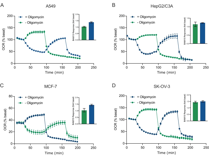
Figure 5: Single or sequential injection of mitochondrial respiratory compounds. Cancer-cell-derived spheroids of MCF-7, HEPG2/C3A, SK-OV-3, and A549 were placed into wells of an XFe96 spheroid microplate in XF RPMI and probed for OCR using the Agilent Seahorse XFe96 analyzer. OCR was measured 5x, after which 2 µg/mL oligomycin (injection Port A: green trace) or 5 µM BAM15 (injection Port A: blue trace or injection port B: green trace) to inhibit the mitochondrial ATP synthase and determine maximal respiratory capacity, respectively. Kinetic OCR data are expressed as % basal (A-D). Maximal respiratory capacity (OCRmax) was calculated as a factor of basal OCR by the equation: OCRmax = OCRBAM15 / OCRbasal. OCRmax was obtained from OCR averages across measurement cycles 8-10 post BAM15 injection with (green bars) and without (blue bars) oligomycin. Data are averages ± SEM from 3-8 individual well replicates across the spheroid assay microplate. Abbreviations: OCR = oxygen consumption rate. Please click here to view a larger version of this figure.
to:
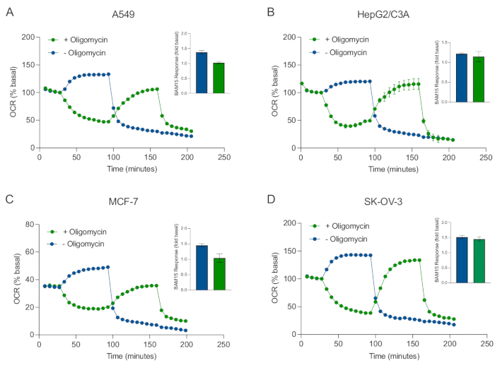
Figure 5: Single or sequential injection of mitochondrial respiratory compounds. Cancer-cell-derived spheroids of MCF-7, HEPG2/C3A, SK-OV-3, and A549 were placed into wells of an XFe96 spheroid microplate in XF RPMI and probed for OCR using the Agilent Seahorse XFe96 analyzer. OCR was measured 5x, after which 2 µg/mL oligomycin (injection Port A: green trace) or 5 µM BAM15 (injection Port A: blue trace or injection port B: green trace) to inhibit the mitochondrial ATP synthase and determine maximal respiratory capacity, respectively. Kinetic OCR data are expressed as % basal (A-D). Maximal respiratory capacity (OCRmax) was calculated as a factor of basal OCR by the equation: OCRmax = OCRBAM15 / OCRbasal. OCRmax was obtained from OCR averages across measurement cycles 8-10 post BAM15 injection with (green bars) and without (blue bars) oligomycin. Data are averages ± SEM from 3-8 individual well replicates across the spheroid assay microplate. Abbreviations: OCR = oxygen consumption rate. Please click here to view a larger version of this figure.
