8,428 Views
•
05:57 min
•
February 10, 2017
DOI:
In clinical practice, esophageal stricture occurrence is a major limitation for extended endoscopic resection of esophageal superficial neoplasia. This video presents an ADSC-sheet transplantation technique that prevents such strictures, making extended endoscopic resection possible in a porcine model. This method can advance a key question in the field of esophageal stricture prevention after endoscopic extended resection.
The main advantage of this technique is that endoscopic ADSC sheet transplantation is meaning-lative, effective, and not time consuming in order to prevent such esophageal stricture. Demonstrating this procedure will be Gabriel Rahmi, a physician from my laboratory, and myself. Collect around 24 million ADSCs per pig using standard cell expansion procedures and T150 culture flasks.
Harvest the ADSCs using 10 milliliters of standard trypsin solution per flask. Then, count the cells using an automatic cytometer. Load 1.5 million cells into each well of a 12-well temperature responsive cell culture dish.
Now, top up each well to four milliliters with culture medium. Then, incubate the plates for 12 hours overnight for the transplantation on the following day. On the day of the transplantation, detach the confluent cell sheets in the plate wells by incubating them at room temperature for 30 minutes.
After half an hour, gently pipette one ADSC sheet and layer it on a 1.5 centimeter diameter hydrophobic paper within a plastic dish. Use this paper as a support membrane. Be patient and take caution when handling the ADSC sheets.
They are very fragile. Next, grasp an ADSC sheet and gently layer it over another to make a double layer construct. Make four double layer constructs per animal.
Under general anesthesia, perform an ESD on the pig. Use a video gastroscope and an electrosurgery unit. First, using an endoscopic knife with soft coagulation, create hemicircumferencial white marks 40 to 45 centimeter dorsal from the dental arch to create a virtual lesion.
Then, separate the mucosal layer from the muscular layer by injecting glycerol solution containing indigo carmine dye into the submucosal layer. Now, make a circumferential incision of the mucosal layer around the dorsal marks, using an endoscopic knife with forced coagulation mode. Next, perform the submucosal dissection from the proximal incision to the distal incision.
Then, complete the resection by ESD, a hemicircumferential, five centimeter long esophageal scar exposing the muscular layer. Now, endoscopically transplant the four double ADSC sheet constructs. Grip the construct with endoscopic forceps and cover it with a large transparent endoscopic cap to protect it during transportation to the wound site.
Then, spread the entire surface of the ADSC sheet construct onto the ulcer bed. Keep the graft in position for 10 minutes to ensure complete and stable engraftment. Continue to apply the ADSC sheets so the four cover about half of the wounded area.
Transportation can be difficult in an animal under general anesthesia breathing and swallowing, so take great care to avoid damaging the sheets. After performing the described procedure on several animals, a PCLE evaluation was conducted three days later. The positive results confirmed the success of the transplantations.
The follow-up period allowed for a multimodal analysis. Compared to control animals, the transplanted animal group showed less frequent alimentary trouble and greater weight gain on day 28. Endoscopic and radiological findings at the end of the follow-up period did uncover one animal from the transplanted group that did develop a severe esophageal stricture.
Histological analysis showed that compared to the transplanted animals, the control animals developed significant fibrotic tissue. We show that an ESD followed by ADSC sheet transplantation can be performed in approximately one and a half hours. While attempting this procedure, it’s important to remember that ADSC sheets are fragile and need to be manipulated with caution.
The key of success is to take the necessary time for each step of the procedure. After exhibitment, this technique paves the way for research showing the field of cellular therapy To treat endoscopically other digestive disease.
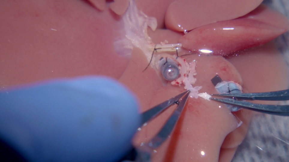
This study reports a successful method of endoscopical adipose tissue-derived stromal cell (ADSC)-sheet transplantation for esophageal stricture prevention after an extended endoscopic submucosal dissection (ESD) in a swine model.
09:40
An Ivor Lewis Esophagectomy Designed to Minimize Anastomotic Complications and Optimize Conduit Function
Related Videos
13204 Views
10:15
Conditional Reprogramming of Pediatric Human Esophageal Epithelial Cells for Use in Tissue Engineering and Disease Investigation
Related Videos
6894 Views
08:06
Creation and Transplantation of an Adipose-derived Stem Cell (ASC) Sheet in a Diabetic Wound-healing Model
Related Videos
11332 Views
08:56
Tissue-Engineered Graft for Circumferential Esophageal Reconstruction in Rats
Related Videos
7065 Views
09:58
Transplantation of Adipose Tissue-Derived Stem Cell Sheet to Reduce Leakage After Partial Colectomy in A Rat Model
Related Videos
7474 Views
08:09
Vessel-sparing Excision and Primary Anastomosis
Related Videos
11268 Views
07:37
Transpupillary-Guided Trans-Scleral Transplantation of Subretinal Grafts in a Retinal Degeneration Mouse Model
Related Videos
1122 Views
06:42
Use of the Scissor-Type Knife During the Peroral Endoscopy Myotomy Procedure for the Treatment of Achalasia
Related Videos
2982 Views
11:19
Robotic Myotomy and Partial Fundoplication for Achalasia
Related Videos
1354 Views
10:41
Robotic D3 Partial Duodenal Resection with Primary Side-to-Side Anastomosis
Related Videos
1628 Views
Read Article
Cite this Article
Perrod, G., Pidial, L., Camilleri, S., Bellucci, A., Casanova, A., Viel, T., Tavitian, B., Cellier, C., Clément, O., Rahmi, G. ADSC-sheet Transplantation to Prevent Stricture after Extended Esophageal Endoscopic Submucosal Dissection. J. Vis. Exp. (120), e55018, doi:10.3791/55018 (2017).
Copy