This study demonstrates the successful establishment of magnetic resonance microscopy imaging as a non-invasive tool to assess the cardiac abnormalities in mice affected with autoimmune myocarditis. The data indicate that the technique can be used to monitor the disease-progression in live animals.
Myocarditis is an inflammation of the myocardium, but only ~10% of those affected show clinical manifestations of the disease. To study the immune events of myocardial injuries, various mouse models of myocarditis have been widely used. This study involved experimental autoimmune myocarditis (EAM) induced with cardiac myosin heavy chain (Myhc)-α 334-352 in A/J mice; the affected animals develop lymphocytic myocarditis but with no apparent clinical signs. In this model, the utility of magnetic resonance microscopy (MRM) as a non-invasive modality to determine the cardiac structural and functional changes in animals immunized with Myhc-α 334-352 is shown. EAM and healthy mice were imaged using a 9.4 T (400 MHz) 89 mm vertical core bore scanner equipped with a 4 cm millipede radio-frequency imaging probe and 100 G/cm triple axis gradients. Cardiac images were acquired from anesthetized animals using a gradient-echo-based cine pulse sequence, and the animals were monitored by respiration and pulse oximetry. The analysis revealed an increase in the thickness of the ventricular wall in EAM mice, with a corresponding decrease in the interior diameter of ventricles, when compared with healthy mice. The data suggest that morphological and functional changes in the inflamed hearts can be non-invasively monitored by MRM in live animals. In conclusion, MRM offers an advantage of assessing the progression and regression of myocardial injuries in diseases caused by infectious agents, as well as response to therapies.
Heart failure is the leading cause of deaths, and myocarditis is one predominant cause of heart failure in young adolescents1. Most patients affected with myocarditis remain asymptomatic and the disease is spontaneously resolved2. However, 10-20% of those affected can develop chronic disease, leading to dilated cardiomyopathy (DCM)3. Various animal models have been developed to study the immune pathogenesis of myocarditis. The disease can be induced in myocarditis-susceptible A/J and Balb/c mice by immunizing the animals with cardiac myosin heavy chain (Myhc)-α or its immunodominant peptide fragments or by infecting with pathogens like coxsackievirus B3 4-9. This study involves Myhc-α 334-352-induced myocarditis in A/J mice. Despite showing myocardial infiltrations, the myocarditis-affected animals appear clinically normal; diagnosis is based on histological evaluation of hearts for inflammation7 and echocardiography10.
Magnetic resonance microscopy (MRM) is a commonly used method to obtain cardiovascular imaging with high resolution three-dimensional planes, permitting assessment of functional details to the level of minute blood vessels (up to 10 μm diameter), but this level of resolving power is not achievable with the routine magnetic resonance imaging (MRI) scanning procedure, in which, the resolution is generally obtained up to 1 mm11-14. MRM offers an advantage as it permits acquisition of high-resolution images and also to derive performance parameters in the early time points of disease process14. Clinically, MRM imaging has been extensively applied to study the functional parameters of diseased heart, lung or brain15-17. In this study, the use of an MRM technique as a non-invasive tool to ascertain cardiac abnormalities in A/J mice affected with autoimmune myocarditis is shown. Specifically, the MRM imaging allows quantification of functional parameters such as left ventricular (LV) end-diastolic volume and ejection fraction (EF) with reasonable accuracy18. The definitions of respective parameters are: LV end diastolic volume, volume of blood in the left ventricle at the end of diastolic cycle, and ejection fraction, stroke volume/end-diastolic volume. The data analysis is performed using the freely available Segment software developed for processing DICOM-compliant cardiovascular images acquired by magnetic resonance scanners19. The data revealed an increase in thickness of the LV wall in the myocarditic animals, corresponding to a decrease in LV end-diastolic volume, stroke volume and ejection fraction, as compared with these functional parameters in healthy mice.
ETHICS STATEMENT:
All animal procedures were conducted in accordance with the guidelines for the Care and Use of Laboratory Animals and approved by the University of Nebraska-Lincoln, Lincoln, NE.
1. Induction of Experimental Autoimmune Myocarditis
2. Animal Handling
3. Image Acquisition
4. Data Analysis of Cardiac Magnetic Resonance Microscopy Imaging
In this report, the utility of MRM technique as a non-invasive modality to determine the structural and functional changes in the hearts of animals affected with EAM is shown. Myocarditis was induced in A/J mice by immunizing the animals with Myhc-α 334-352 in CFA7, and the animals were subjected to MRM experiments on day 21 post-immunization. The MRM imaging was performed on live animals under isoflurane anesthesia at 9.4 T (400 MHz for protons) using an 89 mm vertical-bore magnet equipped with triple axis gradients (maximum strength 100 G/cm). A scout image was acquired to locate and position the mouse heart in the center of the FOV, followed by axial images to obtain the long-axis 4-chamber view. The angle at which the heart was imaged for the 2-chamber view is shown in Figure 2A. Cardiac images were acquired using a 4 cm millipede RF imaging probe with a gradient-echo-based cine pulse sequence. Cardiac function measurements (imaging: LV wall thickness; output: LV end-diastolic volume and ejection fraction) were then analyzed using Segment software. Structural defects in the hearts of EAM-affected mice were evidenced by increase in LV thickness by approximately 1.5-fold (p = 0.018) (Figure 2B and Table 1), with corresponding decrease in LV end-diastolic volume [18.0 ± 4.2 µl vs. 37.5 ± 3.5 µl, Figure 2C(i); p = 0.002] and ejection fraction [49.4 ± 2.3% vs. 71.5 ± 6.0%, p = 0.00066; Figure 2C(ii)] as compared to healthy mice. As expected, histological evaluation of hearts from myocarditic, but not healthy, mice showed multifocal lymphocyte infiltrates, as we have demonstrated previously7; Figure 2D). The data suggest that morphological and functional changes in inflamed hearts can be non-invasively monitored by MRM in live animals.
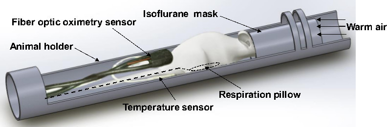
Figure 1. Animal preparation and positioning of probes for acquisition of MRM images from the mouse heart. To acquire images from the heart, the anesthetized mouse is placed in the animal holder specially designed for MRM imaging and connected to the air blow heater to maintain the body temperature. Under continuous anesthesia, the animal is immobilized in the prone position. A pneumatic pillow, fiber optic oximetry sensor and temperature sensor are set up to monitor respiration, pulse and body temperature, respectively, until MRM acquisition of cardiac images is complete. Please click here to view a larger version of this figure.
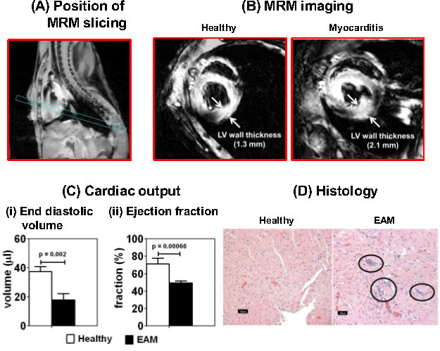
Figure 2. MRM imaging of mice affected with autoimmune myocarditis reveals cardiac abnormalities. Myocarditis was induced in A/J mice by immunizing the animals with Myhc-α 334-352 in CFA. The animals were subjected to MRM imaging on day 21 postimmunization to evaluate cardiac abnormalities. (A) Position of MRM slicing. The angle at which the heart was sliced for image acquisition is shown. (B) MRM imaging. Short-axis slices of heart were captured using echo-based cine pulse sequence in eight time frames with a TR of 500 msec (TE, 5 msec; flip angle, 20°; number of acquisitions, 4; acquisition matrix, 256 x 256) [arrows: LV wall thickness]. (C) Cardiac output. Cardiac output was measured based on (i) LV end-diastolic volume and (ii) ejection fraction in healthy and myocarditic mice using quantitative medical image analysis with Segment software. Mean SEM values for a group of mice are shown (n = 2 to 5 per group). (D) Histology. Hearts from the above treatment groups were evaluated for inflammation by hematoxylin and eosin staining. Circles: multifocal lymphocytic infiltrations. Please click here to view a larger version of this figure.
Animals | Healthy (n = 3) | EAM (n = 5) |
Mouse 1 | 1.03 | 1.48 |
Mouse 2 | 1.3 | 1.59 |
Mouse 3 | 0.94 | 1.44 |
Mouse 4 | 2.11 | |
Mouse 5 | 1.92 | |
Mean ± SEM | 1.09 ± 0.1 | 1.71 ± 0.1 |
Table 1. Comparison of left ventricular (LV) wall thickness between healthy and experimental autoimmune myocarditis (EAM) mice. Three healthy and five EAM-induced mice were subjected to magnetic resonance microscopy (MRM) imaging on day 21 postimmunization. After acquiring the cardiac images by MRM, the thickness of LV wall was measured using Segment software as described in the protocol. The values displayed in the table represent LV wall thickness in mm.
This study describes the MRM procedure and its utility as a non-invasive tool to ascertain cardiac abnormalities in mice affected with autoimmune myocarditis. Since the histologic features of EAM resemble postinfectious myocarditis of humans, mouse models are commonly employed to delineate the immune mechanisms of myocardial injuries23-25. However, the animals affected with myocarditis appear clinically normal, and the diagnosis is made based on histology at the termination of experiments7. The animals are usually sacrificed on day 21 postimmunization. Assessing the disease process in this way at single time points limits use of these models, especially in pharmaceutical research, where monitoring of disease progression in response to treatments is a critical requirement.
To ascertain cardiac abnormalities in living animals, use of non-invasive modalities like MRM is helpful. The MRM technique that described here offers an advantage of obtaining the structural and functional characteristics of hearts without the need to use contrast agents. However, this technique requires acquisition of high resolution 3D anatomical images in strong magnetic fields. Nonetheless, once the images are acquired, functional parameters such as LV end-diastolic volumes and ejection fractions can be analyzed later using commercially available software, without the need to further assemble the MRM apparatus. As shown in Figure 2, MRM examination of animals immunized with Myhc-α 334-352 revealed LV wall thickness to be greater than in healthy mice (Figure 2B), with a corresponding decrease in functional outputs (LV end-diastolic volumes and ejection fractions; Figure 2C). As expected, hearts from immunized, but not healthy, animals had inflammatory infiltrates (Figure 2D). Thus, findings from the MRM technique and histology corroborate each other.
Nonetheless, to obtain reproducible results by MRM, the following three factors need to be addressed. (a) Animals should be placed in the MRM scanner so hearts are positioned in the center of the magnet to expose them to the magnetic field with maximum homogeneity. (b) Motion artifact is a concern in live animal experiments. To diminish image blurring due to respiration and cardiac movements, pulse oximetry and respirometry were used to gate MRM acquisition—that is, to acquire discrete image signals at specific time points within the respiratory and cardiac cycles—which required the use of an animal monitoring system. (c) Acquisition of high-resolution 3D cardiac images is a critical requirement to enable detailed analysis of cardiac abnormalities. To obtain images in all three dimensions, and to increase signal-to-noise ratio, it is important to design more sensitive imaging coils specific to MRM that permit accurate and complete capturing of images in strong magnetic fields in a short-axis orientation within the animal scanners.
In conclusion, development of an MRM technique to evaluate cardiac abnormalities in live animals is challenging. This is especially true for mice, because of their smaller heart sizes (~1/2,000 the mass of a human heart) and higher heart rates (~600 beats per min) compared to humans26. Nonetheless, once developed and validated, the MRM technique can be used to compare the anatomical and functional changes of hearts between healthy and sick animals. Thus, the MRM technique could serve as a valuable, non-invasive tool to assess the longitudinal progression of inflammatory cardiac pathologies related to both the acute and chronic nature of the disease process, and to monitor responses to therapies in living animals.
The authors have nothing to disclose.
This work was supported by the National Institutes of Health (HL114669). CM is a recipient of a postdoctoral research fellowship grant awarded by the Myocarditis Foundation, NJ.
Myhc-a 334-352 (DSAFDVLSFTAEEKAGVYK) | Neopeptide, Cambridge, MA | Store at 4οC | |
CFA | Sigma Aldrich, St Louis, MO | 5881 | Store at 4οC |
MTB H37Rv extract | Difco Laboratories, Detroit, MI | 231141 | Store at 4οC |
PT | List Biologicals Laboratories, Campbell, CA | 181 | Store at 4οC |
1x PBS | Corning, Manassas, VA | 21-040-CV | Store at 4οC |
Isoflurane | Piramal Healthcare, Mumbai, India | NDC66794-013-25 | |
Female A/J mice | Jackson Laboratories, Bar Harbor, ME | 646 | |
Leur-lok sterile 1 ml syrringe | BD, Franklin Lakes, NJ | 309628 | |
Leur-lok sterile 3 ml syrringe | BD, Franklin Lakes, NJ | 309657 | |
Sterile needle, 18 G | BD, Franklin Lakes, NJ | 305195 | |
Sterile needle, 27 1/2 G | BD, Franklin Lakes, NJ | 305109 | |
3-way stopcock | Smiths Medical ASD, Inc. Dublin, OH | MX5311L | |
Kerlix gauze bandage rolls | Covidien, Mansfield, MA | 6720 | |
Kimwipes | Kimberly-Clark Professional, Roswell, GA | 34155 | |
Protouch Stockinette | Medline Industries, Mundelein, IL | 30-1001 | |
Sterile surgical scissors and forceps | INOX tool Corporation | ||
Micro oven | GE Healthcare, | ||
ThermiPAQ hot and cold therapy system | Theramics Corporation, Springfield, IL | ||
Reptile heating lamp | Energy Savers Unlimited, Inc. Carson, CA | ||
3M Transpore tapes | Target Corporation, MN | ||
Up and Up Polymyxin B sulfate/Bacitracin/Neomycin sulfate antibiotic ointment | Target Corporation, MN | ||
North Safety DeciDamp-2PVC foam ear plugs | North Safety Products, Smithfield, RI | ||
Cotton tipped applicator, 6’’ wooden stem | Jorgensen Laboratories, Inc. Loveland, CO | ||
Anesthesia induction chamber | Summit Anesthesia Solutions, Ann Harbor, MI | ||
Summit Anesthesia Support system for regulating flow of anesthesia | Summit Anesthesia Solutions, Ann Harbor, MI | ||
Specially designed animal holder | Agilent Technologies, Santa Clara, CA | ||
Bickford Omnicon F/Air anesthesia gas filter unit | A.M. Bickford, Inc. Wales Center, NY | ||
Pulse-oximeter module, MR compatible small animal monitoring and gating system | Small Animal Instruments, Inc. Stony Brook, NY | ||
Oxygen cylinder | Matheson-Tri Gas, North-Central Zone, Lincoln, NE | ||
Gas regulator | Western Medica, West Lake, OH | ||
Signal breaking module, MR compatible small animal monitoring and gating system | Small Animal Instruments, Inc. Stony Brook, NY | ||
9.4 T (400 MHZ) 89 mm vertical core bore MR scanner | Agilent Technologies, Santa Clara, CA | ||
4-cm millipede micro-imaging RF coil | Agilent Technologies, Santa Clara, CA | ||
SAM PC monitor | Small Animal Instruments, Inc. Stony Brook, NY | ||
Quantitative Medical Image analysis software | http://segment.heiberg.se; Segment v1.8 R1430, Medviso, Oresunds region, Sweden | ||
Matlab software | The Mathworks, Inc. Natick, MA | ||
Computer-Unix operating system |