Waiting
Login processing...

Trial ends in Request Full Access Tell Your Colleague About Jove

Immunology and Infection

A Novel Microdissection Approach to Recovering Mycobacterium tuberculosis Specific Transcripts from Formalin Fixed Paraffin Embedded Lung Granulomas

Published: June 5, 2014 doi: 10.3791/51693

ERRATUM NOTICE

Summary

Microdissection has been extensively employed for the examination of DNA, RNA, and protein within tissue. Laser capture microscopy (LCM) is the most commonly used method, but a new milling technique, mesodissection, is recently available. We demonstrate RNA extraction from mesodissected formalin fixed paraffin embedded tissue slides of Mycobacterium tuberculosis granulomas.

Abstract

Microdissection has been used for the examination of tissues at DNA, RNA, and protein levels for over a decade. Laser capture microscopy (LCM) is the most common microdissection technique used today. In this technique, a laser is used to focally melt a thermoplastic membrane that overlies a dehydrated tissue section1. The tissue section composite is then lifted and separated from the membrane. Although this technique can be used successfully for tissue examination, it is time consuming and expensive. Furthermore, the successful completion of procedures using this technique requires the use of a laser, thus limiting its use. A new more affordable and practical microdissection approach called mesodissection is a possible solution to the pitfalls of LCM. This technique employs the MESO-1/MeSectr system to mill the desired tissue from a slide mounted tissue sample while concurrently dispensing and aspirating fluid to recover the desired tissue sample into a consumable mill bit. Before the dissection process begins, the user aligns the formalin fixed paraffin embedded (FFPE) slide with a hematoxylin and eosin stained (H&E) reference slide. Thereafter, the operator annotates the desired dissection area and proceeds to dissect the appropriate segment. The program generates an archived image of the dissection. The main advantage of mesodissection is the short duration needed to dissect a slide, taking an average of ten minutes from set up to sample generation in this experiment. Additionally, the system is significantly more cost effective and user friendly. A slight disadvantage is that it is not as precise as laser capture microscopy. In this article we demonstrate how mesodissection can be used to extract RNA from slides from FFPE granulomas caused by Mycobacterium tuberculosis (Mtb).

Introduction

Samples have traditionally been manually microdissected from either whole tissue or slides using a needle and scalpel. This necessitates a clear separation between the tissue section of interest and surrounding tissues sections2. With the current advancements in molecular profiling technology, there has been an increasing need to assess tissue at the cellular level. Due to the limitations of manual microdissection, techniques including LCM were established to allow greater isolation precision. This technique allows the researcher to isolate specific cell populations from various cell and slide types, which can then be used for downstream assays such as gene expression profiling. Although very effective for downstream assays, LCM is not without limitations. First, LCM is an expensive and time consuming process. Additionally, due to the unstable nature of RNA, it is often challenging to obtain high quality RNA from LCM samples2. Due to the disadvantages of LCM, new advances in microdissection technology are still needed to make it more accessible to a greater number of researchers in an affordable and time sensitive manner.

One such advancement in microdissection technology now available is a technique known as mesodissection. In this technique a machine is used to mill the annotated tissue section of interest and aspirates it into a consumable mill bit3. Next, this sample can be aspirated into a collection tube and used for downstream applications. The advantages of this system are that it is significantly less expensive and time sensitive. In our experience, the system allows the dissection of a lung granuloma tissue section in ten minutes. On the other hand, traditional LCM would likely require hours to complete the process. The system allows the operator to load a reference slide to use as a comparison as well as tool for annotation. Additionally a report is generated outlining the area of the slide that was dissected. There are two main disadvantages to mesodissection. Although effective at extracting multiple cells, it is challenging to isolate a single cell. In addition, the precision of the image generated is not as clear as if using other imaging microscopes.

Tuberculosis (TB) is a major infectious disease killer of humanity worldwide and results from infection with Mtb. In a majority of individuals exposed to aerosols of Mtb, the infection is latently limited. In at least 10 million people annually, it results in active TB disease4. During latent infection, Mtb is contained within pathological lung lesions known as granulomas. Hence it has been argued that the outcome of Mtb infection is decided at the level of the granuloma5.

Here we demonstrate how mesodissection can be used to microdissect granulomas caused by Mtb. The slides used are from FFPE lung tissue from infected rhesus macaques. For the purpose of this demonstration, we will dissect granulomas in their entirety. We also demonstrate that RNA can be extracted from the recovered tissue. This technique can be applied to tissue samples from various other specimens and then used for a variety of downstream assays.

Subscription Required. Please recommend JoVE to your librarian.

Protocol

1. Calibrate Mesodissection Instrument with 2iD Imaging Software

The 2iD imaging software will be referred to as either the software or the program for the remainder of this article. This step is needed to correctly track the image as well as align multiple slide frames.

  1. Turn on instrument and computer.
  2. Open software and select “calibrate instrument”.
  3. Calibrate stage travel by using the joystick. Move stage to upper left position. Press “set” under step 1 in program.
  4. Move stage to lower right position. Press “set” under step 2 in program.
  5. Calibrate aspiration by first pressing “Z-button” to raise head assembly. Press “aspirate pulse” button and “dissection” button on the joystick until plunger control rod reaches top position. Press “set” under step 3 of program.
  6. Press “aspirate reset” button. Once the plunger stops at the bottom position press “set” in step 4.
  7. Click on the “scale tab”.
  8. Place calibration ruler on blank side.
  9. Focus microscope if needed.
  10. Draw line between the two scale bars using mouse.
  11. Enter distance in box labeled step 2. Press “compute” button.
  12. Click on “crosshair” tab. Turn motor speed to 1.
  13. Insert xScisor into head assembly. Lower head assembly into z-axis ready position by pressing “z-axis” button. NOTE: The head assembly is the large structure on the top the dissection instrument. The xScisor will be referred to as a consumable mill bit for the remainder of this article.
  14. Click “dissection engage” button on joystick. NOTE: This button is located on the top of the joystick.
  15. Define center of rotation visually. To do this, move center of crosshair over center of rotation using defined parameter in step 1 of program. Use the up and down arrows in the “x” and “y-axis” bars to adjust the pixels to align crosshair. Click the “done” tab.
  16. Click on “home” icon. NOTE: This is represented by a house image located in the upper right hand corner.

2. Create Reference Image

  1. Place H&E slide on stage. Place opaque cover on top of slide. Drive the instrument by moving the joystick to the desired area of the H&E slide. Save the image generated. NOTE: Either the “capture reference” tab or “dissect” tab can be used to complete this step.

3. Align Tissue for Dissection

  1. Click the “dissect tissue” option on the home screen.
  2. Fill out the “operator,” “dissection accession number,” “reference accession number,” “xScisor barcode number,” “dissection fluid lot number,” “xScisor size” and “description” in the “setup” tab.
  3. Click on the “find tissue” tab.
  4. Import previously saved reference image.
  5. Place an unstained FFPE slide of 5 micron thickness on the stage. Move stage to the same area as the one depicted in the reference image. Click on “insert image” tab.
  6. Click on “align” tab. Use the mouse and associated arrows to align the reference image to the unstained image.

4. Annotate Slide

  1. Click on the “annotate” tab.
  2. Use the mouse to draw a circle around the desired area of the slide that will be microdissected.

5. Load Consumable Mill Bit

  1. Fill consumable mill bit with desired buffer, here PKD buffer, by pulling the plunger up and down in a fluid motion several times. Make sure to exclude air bubbles.
  2. Raise Z-axis by pressing “Z-axis button”.
  3. Load consumable mill bit into machine. Do this by sliding the consumable mill bit into the top of the machine so that the white line on the instrument lines up with the black line on the consumable mill bit. Press “aspiration reset” button to allow plunger to be lowered into the correct position. NOTE: It should be easy to slide the consumable mill bit. If it is not, make sure the notches are correctly aligned.
  4. Lower Z-axis by again pressing “Z-axis” button.

6. Dissect Tissue (Figure 4)

  1. Open “dissect” tab.
  2. Turn motor and aspiration speeds to 1.
  3. Check “show tracking” box. Note: This allows the user to visualize the dissected area during the dissection process.
  4. Once ready to dissect, continue to hold “engage” button as well as “aspirate” button while moving joystick in a counterclockwise direction to dissect tissue.
  5. Continue to dissect in a counterclockwise direction until the aspirate is “full” tab turns red.

7. Empty and Remove Consumable Mill Bit

  1. Place a 0.5 µl microfuge tube under the tip of the consumable mill bit.
  2. Press “aspiration” control rod quickly to eject sample into microfuge tube.
  3. Press with increased force to eject used consumable mill bit.
  4. Rinse tissue fragment by pulling plunger back. Push plunger forward to express remaining sample into consumable mill bit. The tissue fragment will now be contained within the microfuge tube.

8. Lyse Sample with Proteinase K Digestion Followed by Heat Treatment

  1. Add 70 µl TE pH 8.5 containing 5 µg of proteinase K to recovered tissue sample.
  2. Place sample into programmable heater-shaker.
  3. Complete digestion using programmable heater-shaker at the following setting: 60 ˚C for 30 min at 1,500 rpm; 82 ˚C for 15 min at 1,500 rpm; and 25 ˚C for 1 min at 450 rpm. The sample is now ready for downstream applications.

Subscription Required. Please recommend JoVE to your librarian.

Representative Results

The aforementioned protocol shows how to use a new mesodissection technique to extract RNA from FFPE tissue slides. This protocol’s efficacy is shown through FFPE slides of lung granulomas from NHP infected with Mtb in various infective stages. Figures 1-3 are images of the instrument. Figure 4 depicts how the dissection process occurs and the resulting image generated by the software. Table 1 shows the results of RNA extraction with a granuloma from a NHP at each infective state. RNA was amplified and purified to allow for RT-PCR evidence of 16s (Tables 2-4). Table 4 confirms the presence of Mtb ribosomal subunit 16s, and thus Mtb, in the microdissected samples.

Figure 1
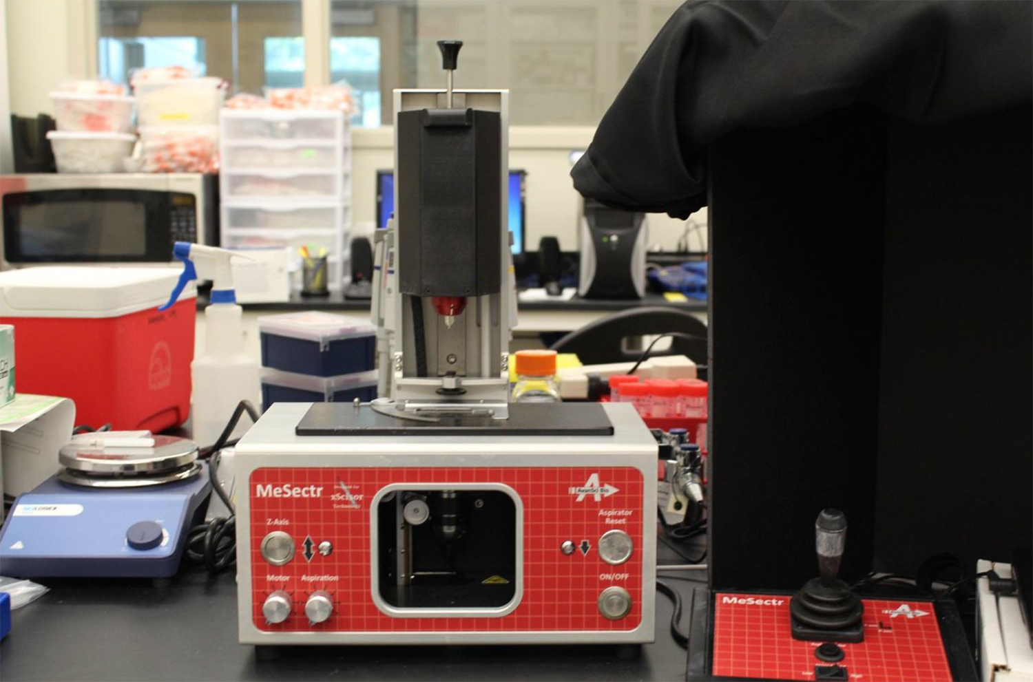
Figure 1. Image of mesodissection instrument including joystick. Please click here to view a larger version of this figure.

Figure 2
Figure 2. Closer view of mesodissection instrument. The “z-axis” button, “motor” speed and “aspiration” speed knobs are shown on the left side of the image. The “aspirator reset button” and “on/off” button are shown on the right side of the image. The slide stage is on the top of the instrument. Please click here to view a larger version of this figure.

Figure 3
Figure 3. Closer view of joystick. The joystick, “aspirate pulse” and “stage speed” buttons shown. Please click here to view a larger version of this figure.

Figure 4
Figure 4. Report of whole granuloma mesodissected from NHP EB23. Animals used in this study were infected with CDC1551 MTb via aerosol. The reference image on the left shows an H&E stained granuloma from an actively infected rhesus macaque. The desired area of interest to be dissected is outlined in green. The image in the middle shows the corresponding unstained FFPE slide of 5 micron thickness before dissection. The areas were aligned using the software and the correlating area of interest is outlined in green. The image on the right depicts the same unstained FFPE slide post dissection. The area dissected is represented in blue. A 400 mm2 consumable mill bit was used for the dissection, which is designed for traverse dissection of medium to large dissection area. Large dissection area is described as less than 100 mm2. Sizes designed for other areas/dissection types are available. Although the report describes the annotated area as 0 mm2, this is inaccurate. When we remove the microdissected slide from the machine and physically look at the area of interest itself, we can see the corresponding area on the slide that no longer has tissue and therefore has been dissected. It should be noted that the authors are renting the instrument and this issue has been fixed on other instruments. Please click here to view a larger version of this figure.

Primate Infective State RNA concentration in ng/µl Total RNA in ng 260/280 260/230
EB23 Active 48.3 724.5 1.71 1.59
HB12 Latent 61.4 921 1.7 1.86
HP41 Reactivated via co infection with SIV 22.3 334.5 1.9 2.18

Table 1. RNA concentrations post extraction. The sample was DNAsed and RNA extraction was performed using the Qiagen RNAeasy FFPE kit. RNA concentration was obtained using a nanodrop. NHP infective stages are also depicted. RNA was eluted in 15 µl RNAse free water.

Primate Infective State cDNA concentration in ng/µl Total cDNA in ng 260/280 260/230
EB23 Active 31.7 951 2.08 2.37
HB12 Latent 156.5 4695 1.94 4.4
HP41 Reactivated via co infection with SIV 41.7 1251 2.19 3.44

Table 2. cDNA concentrations post amplification and purification. NHP infective stages are also depicted. RNA was amplified using Ovation RNA-Seq FFPE System (Part no. 7150) and resulting amplified cDNA was purified using the QIAGEN QIAquick PCR Purification Kit as suggested by Nugen on page 26 of the amplification system’s user guide. cDNA was eluted in 30 µl TE buffer.

Well Reporter Ct Tm value
16S 10-1 Standard 12.35138 79.4
16S 10-2 Standard 16.144611 79.4
16S 10-3 Standard 17.911345 79.4
16S 10-4 Standard 21.762596 79.4
16S 10-5 Standard 25.746624 79.4
16S 10-6 Standard 28.505266 79.1
HB12 Unknown 25.771492 77.8
HP41 Unknown 17.760149 78
EB23 Unknown 24.754618 78.2
Negative Control NTC 33.544422 79.7

Table 3. RT-PCR results with respect to 16s ribosomal subunit. Appropriate amplification observed thus proving the presence of Mtb within samples. Genomic DNA from CDC1551 strain Mtb was used as a standard.

Subscription Required. Please recommend JoVE to your librarian.

Discussion

Mesodissection is a technique that can be applied to RNA extraction from pathologic lesion slides caused by a vast array of pathogens. It is a necessity that the user tracks the image and aligns the image correctly. To accomplish this, the instrument must be calibrated following the directions on the imaging software. When dissecting, if the slide and area of interest being dissected do not align, the user must recalibrate the instrument. Another critical step in the dissection process is to load the consumable mill bit in a manner such that it is devoid of bubbles prior to dissection (Step 5.1). We have found that the presence of bubbles decreases the dissection yield. To correct this problem, we recommend propelling and expressing the plunger multiple times. The user can practice this by adding food coloring to a practice buffer to visualize bubbles, as recommended by the manufacturer. Once effective, the user should then proceed with normal dissection. The last critical step is to dissect the sample in a counterclockwise direction. The machine mills in a counterclockwise direction; therefore, we have found that moving the joystick in a circular motion in a counterclockwise manner increases tissue yield as the leading edge cuts more cleanly than the trailing edge6. Another way to improve tissue yield is to decrease the speed of the dissection process. We have found that setting the aspiration rate and motor speed on the lowest speed while simultaneously setting the stage speed to “low” produce the highest tissue yield. There are two main limitations to this microdissection approach. The first limitation is that we have found point dissection very challenging due to the need to drive the joystick in counterclockwise circles in the dissection process. The second limitation is that the camera on the instrument generates an image that lacks the cellular detail of an image generated from an advanced microscope.

Mesodissection has direct implications with respect to transcriptomic analysis of Mtb. Granuloma formation is a hallmark of TB infection and results from the host’s attempt to contain the pathogen within a confined region of lung5. Conversely, it has been proposed that Mtb has evolved to allow its persistence in granulomatous lesions7. Mtb is subjected to different stresses depending on the stage of infection. Some of these stresses include but are not limited to redox stress, low pH, membrane damage, hypoxia, lack of nutrition, etc7-13. Throughout these various stages, Mtb can still survive in a latent form and even reactivate. This means that different genes are upregulated and downregulated at different infective stages. For example, Mtb encodes for over 200 transcription factors4. In addition, it has been shown that the expression balance between different chemokines, such as α and β chemokines, within the granuloma may be important protection markers8. Evaluation of mycobacterial transcriptomics in granulomatous tissue is likely to further our understanding of the mechanisms involved in their formation and maintenance as well as those genes that are expressed in each state of the infection. This will allow us to evaluate mycobacterial transcriptomics in various TB infective stages including active, latent and reactivated disease as well as various regions within the granuloma itself. In addition to allowing us to further assess mycobacterial transcriptomics, the above procedure also allows for the assessment of host transcriptomics in the same infective states. Furthermore, this approach then can be used to compare, contrast, and analyze the relationship between host and bacteria.

Although an important tool in TB research, the aforementioned protocol can be applied to other pathogens as well. The above protocol uses FFPE slides from infected lung samples, but can also be applied to lesions from other organs. We demonstrate how to use the system to extract RNA, but it can theoretically be used for DNA and protein extraction in an equally effective and time sensitive manner. FFPE blocks are available in most laboratory histology departments; therefore, the technique described here has the possibility of exponentially increasing the knowledge gained from these currently unused samples.

Subscription Required. Please recommend JoVE to your librarian.

Disclosures

The authors disclose that there are no competing financial interests.

Acknowledgments

The authors would like to acknowledge the following NIH awards/subawards for the support of this research: R01HL106790, R01HL106790-S1, R01HL106786, R01AI089323, R21AI091457, R21RR026006, P20RR020159, C06RR017563, 8T32OD011124-08, and P51OD011104.

Materials

Name Company Catalog Number Comments
MeSectr AvanSci Bio Mesodissector
400 nm xScisors AvanSci Bio Other sizes available
THOR AvanSci Bio Programmable Heater-Shaker
NanoDrop2000 ThermoScientific ND-2000
RNeasy FFPE extraction kit  Qiagen 73504
Ovation RNA-Seq FFPE System  Nugen 7150
QIAquick PCR Purification Kit  Qiagen 28104

DOWNLOAD MATERIALS LIST

References

  1. Fend, F., Raffeld, M. Laser capture microdissection in pathology. J Clin Pathol. 53 (9), 666-672 (2000).
  2. Esposito, G. Complementary techniques: laser capture microdissection--increasing specificity of gene expression profiling of cancer specimens. Adv Exp Med Biol. 593, 54-65 (2007).
  3. Adey, N., Bosh, D., Emery, D., Birch, L., Parry, R. Mesodissection of Paraffin Embedded Slide Mounted Tissue Sections. Journal of Molecular Diagnostics. 14 (6), (2012).
  4. Fleischmann, R. D., Alland, D., Eisen, J. A., Carpenter, L., White, O., Peterson, J., et al. Whole-genome comparison of Mycobacterium tuberculosis clinical and laboratory strains. J Bacteriol. 184 (19), 5479-5490 (2002).
  5. Russell, D. G., Barry 3rd, C. E., Flynn, J. L. Tuberculosis: what we don't know can, and does, hurt us. Science. 328 (5980), 852-856 (2010).
  6. Mesodissection of fibrous tissue. AvanSciBio. , (2013).
  7. Paige, C., Bishai, W. R. Penitentiary or penthouse condo: the tuberculous granuloma from the microbe's point of view. Cell Microbiol. 12 (3), 301-309 (2010).
  8. Mehra, S., Alvarez, X., Didier, P. J., Doyle, L. A., Blanchard, J. L., Lackner, A. A., et al. Granuloma correlates of protection against tuberculosis and mechanisms of immune modulation by Mycobacterium tuberculosis. J Infect Dis. 207 (7), 1115-1127 (2013).
  9. Kaushal, D., Schroeder, B. G., Tyagi, S., Yoshimatsu, T., Scott, C., Ko, C., et al. Reduced immunopathology and mortality despite tissue persistence in a Mycobacterium tuberculosis mutant lacking alternative sigma factor, SigH. Proc Natl Acad Sci U S A. (12), 8330-8335 (2002).
  10. Mehra, S., Kaushal, D. Functional genomics reveals extended roles of the Mycobacterium tuberculosis stress response factor sigmaH. J Bacteriol. 191 (12), 3965-3980 (2009).
  11. Rohde, K. H., Veiga, D. F., Caldwell, S., Balazsi, G., Russell, D. G. Linking the transcriptional profiles and the physiological states of Mycobacterium tuberculosis during an extended intracellular infection. PLoS Pathog. 8 (6), (2012).
  12. Fontan, P. A., Voskuil, M. I., Gomez, M., Tan, D., Pardini, M., Manganelli, R., et al. The Mycobacterium tuberculosis sigma factor sigmaB is required for full response to cell envelope stress and hypoxia in vitro, but it is dispensable for in vivo growth. J Bacteriol. 191 (18), 5628-5633 (2009).
  13. Rustad, T. R., Harrell, M. I., Liao, R., Sherman, D. R. The enduring hypoxic response of Mycobacterium tuberculosis. PLoS One. 3 (1), (2008).

Tags

Microdissection Mycobacterium Tuberculosis Formalin Fixed Paraffin Embedded Lung Granulomas Laser Capture Microscopy LCM Mesodissection MESO-1/MeSectr System FFPE Slide H&E Reference Slide

Erratum

Formal Correction: Erratum: A Novel Microdissection Approach to Recovering Mycobacterium tuberculosis Specific Transcripts from Formalin Fixed Paraffin Embedded Lung Granulomas
Posted by JoVE Editors on 07/01/2014. Citeable Link.

The corresponding author was changed for the article, A Novel Microdissection Approach to Recovering Mycobacterium tuberculosis Specific Transcripts from Formalin Fixed Paraffin Embedded Lung Granulomas. The corresponding author was changed from:

Teresa A. Hudock

to:

Deepak Kaushal

A Novel Microdissection Approach to Recovering <em>Mycobacterium tuberculosis </em>Specific Transcripts from Formalin Fixed Paraffin Embedded Lung Granulomas
Play Video
PDF DOI DOWNLOAD MATERIALS LIST

Cite this Article

Hudock, T. A., Kaushal, D. A NovelMore

Hudock, T. A., Kaushal, D. A Novel Microdissection Approach to Recovering Mycobacterium tuberculosis Specific Transcripts from Formalin Fixed Paraffin Embedded Lung Granulomas. J. Vis. Exp. (88), e51693, doi:10.3791/51693 (2014).

Less
Copy Citation Download Citation Reprints and Permissions
View Video

Get cutting-edge science videos from JoVE sent straight to your inbox every month.

Waiting X
Simple Hit Counter