Flowcytometrie is uitgebreid in de immunologie, hematologie en oncologie benut om celpopulaties te definiëren via intrinsieke scatter eigenschappen, celoppervlakantigeen expressie en andere fluorescentie parameters 1-3. Het inzicht in bloedlijn ontwikkeling en ziekte resultaat in belangrijke mate van de continue verfijning van deze methode na de eerste uitvoering 4,5. Toegenomen bewustzijn van de kwantitatieve en de algemene analytische mogelijkheden van flowcytometrie heeft onlangs haar meer wijdverspreid gebruik in stamcelonderzoek aangemoedigd en kan op dezelfde diepe vooruitgang in een korter tijdsbestek 6 mogelijk te maken. De toepassing van flowcytometrie specifiek analyseren en te isoleren neurale populaties is lang beschouwd als een uitdaging. In tegenstelling tot hematopoëtische cellen die van nature voorkomen in suspensie worden neurale celtypes typisch geoogst uit te ingewikkelde bronnen kunnen glia en diverse oTher omringende cellen en een ingewikkeld netwerk van proces dragende neuronen. Bijgevolg neurobiologie heeft nog tot uitvoering van de veelzijdigheid van flowcytometrie om zijn volledige potentieel in de dagelijkse routines onderzoek. Zolang echter als een levensvatbare enkele celsuspensie kan worden gegenereerd (en protocollen ontwikkeld en geoptimaliseerd daartoe 7), flowcytometrie en fluorescentie-geactiveerde celsortering (FACS) kan worden beschouwd als een waardevol bestanddeel van de analytische repertoire neurobiologie 8-11.
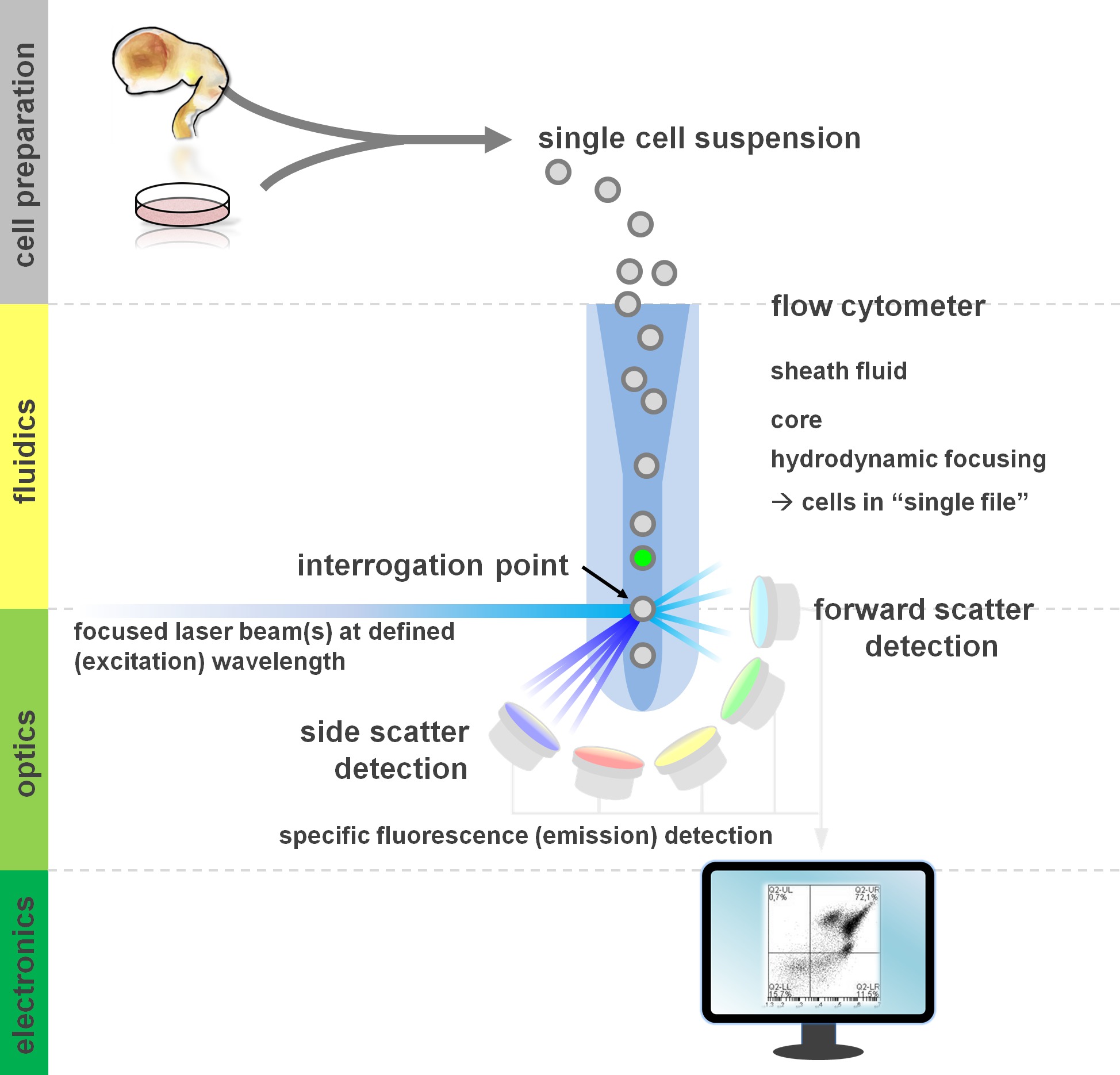
. Figuur 1. Principe van flowcytometrische analyse en componenten van een stromingscytometer flowcytometers bestaan uit drie belangrijke systemen: fluïdica, optica en elektronica. Een gestroomlijnde stroming van cellen in suspensie (bereid uit primair weefsel of in vitro kweek) wordt bewerkstelligd door de huls fluid via hydrodynamische focussen, het beperken van het monster naar het centrum kern. De optische onderdelen zijn samengesteld uit lasers die de stroom van cellen en optische filters die het signaal naar de geschikte detectoren direct verlichten. De lichtsignalen gedetecteerd worden omgezet naar elektronische signalen, vervolgens verwerkt door een computer en gevisualiseerd op een monitor voor data-analyse en gating. Klik hier om een grotere versie van deze afbeelding te bekijken.
Gebruikers van stromingscytometrische methoden winst van ten minste een basiskennis van de onderliggende fundamentals waaronder bouwstenen van een cytometer's (zie voor een overzicht 12,13; zie ook figuur 1). Een laserstraal snijdt een hydrodynamisch gerichte vloeibare stroom die de cellen in suspensie, waardoor pass laserstraal 'bestand' achter elkaar bevat. De interceptiop een cel (of een ander deeltje, wat dat betreft) met de laser resulteert in de verstrooiing van licht van deze ondervragingspunt. Strooilicht kan worden gedetecteerd in het verlengde van de laser richting (voorwaartse verstrooiing, die bij de omvang van het deeltje), alsmede loodrecht op de richting (zijwaartse verstrooiing, die de granulosity van de deeltjes / cel). Deze voornoemde scatter eigenschappen geen specifieke etikettering, dat is de reden waarom een ongemerkt monster (of ook cellulaire puin, luchtbellen, enz.) Een signaal (event) op de bivariate voorwaartse verstrooiing versus kant scatterplot vaak gebruikt voor de initiële venstertijd zal genereren vereisen. Door de geschikte lasers en filters specifiek voor de overeenkomstige excitatie en emissie spectra kan een cel worden geanalyseerd op positiviteit, intensiteit of afwezigheid van fluorescente merkers. De meeste flowcytometrische toepassingen gericht op de karakterisering via celoppervlakte antigenen. In tegenstelling tot de hematopoietische lineage is de neurale stam bleef minder uitgebreid gedefinieerd volgens oppervlak epitoop expressiepatronen 5. Een voordeel exploiteren oppervlakteantigenen is dat levende cellen kunnen worden gesorteerd paradigma zoals FACS cel. In tegenstelling, intracellulaire antigeen kleuring vereist fixatie en permeabilisatie stappen om de epitoop-antilichaam interactie bemiddelen, zich verzet tegen downstream toepassingen die levensvatbare cellen nodig. Van de nota, dergelijke benaderingen nog steeds de mogelijkheid voor tal van kwantitatieve assays 14 als downstream analyses voor RNA en eiwit expressie 15. Hematologie, immunologie en oncologie zijn vaak meer dan een dozijn markers in samenhang met bepaalde subpopulaties 16 definiëren. Daarnaast kan de massa cytometrie of CyTOF nu worden gebruikt voor het analyseren tot 30 parameters tegelijkertijd 17,18.
Voor neurale stamcellen aanvragen evenals primaire culturen 14,19,20 de heterogeniteit van cellen invitro is een veel voorkomend verschijnsel 21-23. De cellen die geen deel uitmaken van de doelgroep van belang belichamen een potentieel verstorende factor voor experimentele uitlezing 24,25. Op geschikte wijze de verschillende cellulaire subsets aanwezig in een heterogene celsuspensie aangebracht onderscheiden (bekend of nog te ontcijferd) antigeen expressie profielen die kunnen worden gebruikt om deze verschillende populaties te definiëren. Flowcytometrie kan dus een cruciale rol spelen bij het oplossen van de cellulaire heterogeniteit en, daardoor, biomedische toepassingen te vergemakkelijken (in vitro testen, celtherapie) en optimaliseren van kwantitatieve uitlezing door te focussen op de meest relevante subset 24,26. Diverse oppervlakte-antigeen combinaties zijn geïdentificeerd in de afgelopen jaren aan de kwantificering en isolatie van specifieke neurale celtypes mogelijk te maken. Dit omvat CD133 voor de verrijking van neurale stamcellen 27, een combinatie van de CD15 / CD24 / CD29 oppervlakte-antigenen voor de isolatie van NSC, differentiated neuron en de neurale lijst cellen 28 of CD15 / CD24 / CD44 / CD184 / CD271 om neurale en gliale subsets 25, onder andere handtekeningen 29,30 isoleren. Beyond neuronen, gliale merkers omvatten A2B5 31, CD44 25, NG2 32 en GLAST 33. Een recente publicatie is gebruik gemaakt van de middenhersenen vloerplaat voorloper marker CORIN 34,35 te verrijken voor dopaminerge voorlopers in Parkinson celtransplantatie paradigma 36. CD moleculen niet alleen markers, maar functioneel relevante mediatoren van cel-cel interacties en het vermogen van een cel om te reageren op signalen van extracellulaire matrix moleculen en groeifactoren 37. Een strategie voor verdere verbetering van de arsenaal van combinatorische CD antigenen neurale stam ontwikkeling karakteriseren bekende intracellulaire merkers gebruikt voor het screenen en bepalen CD antigeen combinaties voor een bepaald celtype. We hebben onlangs uitgebuit een dergelijke aanpak en geïdentificeerd CD49f – / CD200 hoge combinatorische expressie patronen als een nieuwe benadering voor het verrijken van neuronale subsets van neuraal gedifferentieerde geïnduceerde pluripotente stamcellen kweeksystemen 38. Hier nemen we bespreken laatstgenoemde protocol (en optioneel variaties daarvan), waarbij het oppervlak kleuring en intracellulaire kleuring gelijktijdig worden gebruikt voor het definiëren neurale celtypes door flowcytometrie.

Figuur 2. Stroomdiagram van experimentele protocolopties. De figuur toont een schematische weergave van de belangrijkste stappen van het protocol. Optionele stappen (CFSE kleurstof of intracellulaire antigeen etikettering) zijn aangegeven met licht grijze vakken. Na oogsten, is het essentieel om de levensvatbaarheid en aantal cellen van neurale celsuspensies voor celoppervlak kleuring beoordelen. Positiefen negatieve controles dienen te worden opgenomen naast de interessante monsters. Monsters kunnen worden geanalyseerd door flowcytometrische analyse en / of worden gebruikt in cel sortering paradigma's. Klik hier om een grotere versie van deze afbeelding te bekijken.
Hoewel we eerder gebruik primair antilichaam in combinatie met secundaire antilichaam voor intracellulaire kleuring 38, is er nu niet covalente labeling van het primaire antilichaam fluorescente Fab fragmenten (Zenon etikettering) als een lichte variatie, waardoor de stappen celmanipulatieniveau 39 verminderen. Bovendien, als een ander voorbeeld van de veelzijdigheid van het protocol, in dienst nemen we een facultatieve etikettering van een experimentele deelverzameling door carboxyfluoresceïne succinimidylester (CFSE) voorafgaand aan het antigeen kleuring oppervlak. Dergelijke CFSE pre-labeling kan de onmiddellijke directe vergelijking van twee cellijnen of experimentele condities (CFSE-gelabelde vs. ongelabelde) binnen een enkel monster buis, het verminderen van variantie of subtiele verschillen in incubatietijd en opslaan antilichaam. CFSE is een gevestigde fluorescerende kleurstof die vaak wordt gebruikt voor het cell tracking 40, in proliferatie 41,42 en barcoding experimenten 43,44. Tot slot, terwijl de werkelijke sorteer- stappen (FACS, immunomagnetische celscheiding of immunopanning) maken geen deel uit van dit protocol, in principe, de oogst en de etikettering procedures hier beschreven doen opbrengst monsters die kunnen worden onderworpen aan antigeen of intracellulaire-etikettering gebaseerd sorteren toepassingen oppervlak 15 25,28.
Met dit artikel willen we: samenvatten een levensvatbare oppervlakte-antigeen kleuringsprotocol 25,28, vatten een protocol voor de detectie van intracellulaire targets alsmede gecombineerd oppervlak en intracellulaire antigeen analyse 38, presenteren een intracellulaire CFSE kleurstof etikettering stap 41,45 als een experimentele optie voor comvergelijkende analyses van neurale celpopulaties, en samenvatten benaderingen cytometrieanalyse (passende controles 13,46, gating strategie en presentatie van gegevens 47) stromen.
