All procedures were approved by the Institutional Animal Care and Use Committee at the University of Alabama at Birmingham.
1. Orthotopic Pancreatic Tumor Mouse Modeling
- Culture standard human pancreatic-cancer cell lines in Dulbecco’s modified Eagle’s medium (DMEM) supplemented with 10% fetal bovine serum. Maintain all cultures at 37 °C in humidified atmosphere with 5% CO2.
- Use 8-10 week-old female severe combined immunodeficient mice. Place animal cages at 12 hr light and 12 hr darkness cycle at RT (21 ± 2 °C) and 60% humidity.
- Anesthetize all animals using ventilation with 2% of isoflurane mixed with oxygen (2 L/min) throughout surgery. Confirm the depth of anesthesia by toe pinch reflex. Place animals on a heating pad (37 °C) to maintain the body temperature. Apply veterinary ophthalmic ointment on eyes to prevent dryness while under anesthesia.
- Remove the hair in the left upper quadrant of the abdomen of each mouse, and give an analgesic drug (carpofen, 5 mg/kg of body weight subcutaneously) into the area. Apply a betadine solution to the exposed skin. Prepare autoclaved surgical instruments.
- Make a 1 cm incision in the skin and peritoneum using iris straight scissors. Gently remove the pancreas from the abdomen using surgical tweezers.
- Insert 28 G needle of a 0.5 ml insulin syringe into the tail of pancreas and then slowly infuse a solution of 2.5 million human pancreatic cancer cells in 30 µl of DMEM. Confirm that a small bleb is created in the head of pancreas by the solution.
- Gently place the pancreas back into the abdomen using surgical tweezers. Close the peritoneum and the skin in 1 layer with 2 interrupted 5-0 Prolene sutures, and then terminate the anesthesia. Do not return an animal that has undergone surgery to the company of other animals until fully recovered. Remove sutures at 7~10 days post surgery.
- Give another dose of the analgesic drug (carpofen, 5 mg/kg of body weight subcutaneously) at 24 hr after surgery.
- Check tumor size by palpating the surgery area using two fingers. Tumors typically feel denser and bumpier than the surrounding tissues and organs. Usually it takes ~1 – 2 weeks to start feeling a tumor.
- Monitor animals daily for signs of illness. When animals appear ill (lack normal grooming and avoidance behaviors), we terminated them using cervical dislocation while under anesthesia.
2. Magnetic Resonance Imaging
- Apply MRI when tumor size is about 5 – 7 mm in diameter at usually 2~4 weeks after cell implantation. Use an MR scanner dedicated to small animal imaging or a clinical MR scanner equipped with a specialized coil for small animal imaging.
NOTE: We used a 9.4T small animal MR scanner with a combination of a 1H volume resonator/transmitter and a surface coil receiver (30 mm in diameter)(Bruker BioSpin Corp., Billerica, MA). A surface coil provides better signal-to-noise ratio (SNR)14. - Prepare a gadolinium-based MRI contrast agent to inject ~0.1 – 0.2 mmol/kg to each animal in ~0.1 – 0.2 ml PBS (phosphate buffered saline).
NOTE: We used gadoteridol, and injected 0.2 mmol/kg in 0.15 ml PBS over a period of 15 sec (0.1 ml/sec). - Prepare a micro-polyethelene tube (length: 7.62 mm, inner diameter: 0.28 mm, outer diameter: 0.64 mm). Insert a 30 G needle (12.7 mm length) into one end of the tube, and a 30 G blunt tip needle (9.5 mm length) into the other end. Connect a 1 ml syringe containing MR contrast agent to the blunt tip needle, and slowly push the syringe to fill up the entire tube with the MR contrast agent.
- Anesthetize animals using ventilation with ~1 – 2% of isoflurane mixed with oxygen (2 L/min) throughout preparation and imaging. Confirm the depth of anesthesia by toe pinch reflex. Apply veterinary ophthalmic ointment on eyes to prevent dryness while under anesthesia. Dilate the tail vein using a heat lamp before needle insertion. Grab the middle of the 30 G needle using Kelly forceps, and insert it into the tail vein. Tape both the tail and tube onto a piece of plastic or cardboard paper (10 mm width x 100 mm length) to keep the tail straight.
- Place the animal in supine position in an animal bed equipped with circulating warm water (or warm air) to regulate body temperature during imaging. Set the temperature on the bed to 37 °C. Insert a rectal temperature probe to monitor the body temperature during imaging.
- Apply an orthogonally bent plastic board into the abdominal area. Make sure the tumor is located behind the upper end of the board, and then pull down the board slightly (~2 mm) to ensure the tumor is caught by the board. Tape the board to animal bed firmly.
- Tape a respiration pad transducer (SA Instrument, Inc., Stony Brook, NY) on the chest area to monitor animal respiration during imaging. Place a surface coil on the top of the tumor region, and tape it to animal bed firmly. Push the animal bed into the MR scanner to place the tumor region at the center of the volume coil (inner diameter: 72 mm).
- Perform matching and tuning for both the receiver and transmitter, followed by shimming.
- Begin with an anatomical MR sequence to locate the tumor. Use a T2-weighted (T2W) turbo spin-echo sequence to obtain axial images with the following acquisition parameters. Repetition time (TR)/echo time (TE) = 3,000/34 msec, 128 x 128 matrix, 30 x 30 mm field of view, number of averages = 1, echo train length = 4, and 20 contiguous 1 mm thick slices in an interlaced mode to cover the entire tumor region (total scanning time: 1.6 min).
NOTE: Since orthotopic pancreatic tumors are more difficult to be located than subcutaneous ones, conventional localizer images having lower resolution may not be useful. - Acquire T1-weighted (T1W) images with various flip angles to retrieve T1 map. For this purpose, use a gradient echo multiflip angle approach with the following parameters: repetition time (TR)/echo time (TE) = 115/3 msec, 128 x 128 matrix, a 30 x 30 mm field of view, number of averages = 4, ~5 – 7 contiguous 1 mm thick slices in an interlaced mode to cover the tumor region, and seven flip angles of 10, 20, 30, 40, 50, 60, and 70 (total scanning time per flip angle: 1 min).
NOTE: However, multiflip angle approach is efficient only when B1 field homogeneity is high. If not, T1 maps can be obtained with multiple TR approach instead15. - Acquire T1W images before and after gadolinium based MR contrast injection. Use the same acquisition parameters and geometry for T1 mapping but with the fixed flip angle of 30. Use linear encoding to ensure steady state when the center of k-space is obtained, especially when a short TR and a low degree flip angle are used. Acquire 5 baseline images before contrast injection. Then acquire 40 images after contrast injection (total scanning time: 45 min). Use a syringe pump to inject contrast agent at a constant rate (0.01 ml/sec).
- Monitor animal breathing continuously, and adjust isoflurane concentration to keep the respiratory rate to 50 – 100 breaths per min. Monitor animal body temperature throughout imaging.
- After completing DCE-MRI, take off the needle and other probes, and place the animal in an empty cage bedded with paper towels. Massage softly the lower abdominal area. The cage should be placed half under a heat lamp to allow the animal to move in and out of the heat gradient as it recovers. Do not leave an animal unattended until it has regained sufficient consciousness to maintain sternal recumbency.
3. Image Processing and Analysis
- Segment tumor region in T2W images. In T2W images, the signal intensity in tumor region is brighter than that of surrounding tissues, so the tumor boundary can be manually delineated.
NOTE: Semi-automatic segmentation techniques such as global thresholding or active contouring can be used16,17, but uneven background intensity should be corrected especially when a surface coil is used. - Create T1 and proton density maps. In T1W images acquired with a gradient echo sequence, assuming that echo time (TE) is much less than T2* value, the pixel value is determined by

where S0 is proton density, T1 is T1 relaxation time constant, TR is repetition time, and θ is a flip angle. Equation (1) can be rewritten to

when S(θ)/sinθ is replaced with Y, and S(θ)/tanθ is replaced with X. Equation (2) is a linear equation, and its slope and intercept can be used to retrieve T1 and S0 values, respectively. - Calculate MR contrast concentration in DCE-MR images. When gadolinium based MR contrast agent is injected, T1 relaxation time constant is changed over time. So, equation (1) can be rewritten to

T1(t) is related with MR contrast concentration, C(t), as follows,

where r1 is longitudinal relaxivity of MR contrast agent. So, by combining equations (3) and (4), MR contrast concentration is determined by

- Quantitate the pharmacokinetic parameters of MR contrast agent. Cp(t) presents MR contrast concentration in blood plasma at time t after initiating contrast injection. Cp(t) is called arterial input function (AIF). If AIF is available, the pharmacokinetic parameters of MR contrast agent can be calculated by

where Ct(t) is MR contrast concentration in a target tissue, vp is fractional blood plasma volume, ve is fractional extravascular extracellular volume, and Ktrans is volume transfer constant. Flux rate constant, kep, is equal to Ktrans divided by ve. If AIF is not available, then the reference region model can be used instead18,19. The reference region model is based on the flow-limited Kety model20 and uses contrast concentration in a reference region to remove the need for AIF as follows,

where Ct,ROI(t), Ktrans,ROI, and ve,ROI are contrast concentration, volume transfer constant, and fractional extravascular-extracellular volume, respectively, in the region of interest (ROI), while Ct,RR(t), Ktrans,RR, and ve,RR are those in the reference region. Paravertebral muscle is often selected as the reference region, and ve,RR in murine paravertebral muscle is assumed to be constant at 0.08 21. We used the reference region model.
Human pancreatic tumor cells grow successfully in mouse pancreas creating a solid tumor. Figure 1 shows photographs of (A) a normal pancreas where tumor cell solution is injected, and (B) a representative mouse bearing an orthotopic pancreatic tumor xenograft (MIA PaCa-2). Tumor is located in the left upper quadrant of abdomen, next to the spleen. It usually takes 2 – 4 weeks for the tumors to grow up to 5 – 7 mm in diameter after cell implantation.
Motion of orthotopic pancreatic tumor xenografts was substantially suppressed, although motion artifact was present in MR images to a certain magnitude. In T2W MR images, the standard deviation of MR signal in the air above the tumor region was about 2.5 fold larger when pancreatic tumor cells were orthotopically implanted than that when a subcutaneous tumor model was employed. Figure 2A shows the schematic of a setup for DCE-MRI of an orthotopic pancreatic tumor xenograft. The orange circle represents the tumor, caught by the upper end of an orthogonally bent plastic board. Figure 2B shows the schematic of the plastic board with dimensions. The board width (30 mm) was designed to be the same with the width of the animal bed for mice. The board length was designed to be long enough to apply tape firmly. The depth of the board tip (4 mm) was designed for 20 g mice. Figure 2C shows the T2W MR images (sagittal view) of a representative mouse bearing an orthotopic pancreatic tumor xenograft (MIA PaCa-2). Tumor is indicated in a dotted red circle, and the indentation induced by the plastic board is indicated with a white arrow.
Quantitative DCE-MRI is successfully applied for orthotopic pancreatic tumor xenografts using the reference region (RR) model, as described in equation (7). Figure 3A shows contrast maps in tumor region (color scale) at 1 minute before MR contrast (gadoteridol) injection and at 5 and 40 min after injection, respectively, overlapped with T2W MR images (gray scale). The mouse was bearing a MIA PaCa-2 tumor xenograft orthotopically. Figure 3B shows contrast enhancement curves averaged in the tumor region (3 x 3 window) and paravertebral muscle region (9 x 9 window) indicated with two white squares, respectively, in Figure 3A. Figures 3C and 3D show Ktrans and kep maps, respectively.

Figure 1. Photographs of (A) a normal pancreas injected with tumor cell solution and (B) an orthotopic pancreatic tumor xenograft in an immunodeficient mouse. Tumor is located in the left upper quadrant of abdomen, indicated with a white dotted circle. Please click here to view a larger version of this figure.
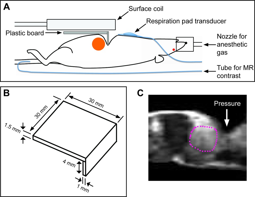
Figure 2. Schematics in DCE-MRI of an orthotopic pancreatic tumor xenograft. (A) Schematic of a setup for DCE-MRI of an orthotopic pancreatic tumor xenograft (orange circular region). A catheter is inserted into the tail vein to infuse MR contrast agent. (B) Schematic of an orthogonally bent plastic board with dimensions. (C) T2W MR images (sagittal view) of a mouse bearing an orthothopic pancreatic tumor xenograft indicated with a dotted red circle, when an orthogonally bent plastic board was applied (indicated with a white arrow). Please click here to view a larger version of this figure.

Figure 3. DCE-MR images and paramatric maps of an orthotopic pancreatic tumor xenograft. (A) Contrast maps at 1 min before or at 5 and 40 min after MR contrast (gadoteridol) injection. (B) Contrast enhancement curves averaged in the tumor region (3 x 3 window) and paravertebral muscle region (9 x 9 window) indicated with 2 white squares, respectively, in Figure 3A. (C) Ktrans map. (D) kep map. Please click here to view a larger version of this figure.
