This study uses a dual-sided illumination light-sheet fluorescence microscopy (LSFM) technique combined with optical clearing to study the murine heart.
Light-sheet fluorescence microscopy has been widely used for rapid image acquisition with a high axial resolution from micrometer to millimeter scale. Traditional light-sheet techniques involve the use of a single illumination beam directed orthogonally at sample tissue. Images of large samples that are produced using a single illumination beam contain stripes or artifacts and suffer from a reduced resolution due to the scattering and absorption of light by the tissue. This study uses a dual-sided illumination beam and a simplified CLARITY optical clearing technique for the murine heart. These techniques allow for deeper imaging by removing lipids from the heart and produce a large field of imaging, greater than 10 x 10 x 10 mm3. As a result, this strategy enables us to quantify the ventricular dimensions, track the cardiac lineage, and localize the spatial distribution of cardiac-specific proteins and ion-channels from the post-natal to adult mouse hearts with sufficient contrast and resolution.
Light-sheet fluorescence microscopy was a technique first developed in 1903 and is used today as a method to study gene expression and also to produce 3-D or 4-D models of tissue samples1,2,3. This imaging method uses a thin sheet of light to illuminate a single plane of a sample so that only that plane is captured by the detector. The sample can then be moved in the axial-direction to capture each layer, one section at a time, and render a 3-D model after the post-processing of the acquired images4. However, due to the absorption and scattering of photons, LSFM has been limited to samples that are either a few microns thick or are optically transparent1.
The limitations of LSFM have led to extensive studies of organisms that have tissues that are optically transparent, such as the zebrafish. Studies involving cardiac development and differentiation are often conducted on zebrafish since there are conserved genes between humans and zebrafish5,6. Although these studies have led to advances in cardiac research related to cardiomyopathies6,7, there is still a need to conduct similar research on higher-level organisms such as mammals.
Mammalian cardiac tissue presents a challenge due to the thickness and opacity of the tissue, the absorption due to hemoglobin in red blood cells, and the striping that occurs due to single-sided illumination of the sample under traditional LSFM methods1,8. To compensate for these limitations, we proposed to use dual-sided illumination and a simplified version of the CLARITY technique9 combined with a refractive index matching solution (RIMS). Therefore, this system allows for the imaging of a sample that is greater than 10 x 10 x 10 mm3 while maintaining a good quality resolution in the axial and lateral planes8.
This system was first calibrated using fluorescent beads arranged in different configurations within the glass tubing. Then, the system was used to image post-natal and adult murine hearts. First, the post-natal mouse heart was imaged at 7 days (P7) to reveal the ventricular cavity, the thickness of the ventricular wall, the valve structures, and the presence of trabeculation. Secondly, a study was conducted to identify cells that would differentiate into cardiomyocytes by using a post-natal mouse heart at 1 day (P1) with Cre-labeled cardiomyocytes and yellow fluorescent protein (YFP). Finally, adult mice at 7.5 months were imaged to observe the presence of renal outer medullary potassium (ROMK) channels after gene therapy8.
All the procedures involving the use of animals have been approved by the Institutional Review Committees (IACUC) at the University of California, Los Angeles, California.
1. Imaging System Setup
Note: See Figure 1 and Figure 2.
2. Imaging System Calibration
3. Sample Preparation
4. Adult Heart Imaging
The technique described here used a dual-sided illumination beam combined with an optical clearing of mouse heart tissue samples to achieve a deeper imaging depth and a larger imaging volume with sufficient imaging resolution (Figure 1). To calibrate the system, fluorescent beads were placed inside the glass tubing and within the imaging system. The beads were then imaged and visualized in the x-y plane, y-z plane, and in the x-z plane, to obtain the point spread function (PSF) for the system8. The system calibration produced a resolution in the lateral direction of dlateral ≈ 2.8 µm and a resolution in the axial directions of dXZ ≈ 17.4 µm and dYZ ≈ 17.9 µm when using glycerol as the embedding medium8.
After the fluorescent beads were used for calibration, the imaging system was used to image mouse hearts at 1 day and at 7 days post-natal (Figure 2)8. The ventricular cavity dimensions, heart valves, and ventricular trabeculation are evident in these images of the mouse heart. To study a cardiomyocyte differentiation within the heart, heterozygous knockin mice with Cre-labeled cardiomyocytes were imaged post-natal. The images of the Cre-labeled cardiomyocytes are in each plane direction along with a 3-D rendering of the heart8 (Figure 3).
In addition to studying post-natal mice, the imaging system was also used to study the adult mouse heart. Initially, gene therapy was used to introduce GFP-tagged ROMK channels into the mouse heart. At 7.5 months, the mouse heart was imaged and these ROMK channels were visualized in the ventricular wall (Figure 4). This step within the procedure illustrates the ability of this system to image large samples and distinguish structures not seen with histological staining within the heart. Figure 4 shows the ROMK channels from each plane direction along with a 3-D rendering of the heart8.
Figure 1: Schematic diagram of the light-sheet fluorescence microscopy system. This is a 3-dimensional rendering of the light sheet fluorescence microscopy system, including the distances between the components and the focal lengths of the components used in the system. This figure has been modified from Yichen Ding et al.8. Please click here to view a larger version of this figure.
Figure 2: Top view of light sheet microscopy system with detection. This is the top view of the dual-sided illumination microscopy system where the darkened blue region represents the beam at each location within the system. Included is a 2-dimensional enlarged section that shows one of the regions consisting of the achromatic doublets and illumination objective. This enlarged section also shows the principle of light sheet formation and how the beams are combined to form a dual-sided illumination. This figure has been modified from Yichen Ding et al.8. Please click here to view a larger version of this figure.
Figure 3: 3-D images of post-natal murine heart. (a) This panel shows an image of the post-natal mouse heart at day 7 showing the ventricle cavity. (b) This panel shows an image of the post-natal mouse heart at day 7 showing the thickness of the ventricle wall. (c) This panel shows the image of the post-natal mouse heart at day 1 showing the location of the valve structures. (d) This image shows an enlarged section from the post-natal mouse heart at day 1 showing the pectinate muscle located in the atrium of the heart. (e) This image shows trabeculation in the ventricle within the post-natal mouse heart at day 1. Scale bar: 1 mm. The red-circled region within the inset shows two translucent mouse hearts after undergoing the CLARITY technique and being inserted into the borosilicate glass tubing. This figure has been modified from Yichen Ding et al.8. Please click here to view a larger version of this figure.
Figure 4: Images of the Cre-labeled cardiomyocytes in a post-natal mouse heart from each plane. The P1 mouse heart was imaged with Cre-labeled cardiomyocytes in each cross-sectional plane: (a) XY, (b) YZ, and (c) XZ. These cross-sectional images show the presence of Cre-labeled cardiomyocytes in the atrium and the ventricle of the P1 mouse. (d) The images were also merged to create a 3-D rendering of the heart. The red-circled region within the inset shows two translucent mouse hearts after undergoing the CLARITY technique and being inserted into the borosilicate glass tubing. Scale bar: 1 mm. This figure has been modified from Yichen Ding et al.8. Please click here to view a larger version of this figure.
Figure 5: Images of the adult mouse heart showing the ROMK channels from each plane. The mouse heart was imaged at 7.5 months of age to show the presence and location of ROMK channels. The heart was imaged in each cross-sectional plane: (a) XY, (b) YZ, and (c) XZ. The gray scale value indicates the optical intensity within the image, with the lighter regions corresponding to more light intensity and the darker regions corresponding to less light intensity. (d) The images were also merged to create a 3-D rendering of the heart, and the original grayscaled images were pseudo-colored so that the ROMK channels appear green. The red-circled region within the inset shows a single translucent adult mouse heart after undergoing the CLARITY technique and being inserted into the borosilicate glass tubing. Scale bar: 1 mm. This figure has been modified from Yichen Ding et al.8. Please click here to view a larger version of this figure.
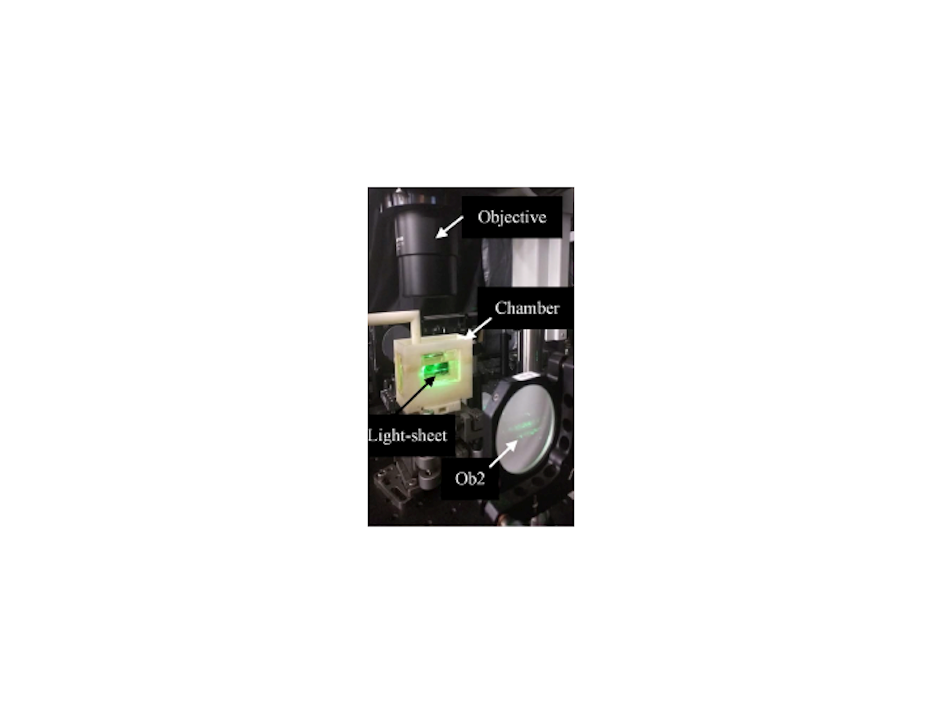
Supplemental Figure 1: Image of the ABS chamber with the borosilicate tubing within the light sheet of the imaging system. Please click here to download this file.
The LSFM system and technique described here utilizes a dual-sided illumination beam combined with an optical clearing to image the mouse heart at post-natal and adult stages of its development. A traditional single illumination beam suffers from photon scattering and absorption through thicker and larger tissue samples1,3. The dual-sided beams provide a more even illumination of the sample, thereby minimizing the effect of striping and other artifacts that are often seen in images produced by a one-sided illumination. This technique also increases the size of the sample that can be imaged by adjusting the position of the dual Gaussian beams8. The maximum separation of these beams allows for imaging a tissue sample several millimeters in size, such as the adult mouse heart, whereas a single illumination limited the size of the sample to smaller hearts such as the hearts of zebrafish and Drosophila.
Fluorescence imaging in tissues that are not optically transparent is more challenging since light scattering may not be uniform throughout the tissue sample. This limits the depth to which a sample can be imaged and the effectiveness of fluorescence imaging. For this reason, it is common to use organisms such as the zebrafish, which naturally have optically transparent tissue5. However, to perform fluorescence imaging studies on organisms that do not naturally have optically transparent skin, there are different optical clearing techniques that have been developed to remove the coloration of the tissue. One such optical clearing technique is CLARITY, where the tissue is placed in a hydrogel, then incubated for a long period of time, and finally placed in a clearing solution that reduces the amount of reflection and refraction between the sample and other media1,9. In this study, this technique was modified to reduce the amount of clearing reagent used and did not require the addition of vacuum pumps or nitrogen gas8.
Although this technique provides an improved image resolution for thicker opaque samples, there are limitations to this strategy that could be improved in future studies. Additional studies have been conducted with Bessel beams and a two-photon nonlinear excitation to provide a better resolution at sufficient imaging depths15,16. These methods have currently been used to study smaller cardiac models but could be adapted to study the dynamic processes within a developing embryo. Also, changes to the CLARITY method for larger tissue samples could improve the effectiveness of fluorescent imaging by reducing the number of fluorophores that can potentially be lost during the clearing process8. Recently, we have integrated this LSFM system with virtual reality to elucidate developmental cardiac mechanics17.
The authors have nothing to disclose.
The authors would like to express gratitude to Thao Nguyen and Atsushi Nakano from UCLA for providing the mice sample to image. This study was supported by grants NIH HL118650 (to Tzung K. Hsiai), HL083015 (to Tzung K. Hsiai), HD069305 (to N. C. Chi and Tzung K. Hsiai.), HL111437 (to Tzung K. Hsiai and N. C. Chi), HL129727 (to Tzung K. Hsiai), and University of Texas System STARS funding (to Juhyun Lee).
CW Laser | Laserglow Technologies | LMM-GBV1-PF3-00300-05 | Excitation of fluorophores |
Neutral density filter | Thorlabs | NDC-50C-4M | Controls amount of light entering system |
Achromatic beam expander | Thorlabs | GBE05-A | Expands the beam of light |
Mechanical slit | Thorlabs | VA100C | Controls width of beam |
Beam splitter | Thorlabs | BS013 | Forms dual-illumination beam |
Stereo microscope with 1X objective lense | Olympus | MVX10 | Used for observation of sample |
ORCA-Flash4.0 LT sCMOS camera | Hamamatsu Photonics | C11440-42U | Used to capture Images |
Acrylonitrile butadiene styrene (ABS) | Stratasys | uPrint | Material used to 3-D print a sample holder |
Fluorescent polystyrene beads | Spherotech Inc | PP-05-10 | Used for imaging system calibration |
Borosilicate glass tubing | Corning | Pyrex 7740 | Tubing for sample embedding |
Glycerol | Fisher Scientific | BP229-4 | Fill for sample chamber |
Phosphate-buffered saline | Fisher Scientific | BP39920 | Rinse solution for mouse hearts |
Paraformaldehyde | Electron microscopy sciences | RT-15700 | First incubation solution |
Acrylamide | Wako Chemicals | AAL-107 | Mixed with 2,2'-Azobis dihydrochloride for second incubation solution for mouse hearts |
2,2'-Azobis dihydrochloride | Wako Chemicals | VA-044 | Mixed with Acrylamide for second incubation solution for mouse hearts |
Sodium dodecyl sulfate | Sigma Aldrich | 71725 | Mixed with Boric acid for third incubtion solution for mouse hearts |
Boric acid | Fischer Scientific | A74-1 | Mixed with Sodium dodecyl sulfate for third incubtion solution for mouse hearts |
Sigma D2158 | Sigma Aldrich | D2158 | Mixed with PB, Tween-20, and Sodium azide as a refractive index matching solution |
Tween-20 | Sigma Aldrich | 11332465001 | Mixed with Sigma D2158, PB, and Sodium azide as a refractive index matching solution |
Sodium azide | Sigma Aldrich | S2002 | Mixed with Sigma D2158, PB, and Tween-20 as a refractive index matching solution |
Adeno-associated virus vector 9 with a cardiac-specific Troponin T promoter tagged with GFP | Vector Biolabs | VB2045 | Expresses GFP when bound to ROMK |
DC Servo Motor Actuator | Thorlabs | Z825B | Used for movement of sample in axial direction within light sheet |
K-Cube Brushed DC Servo Motor Controller | Thorlabs | KDC101 | Connects to motor actuator and controls movement of the actuator |
Amira | FEI Software | N/A | Visualization software for producing 2-D and 3-D images |